Podcast
Questions and Answers
What cellular process directly results in the production of sperm and egg cells during gametogenesis?
What cellular process directly results in the production of sperm and egg cells during gametogenesis?
- Mitosis I
- Mitosis II
- Meiosis I
- Meiosis II (correct)
Primordial germ cells (PGCs) undergo a series of mitotic divisions to form a large number of spermatogonia; depending upon their appearance, what types of spermatogonia are distinguished?
Primordial germ cells (PGCs) undergo a series of mitotic divisions to form a large number of spermatogonia; depending upon their appearance, what types of spermatogonia are distinguished?
- Dark type A, light type A, and type B spermatogonia (correct)
- Dark type A, Dark type B, and type C spermatogonia
- Light type A, light type B, and type C spermatogonia
- Types A, B and C spermatogonia
During spermatogenesis, what type of spermatogonium undergoes mitotic division to form two primary spermatocytes, which are also known as the largest germ cells?
During spermatogenesis, what type of spermatogonium undergoes mitotic division to form two primary spermatocytes, which are also known as the largest germ cells?
- Dark type A spermatogonium
- Light type A spermatogonium
- Type B spermatogonium (correct)
- Type A spermatogonium
Which hormone, secreted by Leydig cells, is responsible for spermatogenesis?
Which hormone, secreted by Leydig cells, is responsible for spermatogenesis?
What is the primary function of the axial filament in a spermatozoon?
What is the primary function of the axial filament in a spermatozoon?
What is the role of the acrosomal cap during fertilization?
What is the role of the acrosomal cap during fertilization?
Which of the following accurately describes the duration of spermatogenesis?
Which of the following accurately describes the duration of spermatogenesis?
The secondary oocyte completes its second meiotic division:
The secondary oocyte completes its second meiotic division:
What is the primary role of the zona pellucida that surrounds the oocyte?
What is the primary role of the zona pellucida that surrounds the oocyte?
What is the term for the cells of cumulus oophoricus that become loosened by the accumulation of intercellular fluid around the time of ovulation?
What is the term for the cells of cumulus oophoricus that become loosened by the accumulation of intercellular fluid around the time of ovulation?
Flashcards
Spermatogenesis
Spermatogenesis
The process of forming spermatozoa from primordial germ cells (PGCs) in the seminiferous tubules of the testes.
Spermatocytogenesis
Spermatocytogenesis
PGCs divide by mitosis to create dark type A spermatogonia, acting as stem cells. These further divide, eventually forming primary spermatocytes.
Spermiogenesis
Spermiogenesis
Transformation of circular spermatids into elongated spermatozoa (sperm).
Oogenesis
Oogenesis
Signup and view all the flashcards
Ovulation
Ovulation
Signup and view all the flashcards
Ovarian Follicle
Ovarian Follicle
Signup and view all the flashcards
Zona Pellucida
Zona Pellucida
Signup and view all the flashcards
Meiotic Divisions
Meiotic Divisions
Signup and view all the flashcards
Sperm Products
Sperm Products
Signup and view all the flashcards
Spermatozoon Structure
Spermatozoon Structure
Signup and view all the flashcards
Study Notes
- Gametogenesis is the process of gamete formation, producing sperm and egg through Meiosis II.
Types of Gametogenesis:
- Spermatogenesis
- Oogenesis
Spermatogenesis
- Spermatogenesis is the formation of spermatozoa from primordial germ cells (PGCs)/spermatogonia located in the seminiferous tubules of the testes.
- PGCs remain dormant in the seminiferous tubules until puberty, when they undergo divisions to form spermatogonia.
- Spermatogenesis occurs in the seminiferous tubules of the testes.
- This process starts after puberty and continues throughout old age.
- The duration is 64-74 days.
Stages of Spermatogenesis:
- Spermatocytogenesis involves PGCs undergoing a series of mitotic divisions to form numerous spermatogonia.
- Three types of spermatogonia distinguished: dark type A, light type A, and type B.
Spermatocytosis
- PGCs divide by mitosis to form dark type A spermatogonia, which act as stem cells.
- Each dark type A spermatogonium undergoes mitosis to form one dark A spermatogonium and one light type A spermatogonium.
- Dark type A spermatogonia are reserved for the repetition of the cycle.
- Light type A spermatogonia undergo mitotic division to form two dark type B spermatogonia.
- Type B spermatogonium undergoes mitotic division to form two primary spermatocytes, which are the largest germ cells.
Meiotic Divisions
- Primary spermatocytes undergo the first meiotic division (reductional division) to form two secondary spermatocytes, each with a haploid number of chromosomes.
- Secondary spermatocytes immediately undergo the second meiotic division (mitotic division) to form two spermatids, each with a haploid number of chromosomes.
- A primary spermatocyte produces four haploid spermatids through meiotic division.
- Spermatids are small cells, about half the size of secondary spermatocytes, with round, darkly stained nuclei located close to the lumen of the seminiferous tubule.
- Each spermatid changes to become spermatozoon or sperm, in a transformation called spermiogenesis.
Hormone Regulation
- Testosterone, secreted by Leydig cells, is the hormone responsible for spermatogenesis.
Spermatozoa Formation
- One primary spermatocyte forms four spermatozoa: two with 22 autosomes and one X chromosome each and two with 22 autosomes and one Y chromosome each.
Spermiogenesis
- Spermiogenesis transforms spermatids into mature spermatozoa.
- During spermiogenesis, the spermatid, a circular cell with a nucleus, Golgi apparatus, centrosome, and mitochondria, transforms into a spermatozoon.
- Nuclear material (chromatin) condenses, and the nucleus moves to one pole to form the head of the spermatozoon.
- The Golgi apparatus forms the acrosomal cap that covers the anterior two-thirds of the nucleus.
- The centrosome divides into two centrioles.
- One centriole moves to the posterior end of the nucleus to occupy the neck region, giving rise to the axial filament.
- The other centriole moves away and becomes ring-shaped, forming an annulus around the distal end of the middle piece.
- The part of the axial filament between the neck and annulus is surrounded by mitochondria to form the middle piece.
- The remaining axial filament elongates to form the principal and end pieces or tail.
- Most of the cytoplasm is shed off, but the cell membrane covers the entire spermatozoon.
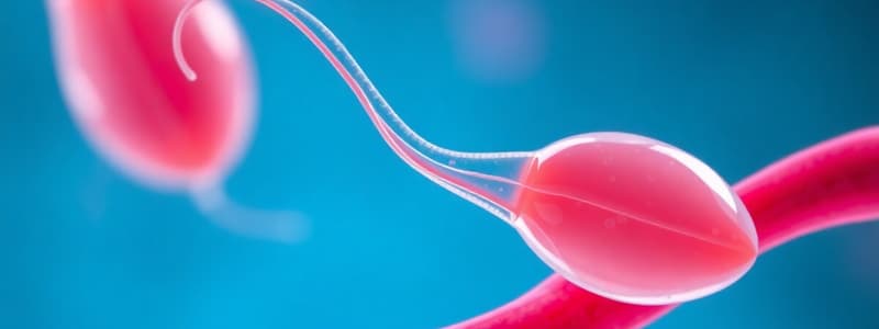
Spermatozoon Structure
- The spermatozoon (50 μm in length) has a head, neck, and tail.
- The tail is further divided into the middle piece, principal piece, and end piece; it forms four-fifths of the total length.
Spermatozoon Head
- The head is shaped like a spearhead and mainly contains a nucleus with condensed chromatin (DNA).
- The anterior two-thirds of the nucleus is covered by an acrosomal cap containing enzymes like hyaluronidase and acrosin.
Spermatozoon Neck
- The neck is narrow and contains a funnel-shaped basal plate and a centriole that gives rise to the axial filament extending through the tail.
Spermatozoon Tail
- The tail has three parts: middle piece, principal piece, and end piece.
- The middle piece contains the axial filament surrounded by a spirally arranged mitochondrial sheath, with the annulus at its distal end.
- The principal piece comprises the axial filament covered by seven outer dense fibers.
- The end piece consists only of the axial filament.
- Axial filament is responsible for the movements of the spermatozoon, and mitochondria supply energy for these movements.
Clinical Correlations: Abnormal Sperms
- Abnormal sperms are more common, has two parts head and tail.
Types of Abnormalities
- Morphological abnormalities include abnormal head and tail shapes two heads or tails, giant or dwarf sperms, and joined sperms.
- For potential fertility, 50% of sperms should be motile after 2 hours of ejaculation, and some should be motile after 24 hours.
- Genetic abnormalities involve sperm having abnormal chromosomal content and are rare compared to oocytes.
Oogenesis
- Oogenesis is the process of forming female gametes (oocytes) from PGCs.
- Oogenesis begins long before birth in the cortex of the ovary and before birth PGCs divide by mitosis to form numerous oogonia.
- Each oogonium enlarges into a primary oocyte, which enters the prophase of the first meiotic division before birth and is arrested due to an oocyte maturation inhibitor (OMI) secreted by follicular cells.
At Puberty
- At puberty, in each ovarian cycle, 5-50 primary oocytes resume the first meiotic division just before ovulation.
- This division forms two daughter cells (secondary oocyte and first polar body), each with a haploid number of chromosomes.
- The first meiotic division is unequal; most cytoplasm goes to the secondary oocyte, while the other forms the first polar body.
After Fertilization
- The secondary oocyte begins the second meiotic division at ovulation, completing it only after sperm penetration (fertilization).
- The second meiotic division is also unequal, forming an ovum (mature oocyte) with most of the cytoplasm and a second polar body with very little cytoplasm.
- One primary oocyte forms one ovum with 22 autosomes and one X chromosome and three polar bodies each with 22 autosomes and one X chromosome.
- The primary oocyte begins the first meiotic division before birth and completes it at puberty just before ovulation.
- Primary oocytes are not formed after birth and the secondary oocyte is in the metaphase stage of the second meiotic division at ovulation, completing it only if fertilization occurs.
- At birth, the ovary contains about two million germ cells.
- By puberty, around 40,000 oocytes remain, as most degenerate and oogenesis is accompanied by the development and growth of ovarian follicles.
Development of Ovarian Follicles
- The oogonium is covered by a single layer of flat epithelial (follicular) cells from the stromal cells of the ovary or from the surface epithelium to form a primordial follicle.
- The oogonium within the follicle contains a single, large nucleus with a prominent eccentric nucleolus.
Steps of Forming Ovarian Follicles
- Flattened follicular cells become columnar, forming a unilaminar primary follicle.
- Follicular cells proliferate to form several layers of membrana granulosa, now called granulosa cells.
- The primary oocyte and granulosa cells secrete a glycoprotein substance forming the zona pellucida, a thick membrane between the cells.
- Granulosa cells rest on a basement membrane separating them from stromal cells, forming a multilaminar (maturing) primary follicle.
- Small fluid-filled cavities appear between follicular cells, fusing to form a large cavity (antral cavity/antrum), and the follicle becomes a secondary (vesicular) follicle.
- The antrum increases in size, pushing the oocyte to one side.
- Granulosa cells surrounding the oocyte are called cumulus oophoricus (or cumulus ovaricus).
- Granulosa cells attaching the oocyte to the follicle wall are called discus proligerus.
- Stromal cells around the granulosa cells condense to form the theca interna (theca = covering).
- Fibrous tissues outside the theca interna condense to form the theca externa, making the ovarian follicle fully matured.
Ovulation
- Ovulation involves shedding an ovum from the ovary.
- The Graafian follicle enlarges, reaching the ovary surface and forming a bulge.
- The theca and stroma on the follicle side become thin and an avascular area (stigma) appears at the most convex superficial position.
- Cells of cumulus oophoricus loosen with intercellular fluid accumulation and ultimately the follicle ruptures when the ovum is released from the ovary (ovulation).
- The expelled secondary oocyte is surrounded by the zona pellucida and radially arranged follicular cells termed corona radiata.
- The fimbriated end of the uterine tube picks up the oocyte and puts it into the lumen of the uterine tube.
- The empty Graafian follicle becomes the corpus luteum which lasts for 10–12 days if the ovum is not fertilized, and 2-3 months if fertilization and pregnancy occurs.
- Graafian follicle cells secrete estrogen, while corpus luteum cells secrete progesterone.
Structure of Secondary Oocyte
- The secondary oocyte is large (more than 100 µm in diameter) and sheds from the ovary.
- It undergoes a second meiotic division to shed the second polar body.
- No nucleus is seen as the nuclear membrane dissolves for the second meiotic division.
- Spindle and chromosomes attach to the equatorial plane (metaphase stage).
- The oocyte is surrounded by the zona pellucida which is then surrounded by corona radiata cells.
- Perivitelline space is present between the cell membrane (vitelline membrane) and zona pellucida; it contains the first polar body from the first meiotic division.
Clinical Correlations: Oocytes
- Oocytes may be binucleated or trinucleated; but they usually degenerate before maturity.
- Oocytes with abnormal chromosomal contents may occur due to nondisjunction in meiosis I or II, resulting in oocytes with 24 or 22 chromosomes instead of 23.
- An oocyte with 24 chromosomes fertilized by a normal sperm (23 chromosomes) yields a zygote with 47 chromosomes (trisomy).
- Trisomy 21 (Down's syndrome) is the most common trisomy and an ovum with 22 chromosomes fertilized by a normal sperm (23 chromosomes) yields a zygote with 45 chromosomes (monosomy), such as Turner's syndrome (45, Xo).
Studying That Suits You
Use AI to generate personalized quizzes and flashcards to suit your learning preferences.