Podcast
Questions and Answers
Which muscle is responsible for elevating the upper eyelid?
Which muscle is responsible for elevating the upper eyelid?
- Inferior oblique
- Medial rectus
- Levator palpebrae superioris (correct)
- Superior rectus
What cranial nerve innervates the extraocular muscles involved in eye movement?
What cranial nerve innervates the extraocular muscles involved in eye movement?
- CN III - Oculomotor nerve (correct)
- CN II - Optic nerve
- CN VI - Abducens nerve
- CN IV - Trochlear nerve
Which extraocular muscle works primarily to depress the eyeball?
Which extraocular muscle works primarily to depress the eyeball?
- Superior rectus
- Superior oblique
- Lateral rectus
- Inferior rectus (correct)
What structure is part of the accessory visual system that aids in tear production?
What structure is part of the accessory visual system that aids in tear production?
Which muscle is the primary mover for adduction of the eyeball?
Which muscle is the primary mover for adduction of the eyeball?
Which extraocular muscle is primarily responsible for elevating the eye?
Which extraocular muscle is primarily responsible for elevating the eye?
What nerve provides motor innervation to the majority of the extraocular muscles?
What nerve provides motor innervation to the majority of the extraocular muscles?
Which cranial nerve is responsible for the sensory innervation of the cornea?
Which cranial nerve is responsible for the sensory innervation of the cornea?
When testing eye movements, which action is primarily associated with the lateral rectus muscle?
When testing eye movements, which action is primarily associated with the lateral rectus muscle?
What is the primary function of the conjunctiva?
What is the primary function of the conjunctiva?
What is the primary function of the eyelids?
What is the primary function of the eyelids?
Which of the following components of the eyelid helps raise the upper eyelid?
Which of the following components of the eyelid helps raise the upper eyelid?
What role does the conjunctiva play in eye health?
What role does the conjunctiva play in eye health?
Which structure is responsible for producing lacrimal fluid?
Which structure is responsible for producing lacrimal fluid?
How does tear drainage occur after being produced by the lacrimal gland?
How does tear drainage occur after being produced by the lacrimal gland?
What is the purpose of goblet cells found in the conjunctiva?
What is the purpose of goblet cells found in the conjunctiva?
What is the palpebral fissure?
What is the palpebral fissure?
Which cranial nerve is primarily responsible for the secretion of lacrimal fluid?
Which cranial nerve is primarily responsible for the secretion of lacrimal fluid?
What type of visual field defect is characterized by loss of vision in the outer half of both visual fields?
What type of visual field defect is characterized by loss of vision in the outer half of both visual fields?
Which condition results in the loss of vision in the same half of both visual fields?
Which condition results in the loss of vision in the same half of both visual fields?
What is the sequence of structures that light passes through in the visual pathway starting from the retina?
What is the sequence of structures that light passes through in the visual pathway starting from the retina?
What visual field defect is characterized by loss of vision in the upper quadrant of the visual field in one eye?
What visual field defect is characterized by loss of vision in the upper quadrant of the visual field in one eye?
What condition allows patients to maintain central vision while peripheral vision is significantly reduced?
What condition allows patients to maintain central vision while peripheral vision is significantly reduced?
Which structure is responsible for synapsing the optic tract signals before they reach the visual cortex?
Which structure is responsible for synapsing the optic tract signals before they reach the visual cortex?
What anatomical structure is directly involved in the processing of visual information that can lead to visual field defects?
What anatomical structure is directly involved in the processing of visual information that can lead to visual field defects?
What visual pathway deficit is typically associated with damage to the optic chiasm?
What visual pathway deficit is typically associated with damage to the optic chiasm?
What is the term for a visual defect resulting from damage along the optic tract?
What is the term for a visual defect resulting from damage along the optic tract?
Which part of the visual pathway is most commonly associated with bitemporal hemianopia?
Which part of the visual pathway is most commonly associated with bitemporal hemianopia?
Which visual pathway structure is directly responsible for carrying visual information to the occipital lobe?
Which visual pathway structure is directly responsible for carrying visual information to the occipital lobe?
What is the visual outcome in one eye if the optic nerve is severed?
What is the visual outcome in one eye if the optic nerve is severed?
Which part of the retina corresponds to the temporal visual fields?
Which part of the retina corresponds to the temporal visual fields?
Which term describes the visual field defect that affects only a quarter of the visual field?
Which term describes the visual field defect that affects only a quarter of the visual field?
What condition results from bilateral damage to the optic nerves?
What condition results from bilateral damage to the optic nerves?
The optic tract carries visual signals to which of the following areas of the brain?
The optic tract carries visual signals to which of the following areas of the brain?
Which muscle is primarily responsible for rotating the eye outward?
Which muscle is primarily responsible for rotating the eye outward?
What is the main function of the oblique muscles of the eye?
What is the main function of the oblique muscles of the eye?
What is the effect of damage to the inferior rectus muscle?
What is the effect of damage to the inferior rectus muscle?
Which cranial nerve is responsible for controlling the movements of the superior oblique muscle?
Which cranial nerve is responsible for controlling the movements of the superior oblique muscle?
What is the primary action of the medial rectus muscle?
What is the primary action of the medial rectus muscle?
What is the primary action of the superior oblique muscle?
What is the primary action of the superior oblique muscle?
Which cranial nerve innervates the lateral rectus muscle?
Which cranial nerve innervates the lateral rectus muscle?
Which action is primarily associated with the lateral rectus muscle?
Which action is primarily associated with the lateral rectus muscle?
What action is performed by the inferior oblique muscle?
What action is performed by the inferior oblique muscle?
Which muscle is not responsible for intorting the eye?
Which muscle is not responsible for intorting the eye?
Which extraocular muscle is primarily responsible for depression of the eyeball?
Which extraocular muscle is primarily responsible for depression of the eyeball?
What is the main role of the superior rectus muscle in eye movement?
What is the main role of the superior rectus muscle in eye movement?
Which extraocular muscle is involved in adduction of the eye?
Which extraocular muscle is involved in adduction of the eye?
What is the primary function of the inferior oblique muscle?
What is the primary function of the inferior oblique muscle?
Which muscle works mainly to depress the eyeball?
Which muscle works mainly to depress the eyeball?
Which of the following muscles is primarily involved in gaze directed upward?
Which of the following muscles is primarily involved in gaze directed upward?
Which extraocular muscle is primarily responsible for adducting the eyeball?
Which extraocular muscle is primarily responsible for adducting the eyeball?
Which muscle aids in looking downward and laterally?
Which muscle aids in looking downward and laterally?
Which extraocular muscle primarily functions to abduct the eye?
Which extraocular muscle primarily functions to abduct the eye?
Which cranial nerve is responsible for the action of the lateral rectus muscle?
Which cranial nerve is responsible for the action of the lateral rectus muscle?
What action is primarily associated with the inferior rectus muscle?
What action is primarily associated with the inferior rectus muscle?
Which muscle is NOT involved in internal rotation of the eye?
Which muscle is NOT involved in internal rotation of the eye?
Which extraocular muscle is primarily responsible for eye elevation?
Which extraocular muscle is primarily responsible for eye elevation?
What muscle is responsible for adduction of the eyeball?
What muscle is responsible for adduction of the eyeball?
How do the oblique muscles of the eye primarily affect eye movement?
How do the oblique muscles of the eye primarily affect eye movement?
Flashcards
Extraocular Muscles
Extraocular Muscles
Muscles that control eye movement within the orbit.
Superior Rectus Muscle
Superior Rectus Muscle
An extraocular muscle that elevates the eye.
Levator Palpebrae Superioris
Levator Palpebrae Superioris
Muscle responsible for raising the upper eyelid.
Oculomotor Nerve (CN III)
Oculomotor Nerve (CN III)
Signup and view all the flashcards
Eyelids
Eyelids
Signup and view all the flashcards
Conjunctiva's role
Conjunctiva's role
Signup and view all the flashcards
Lacrimal gland function
Lacrimal gland function
Signup and view all the flashcards
Extraocular muscles and their actions
Extraocular muscles and their actions
Signup and view all the flashcards
Autonomic innervation of the eye
Autonomic innervation of the eye
Signup and view all the flashcards
Intraocular fluid: formation and flow
Intraocular fluid: formation and flow
Signup and view all the flashcards
Eyelid Function
Eyelid Function
Signup and view all the flashcards
Palpebral Fissure
Palpebral Fissure
Signup and view all the flashcards
Conjunctiva
Conjunctiva
Signup and view all the flashcards
Conjunctiva Function
Conjunctiva Function
Signup and view all the flashcards
Conjunctival Fornices
Conjunctival Fornices
Signup and view all the flashcards
Lacrimal Apparatus Function
Lacrimal Apparatus Function
Signup and view all the flashcards
Lacrimal Gland
Lacrimal Gland
Signup and view all the flashcards
Tear Flow
Tear Flow
Signup and view all the flashcards
Musculos Extraocular
Musculos Extraocular
Signup and view all the flashcards
Actiones del Musculos Extraocular
Actiones del Musculos Extraocular
Signup and view all the flashcards
Le Recto Superior
Le Recto Superior
Signup and view all the flashcards
Le Recto Inferior
Le Recto Inferior
Signup and view all the flashcards
Le Recto Medial
Le Recto Medial
Signup and view all the flashcards
Le Recto Lateral
Le Recto Lateral
Signup and view all the flashcards
Le Oblique Superior
Le Oblique Superior
Signup and view all the flashcards
Le Oblique Inferior
Le Oblique Inferior
Signup and view all the flashcards
Nervo Cranial III (Oculomotor)
Nervo Cranial III (Oculomotor)
Signup and view all the flashcards
Nervo Cranial IV (Trochlear)
Nervo Cranial IV (Trochlear)
Signup and view all the flashcards
Nervo Cranial VI (Abducens)
Nervo Cranial VI (Abducens)
Signup and view all the flashcards
Levation del Palpebra Superior
Levation del Palpebra Superior
Signup and view all the flashcards
Teste del Musculos Extraocular
Teste del Musculos Extraocular
Signup and view all the flashcards
Teste del Movimentos Ocular (MEOMS)
Teste del Movimentos Ocular (MEOMS)
Signup and view all the flashcards
Isolate le Musculos Extraocular
Isolate le Musculos Extraocular
Signup and view all the flashcards
Teste del Recto Medial
Teste del Recto Medial
Signup and view all the flashcards
Teste del Recto Lateral
Teste del Recto Lateral
Signup and view all the flashcards
Teste del Recto Superior
Teste del Recto Superior
Signup and view all the flashcards
Teste del Recto Inferior
Teste del Recto Inferior
Signup and view all the flashcards
Teste del Oblique Superior
Teste del Oblique Superior
Signup and view all the flashcards
Teste del Oblique Inferior
Teste del Oblique Inferior
Signup and view all the flashcards
Defectos del Musculos Extraocular
Defectos del Musculos Extraocular
Signup and view all the flashcards
Strabismo
Strabismo
Signup and view all the flashcards
Diplopia
Diplopia
Signup and view all the flashcards
Traumatismos del Oculo
Traumatismos del Oculo
Signup and view all the flashcards
Malattia de Graves
Malattia de Graves
Signup and view all the flashcards
Paralisia del Nervos Cranial
Paralisia del Nervos Cranial
Signup and view all the flashcards
Bitemporal Hemianopia
Bitemporal Hemianopia
Signup and view all the flashcards
Homonymous Hemianopia
Homonymous Hemianopia
Signup and view all the flashcards
Superior Quadrantanopia
Superior Quadrantanopia
Signup and view all the flashcards
Macular Sparing
Macular Sparing
Signup and view all the flashcards
Optic Chiasm
Optic Chiasm
Signup and view all the flashcards
Optic Nerve
Optic Nerve
Signup and view all the flashcards
Optic Tract
Optic Tract
Signup and view all the flashcards
Optic Radiations
Optic Radiations
Signup and view all the flashcards
What is the role of the optic chiasm?
What is the role of the optic chiasm?
Signup and view all the flashcards
What is the purpose of the lateral geniculate nucleus (LGN)?
What is the purpose of the lateral geniculate nucleus (LGN)?
Signup and view all the flashcards
What is the function of the visual cortex?
What is the function of the visual cortex?
Signup and view all the flashcards
Visual Field Deficits
Visual Field Deficits
Signup and view all the flashcards
Optic Nerve Damage
Optic Nerve Damage
Signup and view all the flashcards
Optic Chiasm Damage
Optic Chiasm Damage
Signup and view all the flashcards
Optic Tract Damage
Optic Tract Damage
Signup and view all the flashcards
Superior Rectus
Superior Rectus
Signup and view all the flashcards
Inferior Rectus
Inferior Rectus
Signup and view all the flashcards
Lateral Rectus
Lateral Rectus
Signup and view all the flashcards
Medial Rectus
Medial Rectus
Signup and view all the flashcards
Inferior Oblique Muscle
Inferior Oblique Muscle
Signup and view all the flashcards
Medial Rectus Muscle
Medial Rectus Muscle
Signup and view all the flashcards
Lateral Rectus Muscle
Lateral Rectus Muscle
Signup and view all the flashcards
Which cranial nerve controls the superior oblique muscle?
Which cranial nerve controls the superior oblique muscle?
Signup and view all the flashcards
What is the action of the lateral rectus muscle?
What is the action of the lateral rectus muscle?
Signup and view all the flashcards
What is the action of the medial rectus muscle?
What is the action of the medial rectus muscle?
Signup and view all the flashcards
What nerve innervates the lateral rectus muscle?
What nerve innervates the lateral rectus muscle?
Signup and view all the flashcards
What action does the superior rectus muscle perform?
What action does the superior rectus muscle perform?
Signup and view all the flashcards
What is the function of the inferior oblique muscle?
What is the function of the inferior oblique muscle?
Signup and view all the flashcards
What is the function of the inferior rectus muscle?
What is the function of the inferior rectus muscle?
Signup and view all the flashcards
Inferior Oblique
Inferior Oblique
Signup and view all the flashcards
Study Notes
Special Senses: Vision and Eye
- The lecture objective is to identify microscopic and macroscopic structures involved in special senses, using virtual microscopy, 2-D, and 3-D images to link these to the larger nervous system.
Lecture Learning Objectives
- Recall the function and components of extraocular structures covered in the lecture, conjunctiva's role, and structures involved in tear production and flow.
- Link the extraocular muscles to their actions and identify their innervation, including which movements test each muscle/nerve.
- List the nerves providing innervation to the eye and extraocular structures, noting the type of innervation they carry (somatic sensory/motor).
- Describe the organization of the eyeball, including structural components of each layer and characteristics; list each when listed.
- Trace the autonomic innervation of the eye to associated muscles, linking it to the autonomic nervous system (responses and sympathetic pathway).
- Recall the spaces of the eye and describe the importance, formation, and flow of intraocular fluid.
- Draw out or write a narrative of the visual pathway, identifying visual fields and portion of retinal signals carried in each structure (optic nerve, chiasm, tract).
- Trace development of the eye focusing on the origin of each structure.
- Visually identify structures of the eye from histology.
Extraocular Anatomy
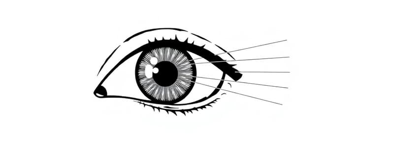
- The 7 Extraocular Muscles, positioning and actions are detailed in images.
- Levator palpebrae superioris raises the eyelid.
- Superior and Inferior oblique muscles act in rotation and depression/elevation
Primary Accessory Visual Structures
- Eyelids, conjunctiva (mucous membrane), Lacrimal apparatus, extraocular muscles, nerves (CNS)
Eyelid
- Function: protect, covers eye anteriorly, spreads lacrimal fluid, from protecting from injury and excess light.
- Forms a wall: between internal and external environments (superior/inferior tarsus, medial/lateral palpebral ligaments, Levator palpebrae superioris muscle and tendon, palpebral fissure).
Conjunctiva
- Transparent mucous membrane covering internal aspect of the eyelid, continuous with sclera.
- Function: contains goblet cells lubricating eye, contain blood vessels supplying sclera, innervation for foreign object identification.
- Superior/inferior conjunctival fornices are pockets formed by conjunctiva, conjunctival sac opens anteriorly at palpebral fissure.
Lacrimal Apparatus
- Function: produce lacrimal fluid (tears) to cleanse and lubricate the eye.
- Tear flow: Lacrimal gland produces tears, excretory ducts, lacrimal canaliculi (canals), lacrimal sac, nasolacrimal duct, and nasal cavity.
Extraocular Muscles (primary action)
- Superior rectus: primarily elevates, adducts, and medially rotates (intorsion).
- Medial rectus: adducts (toward midline).
- Inferior rectus: primarily depresses, adducts, and laterally rotates (extorsion).
- Lateral rectus: abducts (away from midline).
- Superior oblique: primarily medially rotates (intorsion), abducts, and depresses.
- Inferior oblique: primarily laterally rotates (extorsion), abducts, and elevates.
- Levator palpebrae superioris: elevates the superior eyelid.
Innervation
- Somatic motor innervation for extraocular muscles:
- CN III (oculomotor): superior, inferior, medial rectus, inferior oblique, and levator palpebrae superioris.
- CN IV (trochlear): superior oblique.
- CN VI (abducent): lateral rectus.
Somatic Sensory Innervation
- Ophthalmic nerve (V1): frontal nerve, somatic sensory for extraocular structures, conjunctiva and eyelids.
- Nasociliary nerve: somatic sensory to eye and nasal cavity.
- Lacrimal nerve: somatic sensory to lateral extraocular structures.
Autonomic Innervation
- Oculomotor nerve (III): parasympathetic innervation for pupil constriction and lens accommodation, via short ciliary nerves, with sympathetic innervation originating from superior cervical ganglion.
- Facial nerve (VII): parasympathetic motor to lacrimal gland, travels within ophthalmic branch to lacrimal nerve.
- Superior cervical ganglion: sympathetic motor to lacrimal gland from, travels with parasympathetic fibers.
Eye Anatomy
- Adipose cushion present within the orbit.
Organization of the Eyeball
- Three layers: fibrous tunic (sclera, cornea), vascular tunic (choroid, ciliary body, iris), inner layer (retina).
- Contains specific structures such as the optic nerve, optic disc, fovea centralis, lens and vitreous humor.
Fibrous Tunic - Shape and Resistance
- Sclera (posterior 5/6): nearly avascular, white outer layer.
- Cornea (anterior 1/6): completely avascular, receives nutrients from peripheral capillaries and lacrimal & aqueous humor; sensory innervation from ophthalmic branch of CN V.
Vascular Tunic
- Choroid: thin, highly vascularized layer.
- Ciliary Body: ring-like thickening with ciliary processes and muscles; secretes aqueous humor; controls lens thickness.
- Lens: transparent, suspended, in light path.
- Iris: colored part, continuous with ciliary body; contains pupil, sphincter pupillae, and dilator pupillae.
- Pupil: central aperture of the iris; diameter depends on light.
Inner Layer (Retina)
- Retina: sensory neural layer.
- Optic part: sensitive to light (neural and pigmented layers), contains photoreceptors (rods and cones).
- Optic disc: entrance for optic nerve, associated vessels, and blind spot.
- Macula lutea: small oval retinal area with photoreceptor cones—specialized for visual acuity.
- Fovea centralis: located at center of macula, area of most acute vision.
Spaces of the Eye and Humor Production
- Anterior cavity, filled with aqueous humor (Anterior chamber and Posterior chamber); Nutrients and glaucoma.
- Posterior cavity filled with vitreous humor.
Visual Pathway
- Light signals travel from the retina, through the optic nerve, optic chiasm, optic tract, lateral geniculate nucleus, optic radiations, to the occipital lobe's visual cortex, where vision is perceived. The retinal image is inverted. Nasal retinal fibers cross at the optic chiasm, while temporal fibers do not cross.
Development of the Eye
- Development begins in the fourth week of embryonic development, from the developing brain (optic vesicles) and ectoderm (lens).
- Optic vesicles become the retina, connecting to the brain via the optic stalk.
- Lens develops from overlying ectoderm.
- Sclera and choroid form from surrounding mesenchyme.
- Eyelids, conjunctiva, and cornea develop from surrounding mesenchyme and ectoderm.
- Primary vitreous body forms in optic cup.
Eye Histology
- Fibrous Tunic: cornea, sclera.
- Vascular Tunic: lens, iris, ciliary body (ciliary processes and muscles).
- Anterior cavity: contains aqueous humor (anterior chamber and posterior chamber).
- Posterior cavity: contains vitreous humor.
Studying That Suits You
Use AI to generate personalized quizzes and flashcards to suit your learning preferences.

