Podcast
Questions and Answers
What are the five special senses?
What are the five special senses?
- Touch, taste, sight, hearing, balance
- Smell, taste, sight, touch, hearing
- Smell, touch, sight, hearing, balance
- Smell, taste, sight, hearing, balance (correct)
The eyes contain 70% of all sensory receptors in the body.
The eyes contain 70% of all sensory receptors in the body.
True (A)
Which of the following is NOT an accessory structure of the eye?
Which of the following is NOT an accessory structure of the eye?
- Conjunctiva
- Extrinsic eye muscles
- Eyelids
- Retina (correct)
What is the function of the conjunctival membrane?
What is the function of the conjunctival membrane?
What do lacrimal glands produce?
What do lacrimal glands produce?
What is the function of the extrinsic eye muscles?
What is the function of the extrinsic eye muscles?
Which of the following is NOT a layer of the eyeball?
Which of the following is NOT a layer of the eyeball?
What is the function of the cornea?
What is the function of the cornea?
Which of the following structures regulates the amount of light entering the eye?
Which of the following structures regulates the amount of light entering the eye?
What are the two types of photoreceptor cells in the retina?
What are the two types of photoreceptor cells in the retina?
Where is the optic disc located in the eye, and why is it referred to as the blind spot?
Where is the optic disc located in the eye, and why is it referred to as the blind spot?
The fovea centralis is an area of the retina that contains only rods.
The fovea centralis is an area of the retina that contains only rods.
What is the function of the lens in the eye?
What is the function of the lens in the eye?
What is the function of the aqueous humor?
What is the function of the aqueous humor?
What is the purpose of an ophthalmoscope?
What is the purpose of an ophthalmoscope?
Which of the following is NOT a type of refractive error?
Which of the following is NOT a type of refractive error?
What is the name of the reflex that causes the eyes to move medially when focusing on a close object?
What is the name of the reflex that causes the eyes to move medially when focusing on a close object?
The photopupillary reflex causes the pupils to dilate in bright light.
The photopupillary reflex causes the pupils to dilate in bright light.
What is the name of the reflex that causes pupils to constrict when focusing on a close object?
What is the name of the reflex that causes pupils to constrict when focusing on a close object?
Which of the following is NOT a part of the ear?
Which of the following is NOT a part of the ear?
What type of sensory receptors are found in the ear?
What type of sensory receptors are found in the ear?
What is the function of the tympanic membrane?
What is the function of the tympanic membrane?
Which of the following is NOT an auditory ossicle?
Which of the following is NOT an auditory ossicle?
What is the function of the pharyngotympanic tube?
What is the function of the pharyngotympanic tube?
What is the difference between perilymph and endolymph?
What is the difference between perilymph and endolymph?
Which of the following is NOT a part of the bony labyrinth?
Which of the following is NOT a part of the bony labyrinth?
The vestibular apparatus is responsible for the sense of hearing.
The vestibular apparatus is responsible for the sense of hearing.
What is the function of the maculae?
What is the function of the maculae?
What is the function of the otoliths?
What is the function of the otoliths?
Where are the crista ampullaris located?
Where are the crista ampullaris located?
Which of the following is NOT a type of deafness?
Which of the following is NOT a type of deafness?
Sensorineural deafness can be caused by damage to the auditory nerve.
Sensorineural deafness can be caused by damage to the auditory nerve.
Which of the following is NOT a type of taste bud?
Which of the following is NOT a type of taste bud?
Taste buds are constantly replaced by new ones.
Taste buds are constantly replaced by new ones.
Which of the following is NOT a basic taste sensation?
Which of the following is NOT a basic taste sensation?
What is the sense of umami?
What is the sense of umami?
Special senses are formed early during prenatal development.
Special senses are formed early during prenatal development.
What can maternal infections during the first 5 or 6 weeks of pregnancy cause?
What can maternal infections during the first 5 or 6 weeks of pregnancy cause?
Newborn infants have excellent color vision.
Newborn infants have excellent color vision.
What is presbyopia?
What is presbyopia?
Otosclerosis is a type of conduction deafness.
Otosclerosis is a type of conduction deafness.
Smell and taste become less acute as we age.
Smell and taste become less acute as we age.
Flashcards
What are the five special senses?
What are the five special senses?
The sense of smell, taste, sight, hearing and equilibrium.
What are special sense receptors?
What are special sense receptors?
The large, complex sensory organs that are highly specialized and located in specific areas of the body, such as the eyes and ears, are known as special sense receptors.
Where are most of the sensory receptors in our body?
Where are most of the sensory receptors in our body?
Approximately 70% of all sensory receptors are found in the eyes in the human body.
What are the accessory structures of the eye?
What are the accessory structures of the eye?
Signup and view all the flashcards
What are eyelids?
What are eyelids?
Signup and view all the flashcards
What are eyelashes?
What are eyelashes?
Signup and view all the flashcards
What is the conjunctiva?
What is the conjunctiva?
Signup and view all the flashcards
What is the lacrimal apparatus?
What is the lacrimal apparatus?
Signup and view all the flashcards
What are tears composed of?
What are tears composed of?
Signup and view all the flashcards
What are the functions of tears?
What are the functions of tears?
Signup and view all the flashcards
What are extrinsic eye muscles?
What are extrinsic eye muscles?
Signup and view all the flashcards
What are the layers of the eyeball?
What are the layers of the eyeball?
Signup and view all the flashcards
What comprises the fibrous layer?
What comprises the fibrous layer?
Signup and view all the flashcards
What comprises the vascular layer?
What comprises the vascular layer?
Signup and view all the flashcards
What is the iris?
What is the iris?
Signup and view all the flashcards
What comprises the sensory layer?
What comprises the sensory layer?
Signup and view all the flashcards
What are rods?
What are rods?
Signup and view all the flashcards
What are cones?
What are cones?
Signup and view all the flashcards
What is the fovea centralis?
What is the fovea centralis?
Signup and view all the flashcards
What is the optic disc?
What is the optic disc?
Signup and view all the flashcards
What is the lens?
What is the lens?
Signup and view all the flashcards
How is the eye divided?
How is the eye divided?
Signup and view all the flashcards
What is the anterior (aqueous) segment?
What is the anterior (aqueous) segment?
Signup and view all the flashcards
What is the posterior (vitreous) segment?
What is the posterior (vitreous) segment?
Signup and view all the flashcards
What is an ophthalmoscope?
What is an ophthalmoscope?
Signup and view all the flashcards
What is light refraction?
What is light refraction?
Signup and view all the flashcards
What is accommodation?
What is accommodation?
Signup and view all the flashcards
What kind of image is formed on the retina?
What kind of image is formed on the retina?
Signup and view all the flashcards
What is the optic nerve?
What is the optic nerve?
Signup and view all the flashcards
What is the optic chiasma?
What is the optic chiasma?
Signup and view all the flashcards
What are optic tracts?
What are optic tracts?
Signup and view all the flashcards
What are optic radiations?
What are optic radiations?
Signup and view all the flashcards
Study Notes
Special Senses Overview
- Special senses include smell, taste, sight, hearing, and equilibrium
- Special sense receptors are large, complex sensory organs or localized clusters of receptors
The Eye and Vision
- 70% of all sensory receptors are in the eyes
- Each eye has over 1 million nerve fibers carrying information to the brain
Anatomy of the Eye: Accessory Structures
- Extrinsic eye muscles: Six muscles, attached to the outer surface of the eye, controlling gross eye movements
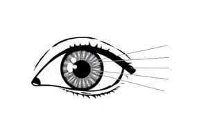
- Eyelids: Meet at the medial and lateral commissure (canthus), protecting the eye
- Eyelashes: Prevent foreign particles from entering the eye
- Tarsal glands: Produce oily secretions lubricating the eye
- Ciliary glands: Located between eyelashes
- Conjunctiva: Membrane lining the eyelids and eyeball, connecting with the cornea and secreting mucus to keep the eye moist
- Lacrimal apparatus: Lacrimal gland (produces tears) and associated ducts. Tears drain across the eye, the lacrimal canaliculi, the lacrimal sac, and into the nasolacrimal duct, emptying into the nasal cavity. Tears contain dilute salt solution, mucus, antibodies, and lysozyme (bacteria-destroying enzyme).
Internal Structures: The Eyeball
- Fibrous layer: Sclera (white connective tissue layer) and Cornea (transparent, central anterior part allowing light passage, easily repairs itself)
- Vascular layer: Choroid (blood-rich nutritive layer), Ciliary body (smooth muscle structures), and Iris (pigmented layer controlling light entering the pupil). The pupil is a rounded opening in the iris.
- Sensory layer: Retina (outer pigmented layer absorbing light and preventing scattering, and inner neural layer with receptor cells, Rods (gray scale vision in dim light), and Cones (color vision, concentrated in the fovea centralis)). The optic disc (blind spot) is where the optic nerve exits the eyeball.
- Humors: Aqueous humor (clear, watery fluid anterior to the lens, maintains intraocular pressure,) and Vitreous humor (gel-like substance posterior to the lens, prevents collapsing).
- Lens: Flexible, biconvex crystal-like structure, held in place by a suspensory ligament attached to the ciliary body
- Ophthalmoscope: Instrument illuminating the interior of the eyeball, detecting various eye conditions.
Physiology of Vision
- Pathway of light through the eye: Light is bent (refracted) by the cornea, aqueous humor, lens, and vitreous humor. The eye is set for distant vision (over 20ft) and must change shape (accommodation) to focus on close objects (less than 20ft).
- Real images formed on retina: Reversed left-to-right and upside down, smaller than the object.
- Visual fields and pathways: Optic nerve (bundle of axons carrying impulses from retina), optic chiasma (location where optic nerves cross), optic tracts (contain fibers from lateral side of same eye and medial side of opposite eye, synapses with neurons in thalamus), optic radiation (axons from thalamus, synapse with cortical cells in occipital lobe), optic cortex (visual interpretation occurs).
- Visual fields and binocular vision: Each eye sees a slightly different view, but field of view overlaps, resulting in binocular vision (depth perception).
- Eye reflexes: Convergence (reflexive movement of eyes medially to focus on near objects), Photopupillary reflex (bright light causing pupils to constrict), Accommodation pupillary reflex (viewing close objects causing pupils to constrict).
- Eye conditions: Emmetropia (normal vision), Myopia (nearsightedness, eyeball is too long, distant objects are blurry), Hyperopia (farsightedness, eyeball is too short, near objects are blurry), Astigmatism (light focuses as lines not points due to unequal curvature of cornea/lens, blurry images)
The Ear: Hearing and Balance
- Ear houses hearing and equilibrium (balance)
- Receptors are mechanoreceptors
- Different organs house receptors for each sense
Anatomy of the Ear
- Three areas: External (outer) ear, Middle ear, Internal (inner) ear
- External ear: Auricle (pinna), External acoustic meatus (auditory canal), Tympanic membrane (eardrum).
- Middle ear: Middle ear cavity (air-filled, mucosa-lined), Pharyngotympanic tube (auditory tube, equalizes pressure), Auditory ossicles (malleus, incus, stapes, transmit vibrations from eardrum to the inner ear).
- Internal ear: Bony labyrinth (osseous labyrinth, cochlea, vestibule, semicircular canals, filled with perilymph), Membranous labyrinth (suspended in perilymph, contains endolymph)
Equilibrium
- Vestibular apparatus: Equilibrium receptors in the inner ear, consisting of static and dynamic equilibrium components.
- Static equilibrium: Maculae (receptors in vestibule) report on head position, help maintain head erectness, send info via vestibular nerve to cerebellum.
- Dynamic equilibrium: Crista ampullaris (receptors in ampulla of each semicircular canal). Cupula (gelatinous cap) drags against endolymph during head movements, stimulating hair cells, impulses travel to vestibular nerve and cerebellum.
Hearing
- Spiral organ of Corti (located in cochlear duct): Hair cells on the basilar membrane, gel-like tectorial membrane bending cells when basilar membrane vibrates, impulses carried to auditory cortex on temporal lobe via cochlear nerve.
- Pathway of vibrations from sound waves: Vibrations move through ossicles to oval window, amplified, causing pressure waves in the basilar membrane and bending hair cells, generating action potentials. High-pitched sounds disturb short, stiff basilar membrane hairs; low-pitched sound disturb long, floppy basilar membrane hairs.
- Hearing and equilibrium deficits: Deafness (any degree of hearing loss), Conduction deafness (transmission of vibrations impaired), Sensorineural deafness (damage to ear/nervous system), Ménière's syndrome (inner ear problem causing deafness and vertigo).
Chemical Senses: Smell and Taste
- Receptors for smell and taste are chemoreceptors, responding to chemicals in solution
- Smell can differentiate wider range of chemicals than taste, both complementing each other in response to many same stimuli.
- Olfactory receptors: Located in the roof of the nasal cavity, detect chemicals dissolved in mucus by olfactory hairs (cilia of neurons), impulses are transmitted to olfactory cortex via olfactory nerve (cranial nerve I). Smell is interpreted in olfactory cortex.
- Taste buds: Receptor organs in taste papillae, on tongue, soft palate, and superior part of pharynx, cheeks. Gustatory cells (taste receptors) possess gustatory hairs. Impulses carried to gustatory complex by several cranial nerves (facial, glossopharyngeal, vagus). Taste buds are replaced by basal cells.
- Basic taste sensations: Sweet, sour, bitter, salty, umami.
Developmental Aspects of Special Senses
- Special sense organs form early in embryonic development
- Maternal infections during first 5-6 weeks of pregnancy can cause visual abnormalities and sensorineural deafness.
- Vision requires learning; infants have poor visual acuity, farsightedness, lack color vision, lack depth perception. Eyes continue growing and maturing until age 8-9. Age-related eye issues (Presbyopia, decreased lens elasticity, difficulty with close-vision, lens discoloration, Constricted pupils).
- Hearing is present at birth, but responses are reflexive. Critical listening and language imitation develop during toddler stage. Age-related issues (Presbycusis, otosclerosis). Taste and smell are acute at birth, but sensitivity decreases after age 40.
Studying That Suits You
Use AI to generate personalized quizzes and flashcards to suit your learning preferences.