Podcast
Questions and Answers
What type of sensory information is primarily detected by Meissner corpuscles?
What type of sensory information is primarily detected by Meissner corpuscles?
- Light, sustained touch
- Discriminative touch (correct)
- Pain and temperature
- Deep pressure
Which receptor adapts rapidly to changes in stimulus intensity?
Which receptor adapts rapidly to changes in stimulus intensity?
- Pacinian corpuscle (correct)
- Merkel cells
- Ruffini ending
- Free nerve ending
Where is the primary somatosensory cortex located?
Where is the primary somatosensory cortex located?
- Parietal lobes (correct)
- Temporal lobe
- Occipital lobe
- Anterior frontal lobe
Which type of receptor is associated with detecting pain?
Which type of receptor is associated with detecting pain?
Which receptor is best suited for detecting muscle stretch?
Which receptor is best suited for detecting muscle stretch?
Which type of touch do Merkel cells primarily detect?
Which type of touch do Merkel cells primarily detect?
What type of adaptation do Ruffini endings exhibit?
What type of adaptation do Ruffini endings exhibit?
Which receptor type is not responsible for detecting temperature?
Which receptor type is not responsible for detecting temperature?
What is a primary function of the superior parietal lobe?
What is a primary function of the superior parietal lobe?
How does the brain typically control bodily sensations?
How does the brain typically control bodily sensations?
What does the somatotopic mapping in the sensory cortex represent?
What does the somatotopic mapping in the sensory cortex represent?
What does 'contralateral' signify in the context of brain function?
What does 'contralateral' signify in the context of brain function?
Which statement accurately describes the handed functionality of the brain?
Which statement accurately describes the handed functionality of the brain?
Which part of the internal capsule contains descending fibers?
Which part of the internal capsule contains descending fibers?
What type of organization does the internal capsule have?
What type of organization does the internal capsule have?
Which sensory modalities are specifically mentioned in relation to the posterior limb of the internal capsule?
Which sensory modalities are specifically mentioned in relation to the posterior limb of the internal capsule?
What does the genu of the internal capsule primarily convey?
What does the genu of the internal capsule primarily convey?
What is the main role of the internal capsule in the brain?
What is the main role of the internal capsule in the brain?
Which of the following statements about the internal capsule is false?
Which of the following statements about the internal capsule is false?
Which of the following structures is NOT mentioned as contributing to the internal capsule?
Which of the following structures is NOT mentioned as contributing to the internal capsule?
What does the term 'lentiform nucleus' refer to in relation to the internal capsule?
What does the term 'lentiform nucleus' refer to in relation to the internal capsule?
Which statement accurately describes the Spinothalamic Tract (STT)?
Which statement accurately describes the Spinothalamic Tract (STT)?
What would a patient with left side STT damage most likely experience?
What would a patient with left side STT damage most likely experience?
What characterizes 'sacral sparing' in the context of spinal lesions?
What characterizes 'sacral sparing' in the context of spinal lesions?
How does syringomyelia develop?
How does syringomyelia develop?
Which of the following accurately describes the pathway of the Spinothalamic Tract from the periphery to the sensory cortex?
Which of the following accurately describes the pathway of the Spinothalamic Tract from the periphery to the sensory cortex?
In the context of the STT, what is the significance of the somatotopic organization?
In the context of the STT, what is the significance of the somatotopic organization?
Which statement about the Spinothalamic Tract is incorrect?
Which statement about the Spinothalamic Tract is incorrect?
What is the clinical manifestation of a patient suffering from syringomyelia?
What is the clinical manifestation of a patient suffering from syringomyelia?
What is a likely symptom of damage to the spinocerebellar tract?
What is a likely symptom of damage to the spinocerebellar tract?
In which condition might pure lesions to the spinocerebellar tract be observed?
In which condition might pure lesions to the spinocerebellar tract be observed?
What characteristic gait is associated with damage to the spinocerebellar tract?
What characteristic gait is associated with damage to the spinocerebellar tract?
Why are symptoms associated with damage to the spinocerebellar tract often masked?
Why are symptoms associated with damage to the spinocerebellar tract often masked?
What type of proprioception is associated with the spinocerebellar tract?
What type of proprioception is associated with the spinocerebellar tract?
Which type of sensory pathway is responsible for pain and temperature perception?
Which type of sensory pathway is responsible for pain and temperature perception?
Which receptors are involved in the dorsal column pathway?
Which receptors are involved in the dorsal column pathway?
Which of the following best describes the spinocerebellar tract?
Which of the following best describes the spinocerebellar tract?
What might be a clinical implication of isolated damage to the spinocerebellar tract?
What might be a clinical implication of isolated damage to the spinocerebellar tract?
What role do muscle spindles and Golgi tendon organs play in the nervous system?
What role do muscle spindles and Golgi tendon organs play in the nervous system?
What is the first order trigeminal neuron responsible for in the sensory pathway from the face?
What is the first order trigeminal neuron responsible for in the sensory pathway from the face?
Which structure does the trigeminothalamic tract target after decussating?
Which structure does the trigeminothalamic tract target after decussating?
What type of sensations does the trigeminal nerve pathway convey from the face?
What type of sensations does the trigeminal nerve pathway convey from the face?
What effect does dorsal column damage in the spinal cord have on sensory perception?
What effect does dorsal column damage in the spinal cord have on sensory perception?
What is the role of the 2nd order trigeminal neurons in the sensory pathway?
What is the role of the 2nd order trigeminal neurons in the sensory pathway?
Which clinical sign is associated with sensory ataxia due to dorsal column damage?
Which clinical sign is associated with sensory ataxia due to dorsal column damage?
What is pseudoathetosis?
What is pseudoathetosis?
Which of the following accurately describes the function of the Fasciculus Cuneatus?
Which of the following accurately describes the function of the Fasciculus Cuneatus?
What structures are primarily involved in processing sensory information from the face?
What structures are primarily involved in processing sensory information from the face?
Which of the following is NOT a function of the trigeminal nerve pathway?
Which of the following is NOT a function of the trigeminal nerve pathway?
Which of the following best describes the role of 3rd order neurons in this sensory pathway?
Which of the following best describes the role of 3rd order neurons in this sensory pathway?
What indicates a potential B12 deficiency related to dorsal column damage?
What indicates a potential B12 deficiency related to dorsal column damage?
Which part of the CNS houses the Nucleus Gracilis and Nucleus Cuneatus?
Which part of the CNS houses the Nucleus Gracilis and Nucleus Cuneatus?
What is the primary function of the trigeminal lemniscus?
What is the primary function of the trigeminal lemniscus?
What is the primary consequence of damage to the dorsal column?
What is the primary consequence of damage to the dorsal column?
Flashcards
Meissner Corpuscle
Meissner Corpuscle
A sensory receptor located in the skin that detects light touch and vibrations. They are rapidly adapting, meaning they quickly stop firing when a stimulus is constant.
Pacinian Corpuscle
Pacinian Corpuscle
A sensory receptor located deep in the skin that detects deep pressure and vibrations. They are rapidly adapting, meaning they quickly stop firing when a stimulus is constant.
Ruffini Ending
Ruffini Ending
A sensory receptor located within the dermis of the skin that detects sustained pressure and stretching. They are slowly adapting, meaning they keep firing as long as the stimulus is present.
Merkel Disc
Merkel Disc
Signup and view all the flashcards
Free nerve endings
Free nerve endings
Signup and view all the flashcards
Muscle Spindle
Muscle Spindle
Signup and view all the flashcards
Sensory Receptor Adaptation
Sensory Receptor Adaptation
Signup and view all the flashcards
Primary Somatosensory Cortex
Primary Somatosensory Cortex
Signup and view all the flashcards
What is the superior parietal lobe's function?
What is the superior parietal lobe's function?
Signup and view all the flashcards
How are brain hemispheres and body sides connected?
How are brain hemispheres and body sides connected?
Signup and view all the flashcards
What is somatotopic mapping?
What is somatotopic mapping?
Signup and view all the flashcards
How is the body mapped onto the sensory cortices?
How is the body mapped onto the sensory cortices?
Signup and view all the flashcards
What is the relationship between sensory cortex and body parts?
What is the relationship between sensory cortex and body parts?
Signup and view all the flashcards
What is the Corpus Callosum?
What is the Corpus Callosum?
Signup and view all the flashcards
What is the Internal Capsule?
What is the Internal Capsule?
Signup and view all the flashcards
How is the Internal Capsule organized?
How is the Internal Capsule organized?
Signup and view all the flashcards
What does the Anterior Limb of the Internal Capsule carry?
What does the Anterior Limb of the Internal Capsule carry?
Signup and view all the flashcards
What does the Genu of the Internal Capsule carry?
What does the Genu of the Internal Capsule carry?
Signup and view all the flashcards
What does the Posterior Limb of the Internal Capsule carry?
What does the Posterior Limb of the Internal Capsule carry?
Signup and view all the flashcards
What does the Retrolenticular part of the Internal Capsule carry?
What does the Retrolenticular part of the Internal Capsule carry?
Signup and view all the flashcards
What is the 'lenticular nucleus?'
What is the 'lenticular nucleus?'
Signup and view all the flashcards
What is the somatotopic organization of the spinothalamic tract?
What is the somatotopic organization of the spinothalamic tract?
Signup and view all the flashcards
How does an expanding ventral horn tumor cause 'sacral sparing'?
How does an expanding ventral horn tumor cause 'sacral sparing'?
Signup and view all the flashcards
Describe syringomyelia and its presentation.
Describe syringomyelia and its presentation.
Signup and view all the flashcards
Describe the pathway of the spinothalamic tract.
Describe the pathway of the spinothalamic tract.
Signup and view all the flashcards
What is the primary function of the spinothalamic tract?
What is the primary function of the spinothalamic tract?
Signup and view all the flashcards
What is the medulla oblongata's function?
What is the medulla oblongata's function?
Signup and view all the flashcards
What are the gracile & cuneate fasiculi?
What are the gracile & cuneate fasiculi?
Signup and view all the flashcards
What sensory information does the Trigeminal nerve (CN V) carry?
What sensory information does the Trigeminal nerve (CN V) carry?
Signup and view all the flashcards
What is the role of the Trigeminothalamic Tract?
What is the role of the Trigeminothalamic Tract?
Signup and view all the flashcards
Where does the trigeminothalamic tract carry sensory information to?
Where does the trigeminothalamic tract carry sensory information to?
Signup and view all the flashcards
Where do the first-order trigeminal neurons synapse?
Where do the first-order trigeminal neurons synapse?
Signup and view all the flashcards
What is the effect of dorsal column damage?
What is the effect of dorsal column damage?
Signup and view all the flashcards
What is a potential symptom of dorsal column damage?
What is a potential symptom of dorsal column damage?
Signup and view all the flashcards
What is another potential symptom of dorsal column damage?
What is another potential symptom of dorsal column damage?
Signup and view all the flashcards
What causes damage to the dorsal column?
What causes damage to the dorsal column?
Signup and view all the flashcards
What is the function of the dorsal column?
What is the function of the dorsal column?
Signup and view all the flashcards
What are third-order neurons in the face sensory pathway?
What are third-order neurons in the face sensory pathway?
Signup and view all the flashcards
Where is sensory information from the face processed?
Where is sensory information from the face processed?
Signup and view all the flashcards
What is the role of the thalamus in the trigeminothalamic tract?
What is the role of the thalamus in the trigeminothalamic tract?
Signup and view all the flashcards
What is the role of second-order neurons in the face sensory pathway?
What is the role of second-order neurons in the face sensory pathway?
Signup and view all the flashcards
Spinocerebellar Tract Damage
Spinocerebellar Tract Damage
Signup and view all the flashcards
Spinocerebellar Tract Function
Spinocerebellar Tract Function
Signup and view all the flashcards
Unconscious Proprioception
Unconscious Proprioception
Signup and view all the flashcards
Consequences of Damage
Consequences of Damage
Signup and view all the flashcards
Dorsal Column Pathway
Dorsal Column Pathway
Signup and view all the flashcards
Spinothalamic Tract
Spinothalamic Tract
Signup and view all the flashcards
Conscious vs Unconscious Proprioception
Conscious vs Unconscious Proprioception
Signup and view all the flashcards
Sensory Receptors
Sensory Receptors
Signup and view all the flashcards
Ascending Sensory Pathways
Ascending Sensory Pathways
Signup and view all the flashcards
Somatosensory Cortex
Somatosensory Cortex
Signup and view all the flashcards
Study Notes
Spinal Cord, Ascending Tracts & Sensation
- The spinal cord's grey matter is mostly surrounded by white matter.
- Axial sections of the spinal cord look different when viewing from above (compared to typical CT/MRI).
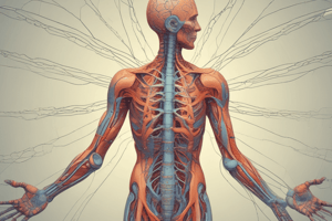
- Sensory receptors detect various sensations, are located at multiple levels within tissues, and come in different types with diverse functions and adaptation speeds.
- Different types of sensory receptors detect different types of sensation.
- Receptors are found at various levels of tissues.
- The major ascending sensory pathways include the dorsal column pathway, the spinothalamic tract, and the spinocerebellar tracts.
- The dorsal column pathway carries discriminative touch, vibration, and conscious proprioception.
- The spinothalamic tract carries pain, temperature, and simple touch.
- The spinocerebellar tracts carry unconscious proprioception.
- The sensory cortexes sit in the left and right parietal lobes.
- The primary somatosensory cortex (postcentral gyrus) gets sensory input from the body.
- Many brain functions are crossed over (contralateral).
- In general, each cerebral hemisphere perceives sensations and controls movements on the opposite side of the body.
- The sensory cortexes on both the left and right sides have a map of body parts (homunculus).
- The amount of cortical area dedicated to a region is proportional to that body part's sensitivity.
- The internal capsule is a dense collection of white matter carrying sensory and motor tracts to and from the cortex.
- The internal capsule is somatotopically organized, containing ascending and descending white matter tracts.
- Sensory tracts are composed of a three-neuron chain from the periphery to the primary sensory cortex.
- The thalamus is an organized collection of subcortical relay nuclei, with two main nuclei for somatosensory input (VPL and VPM).
- The grey matter of the spinal cord is organised into zones with different functions (Rexed Lamina).
- These zones contain cell bodies for neurons involved in ascending (sensory) and descending (motor) pathways.
- White matter in the spinal cord includes multiple fiber bundles, or funiculi (dorsal, lateral, ventral).
- The spinal cord has ascending tracts on both sides.
- Axons in white matter are bundled into fasciculi based on similar functions.
- The dorsal column pathway has fasciculus gracilis and fasciculus cuneatus.
- The spinothalamic tract has neurons that synapse with third-order neurons in the ventral posterior lateral (VPL) thalamic nucleus.
- The spinocerebellar tracts mainly carry unconscious proprioception.
- Damage to the dorsal column pathway causes ipsilateral loss of discriminative touch, vibration, and conscious proprioception below the level of the lesion.
- Damage to the spinothalamic tract results in ipsilateral loss of pain and temperature below the level of the lesion.
- Damage to one side of the cerebellum (involving the spinocerebellar tracts) results in ipsilateral impairments in the body.
- The trigeminothalamic tract carries sensory information from the face via the trigeminal nerve to the contralateral ventral posterior medial nucleus in the thalamus.
- The trigeminal nerve transmits pain, temperature, touch, and proprioceptive signals from the face.
- The medial lemniscus and the trigeminal lemniscus are crucial for conveying sensory information to the brain.
- Damaged tracts often reveal a body-side pattern for associated sensory deficits.
- Follow-up questions cover specific details and aspects of the various tracts and structures.
Studying That Suits You
Use AI to generate personalized quizzes and flashcards to suit your learning preferences.
Related Documents
Description
Test your knowledge on the types and functions of somatosensory receptors in the human body. This quiz covers key concepts such as sensory detection, adaptation, and the location of the primary somatosensory cortex. Perfect for students studying physiology or anatomy!