Podcast
Questions and Answers
Which part of the hard palate is formed by the palatine processes of the maxilla?
Which part of the hard palate is formed by the palatine processes of the maxilla?
- Horizontal plate of palatine bone
- Alveolar arch
- Posterior 1/3
- Anterior 2/3 (correct)
Which feature is a projection from the posterior end of the alveolar arch?
Which feature is a projection from the posterior end of the alveolar arch?
- Pyramidal process of palatine bone
- Posterior nasal spine
- Maxillary tuberosity (correct)
- Greater palatine foramen
What is the conical projection found at the sharp free posterior border of the hard palate called?
What is the conical projection found at the sharp free posterior border of the hard palate called?
- Alveolar arch
- Pyramidal process of palatine bone
- Maxillary tuberosity
- Posterior nasal spine (correct)
What divides the anterior and middle parts of the norma basalis externa?
What divides the anterior and middle parts of the norma basalis externa?
Which foramina transmit the right and left greater palatine nerves and vessels?
Which foramina transmit the right and left greater palatine nerves and vessels?
Which anatomical feature creates the U-shaped ridge of bone in the hard palate?
Which anatomical feature creates the U-shaped ridge of bone in the hard palate?
What divides the anterior part of the hard palate into right and left halves?
What divides the anterior part of the hard palate into right and left halves?
What lies medial to the last molar tooth?
What lies medial to the last molar tooth?
What is the function of the choanae in the respiratory system?
What is the function of the choanae in the respiratory system?
Which structure is located at the base of the skull and allows passage for the spinal cord?
Which structure is located at the base of the skull and allows passage for the spinal cord?
Which part of the sphenoid bone is described by the lateral pterygoid plate?
Which part of the sphenoid bone is described by the lateral pterygoid plate?
What is the role of the stylomastoid foramen?
What is the role of the stylomastoid foramen?
Where would you find the mandibular fossa?
Where would you find the mandibular fossa?
What does the lesser palatine foramina transmit?
What does the lesser palatine foramina transmit?
Which foramen is located adjacent to the carotid canal?
Which foramen is located adjacent to the carotid canal?
Which structure is transmitted through the foramen ovale?
Which structure is transmitted through the foramen ovale?
What is transmitted by the foramen spinosum?
What is transmitted by the foramen spinosum?
Which bone forms a major part of the bony nasal septum?
Which bone forms a major part of the bony nasal septum?
What is the significance of the pterygoid fossa?
What is the significance of the pterygoid fossa?
Which part of the temporal bone includes the mastoid process?
Which part of the temporal bone includes the mastoid process?
What does the jugular foramen transmit?
What does the jugular foramen transmit?
Which of the following bones is located anteriorly to the middle part?
Which of the following bones is located anteriorly to the middle part?
Which features are located at the lower end of the medial pterygoid plate?
Which features are located at the lower end of the medial pterygoid plate?
Which nerve is transmitted through the stylomastoid foramen?
Which nerve is transmitted through the stylomastoid foramen?
Where does the groove for the cartilaginous part of the pharyngotympanic tube lie?
Where does the groove for the cartilaginous part of the pharyngotympanic tube lie?
Which structure is NOT transmitted through the foramen magnum?
Which structure is NOT transmitted through the foramen magnum?
What feature is found on the infratemporal surface of the greater wing of the sphenoid?
What feature is found on the infratemporal surface of the greater wing of the sphenoid?
Which of the following is true about the foramen lacerum?
Which of the following is true about the foramen lacerum?
What separates the choanae from each other?
What separates the choanae from each other?
Which artery is associated with the inferior orbital fissure?
Which artery is associated with the inferior orbital fissure?
Which structure is associated with the mandibular fossa?
Which structure is associated with the mandibular fossa?
What structure passes through the carotid canal?
What structure passes through the carotid canal?
What is the function of the hypophysial fossa?
What is the function of the hypophysial fossa?
Which structure is located anterior to the hypophysial fossa?
Which structure is located anterior to the hypophysial fossa?
What does the tegmen tympani primarily form?
What does the tegmen tympani primarily form?
Which foramen allows the passage of the maxillary nerve?
Which foramen allows the passage of the maxillary nerve?
Which structure is found posterior to the medial end of the superior orbital fissure?
Which structure is found posterior to the medial end of the superior orbital fissure?
What structure does the arcuate eminence correspond to?
What structure does the arcuate eminence correspond to?
Where is the prechiasmatic sulcus located?
Where is the prechiasmatic sulcus located?
Which of the following structures does NOT transmit the oculomotor nerve?
Which of the following structures does NOT transmit the oculomotor nerve?
What structure is transmitted through the foramen spinosum?
What structure is transmitted through the foramen spinosum?
Which bony feature is formed by the body of the sphenoid and the basilar part of the occipital bone?
Which bony feature is formed by the body of the sphenoid and the basilar part of the occipital bone?
The internal auditory meatus is located at which part of the temporal bone?
The internal auditory meatus is located at which part of the temporal bone?
The sigmoid sulcus extends as what kind of shape from the transverse sulcus?
The sigmoid sulcus extends as what kind of shape from the transverse sulcus?
Which cranial nerves are transmitted through the internal auditory meatus?
Which cranial nerves are transmitted through the internal auditory meatus?
What is a characteristic feature of the internal occipital protuberance?
What is a characteristic feature of the internal occipital protuberance?
Which structure forms a groove along the superior border of the petrous part of the temporal bone?
Which structure forms a groove along the superior border of the petrous part of the temporal bone?
The posterior cranial fossa is formed by which of the following bones?
The posterior cranial fossa is formed by which of the following bones?
Flashcards
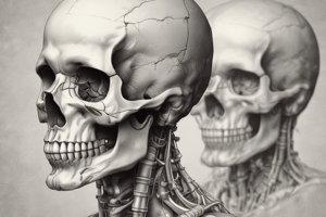
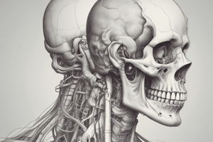
Norma Basalis Externa
Norma Basalis Externa
The outer surface of the base of the skull.
Hard Palate
Hard Palate
Anterior part of the roof of the mouth, formed by two bones.
Palatine Processes
Palatine Processes
Anterior 2/3 of the hard palate, formed from maxilla.
Horizontal Plate of Palatine Bone
Horizontal Plate of Palatine Bone
Signup and view all the flashcards
Greater Palatine Foramen
Greater Palatine Foramen
Signup and view all the flashcards
Incisive Fossa
Incisive Fossa
Signup and view all the flashcards
Alveolar Arch
Alveolar Arch
Signup and view all the flashcards
Posterior Nasal Spine
Posterior Nasal Spine
Signup and view all the flashcards
Lesser Palatine Foramina
Lesser Palatine Foramina
Signup and view all the flashcards
Choanae
Choanae
Signup and view all the flashcards
Vomer
Vomer
Signup and view all the flashcards
Pterygoid Processes
Pterygoid Processes
Signup and view all the flashcards
Infratemporal Surface
Infratemporal Surface
Signup and view all the flashcards
Foramen Ovale
Foramen Ovale
Signup and view all the flashcards
Inferior Orbital Fissure
Inferior Orbital Fissure
Signup and view all the flashcards
Foramen Spinosum
Foramen Spinosum
Signup and view all the flashcards
Stylomastoid Foramen
Stylomastoid Foramen
Signup and view all the flashcards
Jugular Foramen
Jugular Foramen
Signup and view all the flashcards
Foramen Magnum
Foramen Magnum
Signup and view all the flashcards
Sella turcica
Sella turcica
Signup and view all the flashcards
Hypophysial fossa
Hypophysial fossa
Signup and view all the flashcards
Petrous part of temporal bone
Petrous part of temporal bone
Signup and view all the flashcards
Trigeminal impression
Trigeminal impression
Signup and view all the flashcards
Optic canal
Optic canal
Signup and view all the flashcards
Foramen rotundum
Foramen rotundum
Signup and view all the flashcards
Foramen Lacerum
Foramen Lacerum
Signup and view all the flashcards
Carotid canal
Carotid canal
Signup and view all the flashcards
Clivus
Clivus
Signup and view all the flashcards
Internal Occipital Protuberance
Internal Occipital Protuberance
Signup and view all the flashcards
Internal Occipital Crest
Internal Occipital Crest
Signup and view all the flashcards
Sigmoid Sulcus
Sigmoid Sulcus
Signup and view all the flashcards
Internal Auditory Meatus
Internal Auditory Meatus
Signup and view all the flashcards
Study Notes
Skull II - Norma Basalis Externa & Interna
- The external base of the skull (Norma Basalis Externa) is the outer surface of the skull's base.
- The inner surface of the skull's base (Norma Basalis Interna) is divided into three cranial fossae: anterior, middle, and posterior.
- Norma basalis externa is divided into three parts: Anterior, Middle, and Posterior. These three parts are divided by two imaginary transverse lines:
- Line 1: Runs along the posterior border of the hard palate.
- Line 2: Runs along the posterior border of the foramen magnum.
Anterior Part
- Bones: Composed of the hard palate, split into right and left halves separated by the median palatine suture. Each half is further divided:
- Anterior 2/3: Formed by the palatine processes of the maxilla.
- Posterior 1/3: Formed by the horizontal plate of the palatine bone.
- Bony features:
- Alveolar arch: A U-shaped ridge holding the sockets for upper teeth roots.
- Posterior nasal spine: A conical projection on the hard palate's posterior border.
- Maxillary tuberosity: A projection from the posterior alveolar arch.
- Pyramidal process of palatine bone: Projects behind the maxillary tuberosity, on the palatine bone.
- Foramina:
- Incisive fossa: A small pit with four foramina inside, including two median foramina. These transmit the sphenopalatine nerves. Two lateral foramina transmit greater palatine nerves and vessels.
- Greater palatine foramen: A single foramen on each side, located medial to the last molar tooth, transmits the greater palatine nerve and vessels.
- Lesser palatine foramina: One to three foramina on each side, within the pyramidal process of the palatine bone, transmitting the lesser palatine nerve and vessels.
Middle Part
- Bones:
- Vomer and body of the sphenoid bone (anteriorly).
- Pterygoid process of the sphenoid bone, the infratemporal surface of greater wing of sphenoid & squamous part of temporal bone (anterolaterally).
- Petrous, tympanic, and mastoid parts of temporal bone (posterolaterally).
- Basilar and two lateral parts of the occipital bone (posteriorly).
- Bony features:
- Choanae (posterior nasal apertures): Separated by the vomer, forming a major part of the bony nasal septum.
- Pterygoid processes: Each has lateral and medial pterygoid plates with a pterygoid fossa between them. The upper end of the medial pterygoid plate has the scaphoid fossa and pterygoid tubercle; the lower end forms the pterygoid hamulus.
- Groove for pharyngotympanic (Eustachian) tube: Lies between the greater wing of the sphenoid and the petrous part of the temporal bone.
- Squamous part of temporal bone: Has the mandibular fossa and articular eminence, crucial for the temporomandibular joint.
- Infratemporal surface of the greater wing of sphenoid bone: Has a sphenoidal spine at its apex and two foramina (foramen ovale and foramen spinosum).
- Inferior orbital fissure: Separates the greater wing of sphenoid bone from the maxilla, contributing to the orbital cavity.
- Petrous part of temporal bone: A wedge-shaped bone with foramen lacerum, rough quadrate area, and carotid canal. The jugular foramen is positioned posterolaterally to the carotid canal.
- Mastoid part of temporal bone: Has a styloid process, mastoid process, mastoid notch, and occipital groove. It includes the stylomastoid foramen, an opening between the mastoid and styloid processes.
Posterior Part
- Bones:
- Squamous part of the occipital bone.
- Bony features:
- External occipital protuberance
- External occipital crest
- Superior nuchal line
- Inferior nuchal line
- Mastoid emissary foramen
Description of Norma Basalis Interna
- The inner surface of the skull's base is divided into three cranial fossae:
- Anterior cranial fossa
- Middle cranial fossa
- Posterior cranial fossa
Anterior Cranial Fossa
- Bones: Cribriform plate of ethmoid bone and orbital plates of frontal bone (anteriorly).
- Bones: Body and lesser wings of the sphenoid bone (posteriorly)..
- Bony features: Frontal crest, cribriform plate, crista galli, body of sphenoid bone(jugum sphenoidale), and anterior clinoid process.
- Foramina: Foramen cecum; pores of cribriform plate; Anterior ethmoidal foramen; Posterior ethmoidal foramen.
Middle Cranial Fossa
- Bones: Greater wing of sphenoid bone, squamous part of temporal bone, and anterior surface of petrous part of temporal bone.
- Bones: Sella turcica (body of sphenoid); made of hypophysial fossa(pituitary fossa); tuberculum sellae and dorsum sellae:
- Bony features: Prechiasmatic sulcus, posterior clinoid processes, and petrous part of temporal bone (trigeminal impression, arcuate eminence, and tegmen tympani).
- Foramina: Superior orbital fissure, Optic canal, Foramen lacerum, Foramen rotundum, Foramen ovale, Foramen spinosum.
Posterior Cranial Fossa
- Bones: Basilar part of the occipital bone, two lateral parts of the occipital bone, posterior surface of petrous part of temporal bone, and mastoid part of temporal bone.
- Bony features: Clivus, internal occipital protuberance, internal occipital crest, transverse sulcus, sigmoid sulcus, superior petrosal sulcus, inferior petrosal sulcus.
- Foramina: Internal auditory meatus, jugular foramen, anterior condylar foramen, and foramen magnum.
Studying That Suits You
Use AI to generate personalized quizzes and flashcards to suit your learning preferences.