Podcast
Questions and Answers
What primary tissue type overlies connective tissue in the skin?
What primary tissue type overlies connective tissue in the skin?
- Nervous tissue
- Epithelial tissue (correct)
- Muscle tissue
- Adipose tissue
Which of the following best describes the role of the basement membrane in the skin?
Which of the following best describes the role of the basement membrane in the skin?
- It is the primary layer for fat storage in the skin.
- It separates the dermis from the hypodermis.
- It is the outer layer composed of stratified squamous epithelium.
- It lies between the epidermis and dermis providing support and structure. (correct)
How does the subcutaneous layer contribute to the skin's function?
How does the subcutaneous layer contribute to the skin's function?
- By forming the protective outer layer of the skin.
- By actively participating in keratinization.
- By offering an insulating layer due to the presence of adipose tissue. (correct)
- By providing the skin with a direct blood supply.
Which epidermal layer contains cells that appear clear, with nuclei, organelles, and cell membranes no longer visible?
Which epidermal layer contains cells that appear clear, with nuclei, organelles, and cell membranes no longer visible?
How do keratinocytes change as they migrate through the epidermis?
How do keratinocytes change as they migrate through the epidermis?
What is the primary function of keratin in the epidermis?
What is the primary function of keratin in the epidermis?
In which epidermal layer are dendritic (Langerhans) cells primarily found, and what is their function?
In which epidermal layer are dendritic (Langerhans) cells primarily found, and what is their function?
How are tactile (merkel) cells involved in sensory function?
How are tactile (merkel) cells involved in sensory function?
What cellular process helps protect the skin from UV radiation?
What cellular process helps protect the skin from UV radiation?
What accounts for the differences in skin color among individuals?
What accounts for the differences in skin color among individuals?
What is the difference between eumelanin and pheomelanin?
What is the difference between eumelanin and pheomelanin?
How do environmental factors influence skin color?
How do environmental factors influence skin color?
What is the primary difference between basal cell carcinoma and melanomas?
What is the primary difference between basal cell carcinoma and melanomas?
What is the role of dermal papillae in the dermis?
What is the role of dermal papillae in the dermis?
How do lamellated (Pacinian) corpuscles contribute to the function of the skin?
How do lamellated (Pacinian) corpuscles contribute to the function of the skin?
What distinguishes the papillary layer from the reticular layer of the dermis?
What distinguishes the papillary layer from the reticular layer of the dermis?
How do accessory skin structures benefit the repair of damaged skin?
How do accessory skin structures benefit the repair of damaged skin?
What part of the nail is responsible for nail growth?
What part of the nail is responsible for nail growth?
What causes the pale, half-moon-shaped appearance of the lunula?
What causes the pale, half-moon-shaped appearance of the lunula?
How does the arrector pili muscle contribute to thermoregulation?
How does the arrector pili muscle contribute to thermoregulation?
Which of the following is the cause of androgenic alopecia?
Which of the following is the cause of androgenic alopecia?
What is the difference between how eccrine and apocrine sweat glands secrete their products?
What is the difference between how eccrine and apocrine sweat glands secrete their products?
How do sebaceous glands contribute to the health of the skin and hair?
How do sebaceous glands contribute to the health of the skin and hair?
Which component of sweat is responsible for lowering body temperature?
Which component of sweat is responsible for lowering body temperature?
What distinguishes apocrine sweat glands from eccrine sweat glands in terms of location and function?
What distinguishes apocrine sweat glands from eccrine sweat glands in terms of location and function?
How do eccrine sweat glands contribute to thermoregulation?
How do eccrine sweat glands contribute to thermoregulation?
What is the primary way the skin plays a key role in homeostasis?
What is the primary way the skin plays a key role in homeostasis?
If body temperature becomes too low, what physiological response is initiated?
If body temperature becomes too low, what physiological response is initiated?
How might prolonged exposure to a very hot environment lead to hyperthermia?
How might prolonged exposure to a very hot environment lead to hyperthermia?
How does the hypothalamus respond to maintain normal body temperature when the body begins to overheat?
How does the hypothalamus respond to maintain normal body temperature when the body begins to overheat?
What is the role of pyrogens in the development of a fever?
What is the role of pyrogens in the development of a fever?
What is the initial response of the body to inflammation?
What is the initial response of the body to inflammation?
What best describes the process of healing a deep cut?
What best describes the process of healing a deep cut?
How does the 'rule of nines' help in assessing burn injuries?
How does the 'rule of nines' help in assessing burn injuries?
What physiological changes occur in the skin as a result of aging?
What physiological changes occur in the skin as a result of aging?
How is a first-degree burn different from a third-degree burn?
How is a first-degree burn different from a third-degree burn?
Why does the skin become drier with age?
Why does the skin become drier with age?
How does the distribution of melanin granules and melanocyte activity affect skin pigmentation among different individuals?
How does the distribution of melanin granules and melanocyte activity affect skin pigmentation among different individuals?
Why is the arrangement of collagen and elastic fibers in the reticular layer of the dermis important for the skin's overall function?
Why is the arrangement of collagen and elastic fibers in the reticular layer of the dermis important for the skin's overall function?
A person's skin appears yellow; which of the following factors is most likely responsible for this?
A person's skin appears yellow; which of the following factors is most likely responsible for this?
How do accessory skin structures support the repair process in damaged skin, particularly after burns or deep wounds?
How do accessory skin structures support the repair process in damaged skin, particularly after burns or deep wounds?
If a person is exposed to a very hot environment resulting in hyperthermia, which of the following mechanisms will the body use in an attempt to maintain homeostasis?
If a person is exposed to a very hot environment resulting in hyperthermia, which of the following mechanisms will the body use in an attempt to maintain homeostasis?
Flashcards
Integumentary System
Integumentary System
The skin and its accessory structures (hair, nails, and glands) form this system.
The Skin
The Skin
This is the largest organ in the body by weight.
Epidermis
Epidermis
Outer layer, composed of stratified squamous epithelium.
Dermis
Dermis
Signup and view all the flashcards
Hypodermis
Hypodermis
Signup and view all the flashcards
Stratum Basale
Stratum Basale
Signup and view all the flashcards
Keratinization
Keratinization
Signup and view all the flashcards
Keratin
Keratin
Signup and view all the flashcards
Stratum Corneum
Stratum Corneum
Signup and view all the flashcards
Epidermis Functions
Epidermis Functions
Signup and view all the flashcards
Dendritic (Langerhans) Cells
Dendritic (Langerhans) Cells
Signup and view all the flashcards
Tactile (Merkel) Cells
Tactile (Merkel) Cells
Signup and view all the flashcards
Melanocytes
Melanocytes
Signup and view all the flashcards
Eumelanin
Eumelanin
Signup and view all the flashcards
Pheomelanin
Pheomelanin
Signup and view all the flashcards
Albinism
Albinism
Signup and view all the flashcards
Exposure to Sunlight
Exposure to Sunlight
Signup and view all the flashcards
Oxygenation of blood
Oxygenation of blood
Signup and view all the flashcards
Jaundice
Jaundice
Signup and view all the flashcards
Basal/ Squamous Cell Carcinoma
Basal/ Squamous Cell Carcinoma
Signup and view all the flashcards
Melanomas
Melanomas
Signup and view all the flashcards
Dermal Papillae
Dermal Papillae
Signup and view all the flashcards
Papillary Layer
Papillary Layer
Signup and view all the flashcards
Reticular Layer
Reticular Layer
Signup and view all the flashcards
Accessory Structures
Accessory Structures
Signup and view all the flashcards
Nails
Nails
Signup and view all the flashcards
Nail Plate (Body)
Nail Plate (Body)
Signup and view all the flashcards
Nail Matrix
Nail Matrix
Signup and view all the flashcards
Lunula
Lunula
Signup and view all the flashcards
Hair Follicle
Hair Follicle
Signup and view all the flashcards
Hair Root
Hair Root
Signup and view all the flashcards
Hair Bulb
Hair Bulb
Signup and view all the flashcards
Hair Shaft
Hair Shaft
Signup and view all the flashcards
Arrector Pili Muscle
Arrector Pili Muscle
Signup and view all the flashcards
Sebaceous Glands
Sebaceous Glands
Signup and view all the flashcards
Sebum
Sebum
Signup and view all the flashcards
Sweat (Sudoriferous) Glands
Sweat (Sudoriferous) Glands
Signup and view all the flashcards
Eccrine (Merocrine) Glands
Eccrine (Merocrine) Glands
Signup and view all the flashcards
Apocrine Sweat Glands
Apocrine Sweat Glands
Signup and view all the flashcards
Ceruminous Glands
Ceruminous Glands
Signup and view all the flashcards
Protective Barrier
Protective Barrier
Signup and view all the flashcards
Radiation
Radiation
Signup and view all the flashcards
Conduction
Conduction
Signup and view all the flashcards
Convection
Convection
Signup and view all the flashcards
Evaporation
Evaporation
Signup and view all the flashcards
Study Notes
Layers of the Skin
- The skin and its accessory structures constructs the integumentary system
- The skin is the largest organ by weight.
- The skin includes epithelial tissues and connective tissues
- It contains the epidermis and dermis
Key layers
- The epidermis is the outer layer and is composed of stratified squamous epithelium
- A basement membrane separates the epidermis and dermis
- The dermis is the inner layer, and is thicker than the epidermis
- It contains connective tissue, collagenous and elastic fibers, muscle, blood, and nervous tissue
- The subcutaneous layer (hypodermis) is beneath the dermis and provides insulation.
- It consists of areolar and adipose connective tissue
- The subcutaneous layer is not considered part of the skin
- It contains blood vessels that supply skin
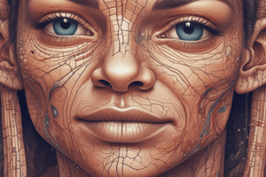
Epidermis Details
- The epidermis lacks blood vessels
- The deepest layer, known as the stratum basale, receives nourishment from blood vessels in the dermis.
- Keratinocytes flatten and die as they migrate
- Keratinization is the process where epidermal cells harden, dehydrate, and accumulate keratin as they migrate outward.
- Keratin, a strong fibrous protein, is produced and stored within the cells.
- Cells become tightly packed as they reach the outer surface, and develop desmosomes forming the stratum corneum, the outermost layer.
- Stratum corneum cells slough off from the skin surface
- The epidermis is thickest on palms and soles (0.8-1.4 mm)
- Most of the body has a thinner epidermis, 0.07 - 0.12 mm
- Its functions include protection against water loss, harmful chemicals, mechanical injury, and pathogens
Layers of the Epidermis
- Stratum corneum is the outermost layer, containing many layers of keratinized, dead epithelial cells, that are flattened and non-nucleated
- Stratum lucidum is between the stratum corneum and stratum granulosum (soles and palms only), and cells appear clear; nuclei, organelles, and cell membranes are no longer visible
- Stratum granulosum is beneath the stratum corneum, containing three to five layers of flattened granular cells with shrunken fibers of keratin and shriveled nuclei.
- Stratum spinosum is beneath the stratum granulosum, containing many layers of cells with centrally located, large, oval nuclei and developing fibers of keratin; cells are becoming flattened
- Stratum basale is the deepest layer. It consists of a single row of cuboidal or columnar cells that divide and grow, this layer includes melanocytes
Special Cells of the Epidermis
- Dendritic (Langerhans) cells are found in the stratum spinosum
- They are phagocytes protecting skin and underlying tissues from infection
- Tactile (Merkel) cells are found in the stratum basale
- These cells work with sensory nerve endings for tactile discs in dermis
- They act as sensory receptors for light touch
- Melanocytes are found in the stratum basale
- They produce the pigment melanin
- Melanocytes absorb UV light from sunlight and provide skin color
- Melanin is distributed to keratinocytes, protecting from UV radiation, like DNA or fibroblast damage, or skin cancer.
Skin Color
- Skin color mainly results from melanin pigment
- Eumelanin: Brownish-black pigment of epidermis
- Pheomelanin: Reddish yellow pigment found in certain areas
Factors Affecting Skin Color
- All people have a similar number of melanocytes, but levels of melanin production vary.
- This is under genetic control, resulting in varying amounts of melanin produced
- Differences in distribution and size of melanin granules can affect the skin color
- Albinism: Inherited mutation in melanin genes, resulting in melanin deficiency
- Exposure to environmental factors such as sunlight, UV light from sunlamps, and X-rays can impact skin color.
- Physiological factors such as oxygenation in blood of dermal blood vessels can cause pinkish, or cyanosis
- Vasodilation and vasoconstriction of dermal blood vessels
- The accumulation of carotene pigment from diet affects skin color as does jaundice
Indoor Tanning and Skin Cancer
- Sun or tanning bed light exposure causes melanocytes to produce melanin, darkening the skin
- UV radiation produced by tanning beds can overwhelm the body’s natural protections against skin cancer
- Basal cell carcinoma and squamous cell carcinoma originate from skin epithelial cells
- Melanomas arise from melanocytes and account for 4% of all skin cancers, however cause 80% of skin cancer deaths.
Dermis Details
- The dermis is the skin's inner layer
- It averages 1 - 2 mm thick
- It contains dermal papillae which are projections between epidermal ridges
- The dermis binds the epidermis to underlying tissues
- It has a connective tissue layer composed of muscle fibers and nerve cell processes
- Dermal blood vessels provide nutrients to all skin cells
- Skin contains hair follicles, sweat and sebaceous glands
- It also includes sensory receptors: Lamellated (Pacinian) corpuscles for pressure, and Tactile (Meissner's) corpuscles for light touch
Layers of the Dermis
- The dermis consists of two layers
- The Papillary layer is superficial and primarily of areolar connective tissue, and is the thinner of the 2 layers
- It is is the location of dermal papillae, which can form fingerprints
- The Reticular layer is deeper. It consists of dense irregular connective tissue and is the thicker of 2 layers
Accessory Structures
- Accessory structures originate from the epidermis
- They extend into either the dermis or the hypodermis
- These structures include hair follicles, nails, and skin glands (sweat and sebaceous)
- Injured and/or burned dermis can regenerate if the accessory structures remain intact
Nails
- Nails are protective coverings on ends of fingers and toes
- It consists of 3 parts:
- Nail plate (body): Visible portion, consists of keratinized cells, and overlies nail bed
- Nail bed: Surface of skin, under nail plate
- Nail matrix: Active growth region, not visible, at proximal end of nail bed
- Lunula: Pale, half-moon-shaped region which overlies nail matrix, the matrix conceals deeper blood vessels
- Cuticle: Fold at proximal end of nail; part of stratum corneum that extends slightly over nail
Hair Follicles
- Hair is present on all skin surfaces, except: palms, soles, lips, nipples, or parts of external reproductive organs
- Hair follicle: Tube-like depression of epidermal cells where hair develops, extends into dermis or subcutaneous layer
- Parts of hair:
- Hair root: Extends from skin surface to dermis or hypodermis
- Hair bulb: Deepest part of hair root containing dividing cells of hair matrix
- Hair shaft: Portion of hair extending beyond skin surface, consists of dead epidermal cells
- Hair papilla contains blood vessels to nourish hair
- Hair color is determined by type and amount of melanin
- Arrector pili muscle: Attached to hair follicle, contracts in response to cold or fear, and produces goosebumps
Hair Loss
- Alopecia is is a common condition
- The top of the head loses hair in androgenic alopecia, the most common type of bladness
- There is a lowered level of testosterone (men) or estrogen (women)
- Progenitor cells are lacking in bald spots, but stem cells are present
- Alopecia areata: the body produces antibodies attacking the hair follicles; autoimmune hair loss
Skin Glands: Sebaceous Glands
- They are holocrine Glands that are usually connected to hair follicles
- They excrete sebum
- Sebum consists of fatty material and cellular debris
- Sebum keeps hair and skin soft and waterproof
- Excess sebum will cause acne
- These glands are absent on palms and soles
Acne
- Acne vulgaris is a sebaceous gland disorder
- It is common at puberty, because sebaceous glands are androgen-responsive
- Excess sebum and epithelial cells will clog sebaceous glands
- Clogged glands support anaerobic bacteria growth, leading to infection with inflammation
- 80% of people between 11-30 years of age will experience acne
- Treatment includes Vitamin A derivatives, systemic antibiotics, salicylic acid, and benzoyl peroxide
Skin Glands : Sweat Glands
- Sweat glands (sudoriferous glands) are located all over the skin.
- These glands form in deeper layers of the dermis or hypodermis as ball-shaped coils
- There are different types of sweat glands:
- Eccrine (merocrine) glands: Consist primarily of water, plus salts and wastes
- These sweat glands respond to body temperature increases
- These glands open to body surface by way of pores.
- Apocrine sweat glands: Found in the axillary and groin areas
- Open into hair follicles
- Secrete by exocytosis
- Respond to emotions and pain -Specialized glands:
- Includes ceruminous glands, which is ear wax.
- Includes mammary glands produce milk.
- Eccrine (merocrine) glands: Consist primarily of water, plus salts and wastes
Glands of the Skin
- Sebaceous glands consists of specialized epithelial cells grouped together, is present to keep the hair pliable and waterproof, it is located near the hair follicles but is absent from the palms and soles
- Merocrine sweat glands secrete odorless fluids, is present to lower body temperature, and is found deep in the dermis covering the whole body, specifically on the neck and back
- Apocrine sweat glands releases a secretion that releases an odor to the skin in armpits and groin, it wets the skin during pain, fear, emotional upset, and sexual arousal, and is found near air follicles in armpits and the groin
- Ceruminous glands are a gland that is modified to secrete ear wax that protects the external acoustic and meatus
- Mammary glands are a gland that is modified to secrete milk that feeds the baby in breasts
Skin Functions
- Skin is versatile and maintains homeostasis
- It functions as a protective barrier
- The skin's protective barrier protects against harmful substances, UV radiation, microorganisms, and water loss
- The skin can sense sensations in pain, temperature, pressure, and touch
- It excretes some wastes
- The skin contributes to the production of Vitamin D that assists with calcium absorption
- It also regulates body temperature by dilating/constricting blood vessels and sweating.
Heat Production and Loss
- Regulating body temperature is vital, as shifts can affect rates of metabolic reactions
- The hypothalamus monitors the set point in the brain
- Body temperature stays close to the set point of 37° Celcius or 98.6° Fahrenheit
- The skin regulates body temperature through homeostatic mechanisms
- Heat is derived from cellular metabolism and mostly by active cells in the liver, and in skeletal and cardiac muscle
Heat Loss through the Skin
- Radiation: Infrared heat rays emitted from warmer skin to cooler environment
- Conduction: Heat moves from warm skin to cooler objects
- Convection: Heat dissipates from skin into circulating air currents.
- Evaporation: Heat transfer by sweat evaporation from the skin
Body Temperature Regulation Mechanisms
-
When body temperature rises:
- Thermoreceptors signal the hypothalamus
- Vasodilation of dermal blood vessels occurs
- Vasoconstriction of deep blood vessels occurs
- Sweat glands are activated
-
When body temperature falls:
- Thermoreceptors signal the hypothalamus
- Vasoconstriction of dermal blood vessels occur
- Vasodilation of deep blood vessels occurs
- Sweat glands are inactive
- Muscles contract involuntarily (shivering)
Problems in Temperature Regulation
- Hyperthermia is: abnormally high body temperature
- It occurs when sweat cannot evaporate e.g. on humid days
- Air temperature reduces radiation effectiveness
- The body gains heat from hotter air
- Skin becomes dry, resulting in weakness, dizziness, nausea, headache, and rapid pulse
- Hypothermia is: abnormally low body temperature
- It occurs from extreme cold or illness
- The hypothalmus causes involuntary skeletal muscle contractions i.e. shivering
- Hypothermia progresses to confusion, lethargy, loss of reflexes, and consciousness
- Organs shut down without medical intervention
Elevated Body Temperature
- A diminished capacity to control temperature results in a hot environment
- Exposure to high heat can overwhelm homeostatic mechanisms for maintaining temperature
- A faster increase in body heat causes rise in body temperature
- Extreme vasodilation will collapse the cardiovascular system
- Fever:
- The immune system elevates the set point to combat infection
- Phagocytes will release pyrogens due to virus or bacterial presence causing the hypothalmus to increase the set point and raise body temperature.
- Elevated body temperature helps the body to destroy pathogens
Healing of Wounds and Burns
- Inflammation is a defensive response to an injury
- Also Body's attempt to restrict spread of infection
- Blood vessels in affected tissues dilate and leak fluids into the tissue.
- Inflamed skin may become:
- It may become reddened
- It may becoe Swollen
- It may become Warm
- It may become Painful
Cuts
- When a shallow cut affects only the epidermis, more epidermal cells divide along its margin to fill the gap
- When Deep cuts reaches the dermis or subcutaneous layer; blood vessels break and create a clot
- The clot's makeup is fibrin, blood cells, and platelets
- The tissue is covered by a scab, clotted or dried tissue fluid
- Epithelial cells reproduce and fill in the wound
- Fibroblasts secrete collagen fibers to bind wound together
- Growth factors stimulate new tissue formation
- Phagocytic cells remove dead cells and debris; the scab sloughs off
- Excess collagen fibers may form an elevated scar
Burns
- Burns are classified by extent of tissue damage
- Superficial, partial-thickness (first degree) burn: affects only epidermis (e.g., sunburn), with only redness, heat, and inflammation; healing takes days to weeks with no scarring.
- Deep, partial-thickness (second degree) burn: destroys epidermis and some dermis from hot liquids; healing varies with severity of burn and regeneration of stem cells in hair follicles and glands; usually recovers with no scarring.
- Full-thickness (third degree) burn: destroys epidermis, dermis, and accessory structures after prolonged heat, flames, or hot liquids; requires skin grafts or substitutes with some healing from margins.
Rule of Nines
- It divides body surface into regions of 9% or multiples of 9
- Is used to estimate extent of injured body surface for burn treatment, the volume of electrolytes or replacement skin to be used can be figured
Life-Span Changes
- Slower cell cycle results in scaly skin and age spots
- Epidermis and dermis become thinner
- Loss of fat in subcutaneous layer; person feels cold
- Wrinkling and sagging of skin occur
- Sebaceous glands secrete less oil causing skin to become dry
- Melanin production slows; and hair becomes white
- Hair thins
- Number of hair follicles decreases
- Nail growth becomes impaired
- Sensory receptors decline
- Body temperature regulation less effective
- Diminished ability to produce Vitamin D
Studying That Suits You
Use AI to generate personalized quizzes and flashcards to suit your learning preferences.