Podcast
Questions and Answers
Which of the following is NOT a function of the skeleton?
Which of the following is NOT a function of the skeleton?
- Hormone production (correct)
- Protection
- Production of blood cells
- Support
What is the main structural difference between compact bone and spongy bone?
What is the main structural difference between compact bone and spongy bone?
- Compact bone is smooth and homogeneous (correct)
- Compact bone is only found in the skull
- Compact bone is less dense than spongy bone
- Compact bone has larger spaces
Which bone marking is characterized as a small rounded projection?
Which bone marking is characterized as a small rounded projection?
- Tubercle (correct)
- Tuberosity
- Crest
- Trochanter
Which part of the skeleton is included in the appendicular skeleton?
Which part of the skeleton is included in the appendicular skeleton?
In which location would you typically find cartilage in the human body?
In which location would you typically find cartilage in the human body?
What description best fits a 'fossa' in bone terminology?
What description best fits a 'fossa' in bone terminology?
Which of these bone markings is described as a sharp, slender, often pointed projection?
Which of these bone markings is described as a sharp, slender, often pointed projection?
Which of the following statements about bone markings is false?
Which of the following statements about bone markings is false?
What is the primary type of bone tissue found in long bones such as the femur?
What is the primary type of bone tissue found in long bones such as the femur?
Which type of bones are characterized by being cube-shaped and containing more spongy bone than compact bone?
Which type of bones are characterized by being cube-shaped and containing more spongy bone than compact bone?
What structure covers the outer surface of a bone and contains blood vessels and nerves?
What structure covers the outer surface of a bone and contains blood vessels and nerves?
Which cells are responsible for bone formation and are found in the periosteum?
Which cells are responsible for bone formation and are found in the periosteum?
What is the function of the articular cartilage found on the epiphyseal surface of long bones?
What is the function of the articular cartilage found on the epiphyseal surface of long bones?
What is the role of osteoclasts found in the endosteum?
What is the role of osteoclasts found in the endosteum?
What does the epiphyseal plate allow for in growing animals?
What does the epiphyseal plate allow for in growing animals?
What is the medullary cavity primarily responsible for?
What is the medullary cavity primarily responsible for?
Which bones are classified as irregular bones?
Which bones are classified as irregular bones?
Wormian bones can be found in which part of the body?
Wormian bones can be found in which part of the body?
Which structure is the primary passageway for cranial nerves VII and VIII?
Which structure is the primary passageway for cranial nerves VII and VIII?
What does the Foramen Magnum provide an opening for?
What does the Foramen Magnum provide an opening for?
Which feature is associated with the Sphenoid bone?
Which feature is associated with the Sphenoid bone?
Which of the following cranial nerves passes through the Superior Orbital Fissures?
Which of the following cranial nerves passes through the Superior Orbital Fissures?
What is the main role of the Crista Galli?
What is the main role of the Crista Galli?
Which opening is specifically for the Middle Meningeal Artery?
Which opening is specifically for the Middle Meningeal Artery?
What do the Occipital Condyles articulate with?
What do the Occipital Condyles articulate with?
Which bone forms the roof of the nasal cavity?
Which bone forms the roof of the nasal cavity?
What structure is responsible for the passage of the Optic Nerve?
What structure is responsible for the passage of the Optic Nerve?
Which of the following bones contains the Foramen Magnum?
Which of the following bones contains the Foramen Magnum?
Which of the following is NOT a function of the Superior and Middle Nasal Conchae?
Which of the following is NOT a function of the Superior and Middle Nasal Conchae?
Which structure serves as the attachment site for neck and tongue muscles?
Which structure serves as the attachment site for neck and tongue muscles?
Which bones are considered paired bones in the facial skeleton?
Which bones are considered paired bones in the facial skeleton?
Which passage is associated with Infraorbital blood vessels?
Which passage is associated with Infraorbital blood vessels?
Which bone marking is known for articulating with the mandibular fossa?
Which bone marking is known for articulating with the mandibular fossa?
What is the primary function of the intervertebral discs?
What is the primary function of the intervertebral discs?
Which region of the vertebral column comprises 7 vertebrae?
Which region of the vertebral column comprises 7 vertebrae?
What is the consequence of a ruptured disc?
What is the consequence of a ruptured disc?
Which of the following conditions describes an excessive lateral curvature of the spine?
Which of the following conditions describes an excessive lateral curvature of the spine?
What structure connects the body of a vertebra to the laminae?
What structure connects the body of a vertebra to the laminae?
Which of the following is TRUE regarding the superior and inferior articular processes?
Which of the following is TRUE regarding the superior and inferior articular processes?
What is the shape of the vertebral column primarily responsible for?
What is the shape of the vertebral column primarily responsible for?
Which cervical vertebra is uniquely designed to allow for the rotation of the head?
Which cervical vertebra is uniquely designed to allow for the rotation of the head?
What is the primary role of the vertebral foramen?
What is the primary role of the vertebral foramen?
The rounded central portion of a vertebra facing anteriorly is known as what?
The rounded central portion of a vertebra facing anteriorly is known as what?
Flashcards are hidden until you start studying
Study Notes
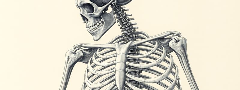
Skeleton Overview
- The skeleton is composed of two tissues: bone and cartilage.
- Cartilage is present in the embryo and persists in specific areas of the adult body, including the nose, larynx, trachea, joints, ribcage, and ear.
- The skeleton supports the body, protects internal organs, provides leverage for movement, stores minerals, and produces blood cells.
Skeleton Subdivisions
- The axial skeleton forms the central axis of the body and includes the skull, sternum, and vertebral column.
- The appendicular skeleton includes the limbs, pelvic girdle, and pectoral girdle.
Bone Markings
- Bone markings are surface features associated with ligament attachments, joint formation, and passage of nerves and blood vessels.
- Projections include tuberosity, crest, trochanter, line, tubercle, epicondyle, spine, process, head, facet, condyle, and ramus.
- Depressions or Cavities include groove, fissure, foramen, notch, meatus, sinus, and fossa.
Bone Classification
- The human body has 206 bones.
- Compact bone is dense and homogeneous.
- Spongy bone contains trabeculae (bone bars) and spaces.
- Long bones are longer than wide, with a shaft and heads (e.g., femur).
- Short bones are cube-shaped with more spongy bone (e.g., tarsals).
- Flat bones have two thin compact bone layers with spongy bone in between (e.g., skull bones).
- Irregular bones have unusual shapes (e.g., vertebrae).
- Sesamoid bones are found within tendons (e.g., patella).
- Wormian/Sutural bones are small bones found between cranial bones.
Anatomy of a Long Bone
- The shaft (diaphysis) is the long axis, primarily composed of compact bone.
- The periosteum is a fibrous membrane surrounding the bone, containing osteoblasts and Sharpey's fibers for blood vessel and nerve passage.
- Osteoclasts are found in the endosteum, the inner lining of the shaft.
- The epiphysis is at the end of long bones, containing mostly spongy bone with a thin layer of compact bone.
- Articular cartilage covers the epiphyseal surfaces, composed of hyaline cartilage for smooth joint movement.
- The epiphyseal plate is hyaline cartilage allowing bone growth in young individuals. The epiphyseal line is a remnant of bone growth.
- The central cavity (medullary cavity) stores fat and contains the endosteum.
Skull Bones
- The Occipital bone forms the posterior portion of the cranium.
- The lambdoid suture articulates the occipital bone with the parietal bones.
- The foramen magnum allows passage of the spinal cord.
- The hypoglossal canal is a passageway for the hypoglossal nerve (cranial nerve XII).
- Occipital condyles articulate with the atlas (C1).
Sphenoid Bone
- The sphenoid bone is bat-shaped and forms the anterior plateau of the middle cranial fossa.
- The greater wings contribute to the orbital socket.
- The superior orbital fissure allows passage of cranial nerves III, IV, V, and VI to the eye.
- The inferior orbital fissure allows passage of infraorbital vessels and cranial nerve V.
- The sella turcica (Turk's saddle) holds the hypophyseal fossa, which houses the pituitary gland.
- The optic canals are openings for the optic nerves.
- The foramen rotundum and ovale are openings for branches of the fifth cranial nerve.
- The foramen spinosum is an opening for the middle meningeal artery.
Ethmoid Bone
- The ethmoid bone is anterior to the sphenoid and forms the roof of the nasal cavity, the upper nasal septum, and part of the medial orbital wall.
- The crista galli is a vertical projection that attaches to the dura mater of the brain.
- The cribriform plates contain olfactory foramina for the passage of olfactory fibers.
- The crista galli and cribiform plates form the horizontal plate.
- The perpendicular plate forms the superior nasal septum.
- The lateral masses contribute to the medial orbital walls.
- The superior and middle nasal conchae are turbinates that help warm and humidify air.
Facial Bones
- The facial bones consist of seven paired bones and two single bones: the vomer and mandible.
Mandible
- The mandibular body forms the chin.
- The mandibular condyle articulates with the mandibular fossa.
- The coronoid process is a site for muscle attachment.
- The mental foramen allows passage of mental blood vessels.
- The alveolar margin contains the inferior teeth sockets.
- The mandibular symphysis marks the site of mandibular fusion.
- The mandibular foramen allows passage of cranial nerve V.
Maxillae
- The alveolar margin contains the superior teeth sockets.
- The palatine processes form the anterior portion of the hard palate.
- The infraorbital foramen allows passage of infraorbital blood vessels.
- The incisive fossa provides a passage for the nasopalatine artery and nerve.
Lacrimal Bone
- The lacrimal fossa allows for tear drainage and contributes to the orbital socket.
Palatine Bone
- The palatine bones form the posterior portion of the hard palate.
Zygomatic Bone
- The zygomatic bones form the cheeks and part of the orbital socket.
Nasal Bone
- The nasal bones form the bridge of the nose and contribute to the orbital socket.
Hyoid Bone
- The hyoid bone serves as an attachment point for neck and tongue muscles.
Vertebral Column
- The vertebral column is formed by 24 vertebrae, the sacrum, and the coccyx.
- The vertebral column protects the spinal cord and allows nerves to exit through openings between the vertebrae.
- The cervical vertebrae (7 bones) are in the neck.
- The thoracic vertebrae (12 bones) are in the upper back.
- The lumbar vertebrae (5 bones) are in the lower back.
- Intervertebral discs separate vertebrae, providing cushioning and shock absorption.
- They consist of an inner nucleus pulposus (gelatinous mass) surrounded by an outer annulus fibrosus (collagen fibers).
- Ruptured discs occur when the nucleus pulposus herniates through the annulus fibrosus, compressing spinal nerves.
- The primary curvature (thoracic and sacral) is present at birth.
- The secondary curvature (cervical and lumbar) develops later.
Spinal Curvature Abnormalities
- Scoliosis is an abnormal lateral curvature.
- Kyphosis is an abnormal dorsal curvature.
- Lordosis is an abnormal anterior curvature.
Structure of a Vertebra
- The body (centrum) is the rounded central portion facing anteriorly.
- The vertebral arch consists of the pedicles (connecting the body to the laminae), laminae (connecting the spinous and transverse processes), and the spinous process.
- The vertebral foramen is an opening between the arch and body, allowing passage of the spinal cord.
- The transverse process projects laterally from the arch.
- The spinous process projects medially/posteriorly from the arch.
- The superior and inferior articular processes are paired projections lateral to the vertebral foramen, articulating between vertebrae.
- The intervertebral foramina are spaces within the pedicels that allow the exit of spinal nerves between vertebrae.
Cervical Vertebrae
- C1 (atlas) has no body and features large depressions to receive the occipital condyles of the skull.
- C2 (axis) acts as a pivot for the atlas' rotation.
Other Important Features
- Mastoid process: Attachment site for muscles.
- External acoustic canal: Passage for sound to the middle ear.
- Internal acoustic canal: Passage for cranial nerves VII and VIII.
- Foramen lacerum: Passage for the internal carotid artery.
- Lambdoid suture: Point of articulation between the occipital and parietal bones.
- Foramen magnum: Passage of the spinal cord.
- Hypoglossal canal: Passage of cranial nerve XII.
- Occipital condyles: Point of articulation between the occipital bone and atlas (C1).
Sphenoid Bone Features
- Greater wings: Part of the orbital socket.
- Superior orbital fissure: Passage for cranial nerves III, IV, V, and VI.
- Inferior orbital fissure: Passage for infraorbital vessels and cranial nerve V.
- Hypophyseal fossa: Holds the pituitary gland.
- Optic canal: Passage for the optic nerve (cranial nerve II).
- Foramen rotundum and ovale: Passage for branches of the fifth cranial nerve.
- Foramen spinosum: Passage for the middle meningeal artery.
Ethmoid Bone Features
- Lateral masses: Part of the orbital socket.
- Crista galli: Attachment site for the dura mater of the skull.
- Cribriform plates: Hold the olfactory foramina for the passage of the olfactory nerve (cranial nerve I).
- Horizontal plate: Formed by the crista galli and cribiform plates.
- Perpendicular plate: Forms the superior nasal septum.
- Superior and inferior nasal conchae: Turbinates that help warm and humidify air.
Mandible Features
- Mandibular body: Forms the chin.
- Mandibular condyle: Articulates with the mandibular fossa.
- Coronoid process: Site of muscle attachment.
- Mental foramen: Passage for mental blood vessels.
- Alveolar margin: Site of inferior teeth sockets.
- Mandibular symphysis: Site of mandibular fusion.
- Mandibular foramen: Passage for cranial nerve V.
Maxillae Features
- Alveolar margin: Site of superior teeth sockets.
- Palatine processes: Anterior portion of the hard palate.
- Infraorbital foramen: Passage for infraorbital blood vessels.
- Incisive fossa: Passage for the nasopalatine artery and nerve.
Lacrimal Bone Features
- Lacrimal fossa: Passage for tears and contributes to the orbital socket.
Palatine Bone Features
- Palatine bones: Forms the posterior portion of the hard palate.
Zygomatic Bone Features
- Zygomatic bones: Forms the cheeks and part of the orbital socket.
Nasal Bone Features
- Nasal bones: Forms the bridge of the nose and contributes to the orbital socket.
Hyoid Bone Features
- Hyoid bone: Attachment site for neck and tongue muscles.
Studying That Suits You
Use AI to generate personalized quizzes and flashcards to suit your learning preferences.


