Podcast
Questions and Answers
Which of the following is an example of bones acting as 'steel girders' and 'reinforced concrete' within the body?
Which of the following is an example of bones acting as 'steel girders' and 'reinforced concrete' within the body?
- Bones protecting soft organs such as the brain and heart.
- The rib cage supporting the thoracic wall. (correct)
- Bones storing minerals like calcium and phosphorus.
- Skeletal muscles using bones as levers for movement.
What is the functional significance of spongy bone being located within the epiphyses of long bones?
What is the functional significance of spongy bone being located within the epiphyses of long bones?
- It provides a smooth surface for joint articulation.
- It facilitates mineral storage within the bone.
- It increases the bone's weight-bearing capacity.
- It helps in blood cell formation. (correct)
How does bone remodeling maintain skeletal strength and calcium balance?
How does bone remodeling maintain skeletal strength and calcium balance?
- By continuously depositing calcium in the bone matrix, regardless of blood calcium levels.
- By adjusting bone mass based on mechanical stress and regulating calcium release or storage in response to blood calcium levels. (correct)
- By solely relying on osteoblast activity to build new bone matrix.
- By halting bone remodeling when blood calcium levels are within homeostatic range.
In fetal development, most of the skeleton is originally made up of what?
In fetal development, most of the skeleton is originally made up of what?
Which statement best describes the role of fontanels in a newborn's skull?
Which statement best describes the role of fontanels in a newborn's skull?
Why is the hyoid bone unique compared to other bones in the body?
Why is the hyoid bone unique compared to other bones in the body?
How do intervertebral discs contribute to the function of the vertebral column?
How do intervertebral discs contribute to the function of the vertebral column?
What is the primary functional advantage of the shoulder (pectoral) girdle's structure?
What is the primary functional advantage of the shoulder (pectoral) girdle's structure?
What is the consequence of a broken clavicle?
What is the consequence of a broken clavicle?
If a person has a joint that allows slight movement; which of the following joint classifications apply?
If a person has a joint that allows slight movement; which of the following joint classifications apply?
Flashcards
Skeletal System
Skeletal System
Internal framework, protects organs, anchors muscles for movement.
Functions of Bones
Functions of Bones
Bones protect organs, support the body and act as levers for movement.
Compact Bone
Compact Bone
Dense, smooth, and homogeneous bone tissue.
Spongy Bone
Spongy Bone
Signup and view all the flashcards
Long Bones
Long Bones
Signup and view all the flashcards
Short Bones
Short Bones
Signup and view all the flashcards
Flat Bones
Flat Bones
Signup and view all the flashcards
Irregular Bones
Irregular Bones
Signup and view all the flashcards
Ossification
Ossification
Signup and view all the flashcards
Axial Skeleton
Axial Skeleton
Signup and view all the flashcards
Study Notes
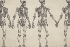
Skeletal System Overview
- Provides an internal framework for the body.
- Protects organs through enclosure.
- Anchors skeletal muscles to facilitate movement via muscle contraction.
- Allows the human body to stand and protects soft organs.
Functions of Bones
- Provides support as the "steel girders" and "reinforced concrete" of the body's internal framework.
- Leg bones act as pillars to support the body trunk during standing.
- The rib cage supports the thoracic wall.
- Protects soft body organs like the brain (skull), spinal cord (vertebrae), and organs in the thorax (rib cage).
- Skeletal muscles, attached by tendons, use bones as levers for movement.
- Stores fat in internal cavities.
- Bone serves as a storehouse for minerals, especially calcium and phosphorus.
- Blood cell formation (hematopoiesis) occurs in marrow cavities.
Osseous Tissue
- Adult skeleton consists of 206 bones composed of osseous tissue, which provides hard, mineralized connective tissues.
- Compact bone is dense, smooth, and homogeneous.
- Spongy bone is composed of small needle-like pieces with lots of open space.
Classification of Bones
- Long bones are typically longer than wide, with a shaft and heads at both ends; mostly compact bone.
- Short bones are generally cube-shaped and contain mostly spongy bone.
- Sesamoid bones form within tendons (e.g., the patella/kneecap); are a special type of short bone.
- Flat bones are thin, flattened, and usually curved, with two thin layers of compact bone sandwiching a layer of spongy bone.
- Irregular bones do not fit into other categories (e.g., vertebrae and hip bones).
- Long bones are composed of a shaft (diaphysis) with two ends (epiphyses).
- The epiphyses are covered with hyaline cartilage and contain spongy bone (where red marrow resides).
- The shaft is compact bone with a cavity containing yellow marrow.
Bone Formation, Growth, and Remodeling
- Ossification (bone formation) has two phases: hyaline cartilage model covered by bone matrix by osteoblasts.
- Bones are remodeled continually in response to calcium levels and gravitational/muscular forces.
- Osteoclasts break down matrix to release calcium into blood when blood calcium levels drop.
- Parathyroid hormone (PTH) stimulates osteoclast activity.
- High blood calcium prompts calcium deposition in bone matrix as hard calcium salts.
- Remodeling (essential for strength during long-bone growth) thickens bones and forms large projections in areas of bulky muscle attachment.
- Osteoblasts lay down new matrix and become osteocytes (mature bone cells).
- Bones become thinner and atrophy without stress.
- Cartilage remains only in isolated areas of the skeleton, e.g., the bridge of the nose and parts of the ribs.
Skeleton Overview
- The skeleton comes from the Greek word meaning "dried-up body.".
Axial Skeleton
- Forms the longitudinal axis of the body and consists of the skull, vertebral column, and bony thorax.
- Cranium encloses and protects fragile brain tissue.
- Facial bones hold the eyes in an anterior position and allow facial muscles to show feelings.
- All but one of the skull bones are joined by sutures, interlocking immovable joints.
- The mandible (jawbone) is the only skull bone attached by a freely movable joint.
Cranial Bones
- The cranial bones are flat, bounded, and fused to protect the brain.
- The frontal bone forms the forehead, bony projections under the eyebrows, and the superior part of each eye's orbit.
- Parietal bones form most of the superior and lateral walls of the cranium and meet at the sagittal suture.
- Temporal bones lie inferior to the parietal bones.
- The occipital bone is the most posterior, forming the floor and back wall of the cranium. It joins the parietal bones at the lambdoid suture, includes the foramen magnum (large hole) for the spinal cord, and contains occipital condyles.
- The butterfly-shaped sphenoid bone spans the width of the skull, forms parts of the cranial cavity floor, includes the sella turcica (Turk's saddle) for the pituitary gland, and features the foramen ovale for cranial nerve V.
- The ethmoid bone is irregularly shaped, lies anterior to the sphenoid, forms the roof of the nasal cavity, includes crista galli, and features cribriform plates that allow olfactory nerve fibers to pass.
Facial Bones
- Provide a framework for facial muscles, form eye sockets, and form jaws for the teeth.
- Maxillae contain sinuses.
- Paired palatine bones form the posterior part of the hard palate.
- Zygomatic bones (cheekbones) form a portion of lateral walls of the orbits.
- Fingernail-sized lacrimal bones form part of the medial walls of each orbit and serve as a passageway for tears.
- Small rectangular nasal bones form the bridge of the nose.
- The single vomer bone in the midline of the nasal cavity forms most of the nasal septum.
- The inferior nasal conchae are thin, curved bones projecting from the lateral walls of the nasal cavity.
- The mandible (lower jaw) is the largest, strongest facial bone, joining the temporal bones to form the only freely movable joints in the skull.
Hyoid Bone
- Not part of the skull but closely related to the mandible and temporal bones.
- Unique as it does not articulate directly with any other bone.
- Horseshoe-shaped with a body and two pairs of horns (cornua).
Fetal Skull
- Newborn skull has fibrous regions called fontanels.
- Fontanels are gradually converted to bone during early infancy.
Vertebral Column/Spine
- Serves as axial support, extending from the skull to the pelvis.
- Consists of 26 irregular bones (vertebrae) connected and reinforced by ligaments.
- Vertebrae are separated by pads of fibrocartilage (intervertebral discs) that cushion and allow flexibility.
- The seven cervical vertebrae (C1-C7) are in the neck region of the spine; C1 (atlas) supports the head, allows nodding, and C2 (axis) acts as a pivot for head rotation.
- Twelve thoracic vertebrae (T1-T12) articulate with the ribs; have somewhat heart-shaped bodies with costal facets.
- Five lumbar vertebrae (L1-L5) have massive, blocklike bodies and are the sturdiest vertebrae.
- The sacrum is formed by the fusion of five vertebrae, articulates with L5, and connects to the coccyx, forming the posterior wall of the pelvis.
- Coccyx (tail bone) is formed from the fusion of three to five tiny, irregularly shaped vertebrae.
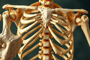
Bony Thorax
- The sternum, ribs, and thoracic vertebrae together are often called the thoracic cage.
- Forms a protective cage around organs of the thoracic cavity (heart, lungs, and major blood vessels).
- The sternum (breastbone) is a typical flat bone resulting from the fusion of the manubrium, body, and xiphoid process.
- The sternum is attached to the first seven pairs of ribs.
- Twelve pairs of ribs form the walls of the bony thorax; all ribs articulate posteriorly with the vertebral column
- True ribs (first seven pairs) attach directly to the sternum by costal cartilages.
- False ribs (next five pairs) either attach indirectly or not at all to the sternum.
- The last two pairs of false ribs are called floating ribs.
Appendicular Skeleton
- Consists of 126 bones of the limbs and girdles that attach the limbs to the axial skeleton.
- The shoulder girdle consists of the clavicle and scapula, allowing exceptionally free but easily dislocated movement.
- The clavicle (collarbone) is a slender bone that acts as a brace and attaches to the manubrium.
- The scapulae (shoulder blades) are triangular that flare when we move our arms posteriorly
Upper Limbs
- Consist of 30 bones that form the frameworks of the arm, forearm, and hand.
- The arm is formed by the humerus, a typical long bone.
- The forearm has two bones: radius (lateral bone) and ulna (medial bone).
- The hand consists of the carpals, metacarpals, and phalanges.
- Eight carpal bones form the wrist; arranged into rows of four bones each.
- The palm has five metacarpals that are numbered 1 to 5 from the thumb toward the little finger.
- There are fourteen phalanges in each hand.
Pelvic Girdle
- Formed by two coxal bones (ossa coxae), which are large, heavy, and securely attached to the axial skeleton.
- Each hip bone is formed by the fusion of the ilium, ischium, and pubis. -The ilium connects posteriorly with the sacrum and forms most of the hip.
- The ischium ("sit-down bone") forms most inferior part of the coxal bone.
- The femur (thigh bone) is the only bone in the thigh containing the patellar surface that forms a joint with the patella
Lower Limbs
- The tibia and fibula form the skeleton of the leg.
- The tibia (shinebone) is larger and more medial, forming the medial malleolus.
- The foot is composed of tarsals, metatarsals, and phalanges, supports body weight, and serves as a lever for locomotion. The tarsus forms the posterior half of the foot containing seven tarsal bones.
- The talus and calcaneus bear the body weight.
- Five metatarsals form the sole, and 14 phalanges form the toes.
Joints
- Every bone (except the hyoid) forms a joint (articulation) with at least one other bone.
- Joints hold bones securely together and give the rigid skeleton mobility.
- Synarthroses are immovable joints.
- Amphiarthroses are slightly movable joints.
- Diarthroses are freely movable joints.
- Fibrous joints consist of bones united by fibrous tissue, best exemplified by the sutures of the skull.
- In cartilaginous joints, bones are connected by cartilage (e.g., pubic symphysis and intervertebral joints connected by fibrocartilage discs).
- Synovial joints have articulating bone ends separated by a joint cavity containing synovial fluid.
Studying That Suits You
Use AI to generate personalized quizzes and flashcards to suit your learning preferences.