Podcast
Questions and Answers
What role does the sarcoplasmic reticulum play in muscle contraction?
What role does the sarcoplasmic reticulum play in muscle contraction?
- It produces ATP for energy.
- It stores calcium ions needed for contraction. (correct)
- It decreases the contraction speed.
- It synthesizes myofilaments.
Which component of the skeletal muscle fiber is responsible for the attachment of myosin during contraction?
Which component of the skeletal muscle fiber is responsible for the attachment of myosin during contraction?
- Troponin
- Tropomyosin
- Calcium ions
- Actin filament (correct)
Which statement correctly describes the role of troponin in muscle contraction?
Which statement correctly describes the role of troponin in muscle contraction?
- It covers the actin-binding sites during contraction.
- It forms thick filaments along with myosin.
- It binds to ATP and releases energy for contraction.
- It binds to calcium ions released during contraction. (correct)
What is a motor unit in skeletal muscle composition?
What is a motor unit in skeletal muscle composition?
What is the primary function of tropomyosin in a relaxed state of skeletal muscle?
What is the primary function of tropomyosin in a relaxed state of skeletal muscle?
Which type of muscular tissue is striated and involuntary?
Which type of muscular tissue is striated and involuntary?
What is the function of tendons in skeletal muscle?
What is the function of tendons in skeletal muscle?
What term describes the connective tissue covering that surrounds individual muscle fibers?
What term describes the connective tissue covering that surrounds individual muscle fibers?
What is myoglobin's primary function in skeletal muscle cells?
What is myoglobin's primary function in skeletal muscle cells?
What does the term 'biceps' indicate about the muscle?
What does the term 'biceps' indicate about the muscle?
Which muscle is named based on its shape, resembling a trapezoid?
Which muscle is named based on its shape, resembling a trapezoid?
Which of the following describes smooth muscle tissue?
Which of the following describes smooth muscle tissue?
What is the typical relationship between the origin and insertion of a muscle?
What is the typical relationship between the origin and insertion of a muscle?
Flashcards are hidden until you start studying
Study Notes
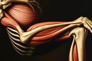
Skeletal Muscle Structure
- Skeletal muscles are attached to bones, striated, and voluntary.
- Tendon: A cord-like structure that attaches a muscle to a bone.
- Aponeurosis: A flattened, sheet-like tendon.
- Tendon Sheaths: Enclose certain muscles in the wrist and ankle.
Types of Muscle Tissue
- Skeletal muscle tissue: Primarily attached to bones; striated and voluntary.
- Cardiac muscle tissue: Forms the heart's wall; striated but involuntary.
- Smooth muscle tissue: Located in the viscera (internal organs); non-striated and involuntary.
Functions of muscles
- Movement of the body: allows for locomotion and motion.
- Positional stabilization: Maintaining posture and body position.
- Managing the volume of organs: Controlling the size of internal organs.
- Heat production: Generating heat to maintain body temperature.
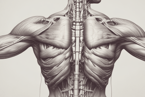
Connective Tissue Coverings of Muscle
- Endomysium: Surrounds individual muscle fibers, binds them together, and supports capillaries and nerve endings within the muscle.
- Perimysium: Binds muscle fibers into bundles (fascicles) and supports the blood vessels and nerve fibers serving them.
- Epimysium: Covers the entire muscle and holds it together.
Naming of Skeletal Muscles
- Location: Names indicating the area where the muscle is located.
- Intercostals: Between the ribs.
- Brachial: On the arm.
- Femoral: On the femur.
- Shape: Shape of the muscle.
- Deltoid: Shaped like the Greek letter delta.
- Trapezius: Shaped like a trapezoid.
- Rhomboid: Shaped like a rhombus.
- Number of Heads of Origin: Number of points where the muscle originates.
- Biceps: 2 heads.
- Triceps: 3 heads.
- Quadriceps: 4 heads.
- Size: Size of the muscle.
- Maximus: Large.
- Minimus: Small.
- Longus: Long.
- Brevis: Short.
- Direction of Muscle Fibers: Direction in which muscle fibers run.
- Rectus: Fibers parallel to the midline (e.g., Rectus abdominis).
- Transverse: Fibers perpendicular to the midline (e.g., Transverse plane).
- Oblique: Fibers run diagonally to the midline.
Location of Attachment
- Sternocleidomastoid Muscle: Originates on the sternum and clavicle and inserts into the mastoid process of the temporal bone.
Actions
- Adductor: Brings a limb closer to the midline.
- Abductor: Moves a limb away from the midline.
- Flexor: Decreases the angle between two bones.
- Extensor: Increases the angle between two bones.
- Pronator: Rotates the palm down.
- Supinator: Rotates the palm up.
- Levator: Lifts a body part.
- Depressor: Lowers a body part.
Skeletal Muscle Cell Structure
- Multinucleated: Each muscle cell has multiple nuclei located on the periphery.
- Sarcolemma: The plasma membrane of a muscle fiber.
- Myoglobin: A protein that binds and stores oxygen within muscle cells.
- T-Tubules (Transverse Tubules): Invaginations of the sarcolemma that extend into the muscle cell.
- Sarcoplasmic Reticulum (SR): A network of membrane-enclosed tubules surrounding myofibrils within the muscle cell; stores calcium ions needed for muscle contraction.
- Terminal Cisternae: Expanded portions of the SR that lie on either side of T-tubules.
Myofibrils and Sarcomeres
- Myofibrils: Smaller fibers within a skeletal muscle fiber.
- Myofilaments: Long strands composed of proteins like actin, myosin, and others that make up myofibrils.
- Sarcomere: The basic contractile unit of a skeletal muscle, composed of myofilaments arranged in a specific pattern.
- Myosin: Thick filaments in the sarcomere.
- Actin: Thin filaments in the sarcomere.
- Troponin and Tropomyosin: Regulatory proteins that control the interaction between actin and myosin.
Contraction of a Skeletal Muscle
- Myosin Molecule: Two heads and a tail; One head binds to actin, and the other binds to ATP.
- Actin Filament: Possesses sites for attaching myosin heads.
- Troponin: Binds to calcium, which is released during muscle contraction.
- Tropomyosin: Covers the actin-binding sites during muscle relaxation.
- Motor Unit: A single motor neuron and all the muscle fibers it innervates.
Neuromuscular Junction (NMJ)
- The synapse between a motor neuron and a skeletal muscle fiber.
- Includes the axon terminals and synaptic end bulbs of a motor neuron, plus the motor end plate of the sarcolemma.
Muscle Twitch
- A single contraction of a muscle fiber.
Studying That Suits You
Use AI to generate personalized quizzes and flashcards to suit your learning preferences.