Podcast
Questions and Answers
Which of the following accurately describes the primary focus of rotator cuff tendons' function?
Which of the following accurately describes the primary focus of rotator cuff tendons' function?
- To provide the primary power for shoulder abduction.
- To facilitate external rotation of the arm.
- To stabilize and compress the humeral head in the glenoid. (correct)
- To control scapular movement during arm elevation.
Which of the following conditions is NOT typically associated with subacromial pain syndrome?
Which of the following conditions is NOT typically associated with subacromial pain syndrome?
- Tendonitis or tendinopathy.
- Glenohumeral instability. (correct)
- Impingement.
- Bursitis.
In the context of rotator cuff tendinopathy, what does the term 'extrinsic' refer to?
In the context of rotator cuff tendinopathy, what does the term 'extrinsic' refer to?
- Tension overload due to excessive muscle use.
- Factors originating within the tendon itself, such as degeneration.
- Inflammatory processes within the subacromial space.
- Mechanical compression from external structures. (correct)
According to Neer's stages of impingement, which stage is characterized by edema and hemorrhage, typically seen in younger, athletic individuals?
According to Neer's stages of impingement, which stage is characterized by edema and hemorrhage, typically seen in younger, athletic individuals?
A patient presents with a painful arc of motion, pain reproduced during tests that compress subacromial tissues, and weakness with isometric resistance. Which condition is most likely?
A patient presents with a painful arc of motion, pain reproduced during tests that compress subacromial tissues, and weakness with isometric resistance. Which condition is most likely?
In the context of rotator cuff tears, what is the general implication of an 'articular side' tear?
In the context of rotator cuff tears, what is the general implication of an 'articular side' tear?
Why might thoracic manipulation improve shoulder function in a patient with rotator cuff tendinopathy?
Why might thoracic manipulation improve shoulder function in a patient with rotator cuff tendinopathy?
Which of the following capsular patterns is most indicative of adhesive capsulitis?
Which of the following capsular patterns is most indicative of adhesive capsulitis?
A patient with adhesive capsulitis limited in external rotation, abduction, and internal rotation is most likely to present with which of the following?
A patient with adhesive capsulitis limited in external rotation, abduction, and internal rotation is most likely to present with which of the following?
According to the Cofield classification system for RTC tears, how would a tear measuring 2 cm be classified?
According to the Cofield classification system for RTC tears, how would a tear measuring 2 cm be classified?
Which of the following is the most common nerve injured in proximal humeral fractures?
Which of the following is the most common nerve injured in proximal humeral fractures?
Which of the following is true regarding SLAP lesions?
Which of the following is true regarding SLAP lesions?
Which of the following is the primary stabilizer of the AC joint in the anterior-posterior plane?
Which of the following is the primary stabilizer of the AC joint in the anterior-posterior plane?
Which of the following is an intrinsic mechanism of rotator cuff tendinopathy?
Which of the following is an intrinsic mechanism of rotator cuff tendinopathy?
According to the presentation, what is a primary benefit of thoracic manipulation in patients with shoulder dysfunction?
According to the presentation, what is a primary benefit of thoracic manipulation in patients with shoulder dysfunction?
Flashcards
Subacromial Pain Syndrome
Subacromial Pain Syndrome
A condition involving pain in the subacromial space, often due to rotator cuff disorders.
Rotator Cuff Disorders
Rotator Cuff Disorders
Various issues that affect the rotator cuff tendons and muscles.
"Impingement-type" Syndromes
"Impingement-type" Syndromes
This involves compression of shoulder structures, especially under the acromion.
RTC Tendinopathy
RTC Tendinopathy
Signup and view all the flashcards
Extrinsic RTC Tendinopathy
Extrinsic RTC Tendinopathy
Signup and view all the flashcards
Intrinsic RTC Tendinopathy
Intrinsic RTC Tendinopathy
Signup and view all the flashcards
Stages of Tendinopathy
Stages of Tendinopathy
Signup and view all the flashcards
RTC: Cause or Effect?
RTC: Cause or Effect?
Signup and view all the flashcards
Posterior Capsule Tightness
Posterior Capsule Tightness
Signup and view all the flashcards
Shoulder Hypermobility
Shoulder Hypermobility
Signup and view all the flashcards
RTC Muscle Performance
RTC Muscle Performance
Signup and view all the flashcards
Classifying Rotator Cuff Tears
Classifying Rotator Cuff Tears
Signup and view all the flashcards
Rotator Cuff Muscles
Rotator Cuff Muscles
Signup and view all the flashcards
Glenohumeral Instability
Glenohumeral Instability
Signup and view all the flashcards
Shoulder Stability
Shoulder Stability
Signup and view all the flashcards
Shoulder Subluxation/Dislocation
Shoulder Subluxation/Dislocation
Signup and view all the flashcards
Bankart Lesion
Bankart Lesion
Signup and view all the flashcards
Positive Apprehension sign
Positive Apprehension sign
Signup and view all the flashcards
Hill Sachs Lesion
Hill Sachs Lesion
Signup and view all the flashcards
Latarjet Procedure
Latarjet Procedure
Signup and view all the flashcards
TUBS acronym
TUBS acronym
Signup and view all the flashcards
AMBRI acronym
AMBRI acronym
Signup and view all the flashcards
SLAP Lesions
SLAP Lesions
Signup and view all the flashcards
Types of SLAP lesions
Types of SLAP lesions
Signup and view all the flashcards
Adhesive Capsulitis
Adhesive Capsulitis
Signup and view all the flashcards
Capsular Pattern
Capsular Pattern
Signup and view all the flashcards
Risk Factors for Adhesive Capsulitis
Risk Factors for Adhesive Capsulitis
Signup and view all the flashcards
Natural History of Adhesive Capsulitis
Natural History of Adhesive Capsulitis
Signup and view all the flashcards
Lateral Rotation
Lateral Rotation
Signup and view all the flashcards
Fracture
Fracture
Signup and view all the flashcards
Two-part: surgical neck
Two-part: surgical neck
Signup and view all the flashcards
Two-part: Greater Tuberosity
Two-part: Greater Tuberosity
Signup and view all the flashcards
Three-part Fracture
Three-part Fracture
Signup and view all the flashcards
Standard four-part
Standard four-part
Signup and view all the flashcards
Nerve Injuries
Nerve Injuries
Signup and view all the flashcards
AC Joint Injury
AC Joint Injury
Signup and view all the flashcards
Types of AC Joint injuries
Types of AC Joint injuries
Signup and view all the flashcards
AC Treatment
AC Treatment
Signup and view all the flashcards
Study Notes
- The presentation covers shoulder pathology, rotator cuff disorders, glenohumeral instability, adhesive capsulitis, fractures, and acromioclavicular joint pathology.
Objectives
- Recognize common shoulder pathologies and impairments.
- Discuss the etiology, risk factors, and biomechanical influences contributing to shoulder pathologies.
- Discuss current diagnostic and medical management of common shoulder pathologies.
- Explain the various forces acting on the upper quadrant and how posture and ergonomics attenuate them.
- Recognize clinical features that assist in identifying different shoulder pathologies.
Conditions to Consider
- Subacromial pain syndrome, glenohumeral instability, adhesive capsulitis, fractures, and acromioclavicular (AC) joint dysfunction are conditions to consider.
Subacromial Pain Syndrome
- Subacromial pain syndrome includes impingement, tendonitis, bursitis, tendinopathy, and tears.
Rotator Cuff Disorders
- Rotator cuff disorders involve issues often occurring in the subacromial space.
- Diagnoses to consider are impingement, subacromial bursitis, RTC issues (posterior), RTC tendinopathy (itis, osis), biceps tendinopathy, and RTC tears (partial to full thickness).
- Similar precautions and prognosis are considered.
- Common key positive findings include a painful arc of motion, pain reproduced with tests that tension or compress subacromial tissues, pain with isometric resistance, weakness, and atrophy.
- Common key negative signs include a significant loss of motion and GH instability.
- Impingement-type syndromes account for 40-60% of all shoulder pain
- Mechanical impingement involves structures in the subacromial space with the anterior undersurface of the acromion.
- RTC tendinopathy and hypertrophy of the AC joint could be considered diagnoses.
- Degenerative and/or mechanical compression can occur.
- Mechanical compression ("extrinsic") and tension overload and degeneration ("intrinsic") are characteristics of RTC tendinopathy.
- Acromion shape plays a role in RTC tendinopathy.
- Normal CA ligaments and thickening of CA ligaments are involved.
- Neer described stages of impingement (tendinopathy) in 1983.
- Stage 1 is edema and hemorrhage, often in younger athletic individuals, which is reversible.
- Stage 2 is tendinitis and fibrosis, typically in the 25-40 age range.
- Stage 3 involves bony changes and tearing, common in older individuals (>40 y).
- Subacromial impingement and RTC tears can be a cause or an effect.
- Subacromial impingement may result from tendon dysfunction, with articular side tears more common than bursal side tears.
- RTC tendinopathy considers the pec muscle length.
- Thoracic manipulation improves shoulder function, kinematics, or thoracic mobility.
- Tight posterior capsules can lead to anterior humeral head translation, with increased pressure on the CA arch = leading to degenerative changes over time.
- Rotator cuff (RTC) muscle performance (weakness or tear) has an impact on impingement-type symptoms.
- Leads to increased pressure of the cuff on the CA arch = degenerative damage over time
- Rotator cuff tears can be pathogenic or include contributing factors.
- Tears are classified by size and severity.
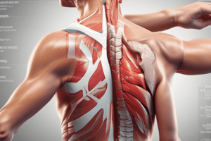
- Tendons form a continuous veil of connective tissue, interdigitate, and collaborate to centralize and compress the HH in the glenoid.
- Impingement, acromion morphology, and RTC tears are structural considerations in RTC tendinopathy.
- True weakness vs motor control is a muscle performance element to consider.
- Rounded shoulder posture and lumbar/thoracic spine issues are addressed.
- Subacromial impingement, GH instability, trauma, and congenital abnormalities can cause RTC "disease."
- Atraumatic tendon tears are rare under 40 years, with partial thickness typically beginning between 40-60 years.
- US studies of 588 patients showed no tears at 48.7 years, UL tears at 58.7 years, and B/L tears at 67.8 years, with a 50% likelihood of B/L > 66 years.
- Rathbun & Macnab described vascular insufficiency in 1970.
- Areas of hypo-vascularity occur in the watershed region and in the insertion at the greater tuberosity.
- A discrepancy between the bursal side rich vascularity and the articular side tenuous vascularity leads to increased incidence of partial tears on the articular side.
- An ultrasound is a standard of care outside of the U.S.
- MRI is the standard of care in the U.S., but has issues like over-utilization and cost.
- A 2003 study by Dinnes found no difference in sensitivity or specificitybetween MRI and Ultrasound.
- MRI, when used as a reference point, found Supraspinatus tendon accuracy around 91.1%, Infraspinatus tendon accuracy around 84.4%, Subscapularis tendon accuracy around 77.8%, and Long head of biceps tendon accuracy around 86.7%.
- In a normal rotator cuff, the tendon is dark and continuous to the greater tuberosity.
- A full-thickness tear appears white and incomplete on an MRI.
- Cofield classification system is generally used.
- A small tear is ≤ 1 cm, medium tear is 1-3 cm; large tear is 3-5 cm; massive tear ≥ 5 cm.
- Proximal attachment of LHB is supraglenoid tubercle.
- Role is not that of a prime mover.
- The diagnosis is often seen as an additional finding during imaging for RTC pathology.
- Diagnoses can be either acute (traumatic) or chronic (degenerative).
Glenohumeral Instability
- Shoulder stability is a coordination of static (bony congruence, cartilage, joint capsule, ligaments) and dynamic (musculature) stabilizers.
- Laxity, subluxation, partial dislocation, and dislocation are spectrums of instability.
- Abnormal symptomatic translation of HH relative to the glenoid can require reduction to restore alignment.
- Younger age, history of dislocation/subluxation, positive apprehension/relocation tests, and generalized laxity are key positive signs.
- Lack of dislocation/subluxation history and apprehension with testing are key negative signs.
- Classification of GH Instability
- Frequency: Single episode, occasional (2-5), frequent (>5)
- Etiology: Traumatic (macrotrauma), atraumatic, congenital, neuromuscular
- Direction: Unidirectional, multidirectional
- Severity: Dislocation, subluxation
- FEDS classification yields 36 combinations, but 6 categories are most meaningful:
- Solitary/occasional/frequent traumatic anterior dislocation (STAD/OTAD/FTAD)
- Solitary/occasional/frequent traumatic anterior subluxation (STAS/OTAS/FTAS)
- Mode of injury:
- Traumatic: TUBS (traumatic unidirectional Bankart lesion surgery)
- Atraumatic: AMBRI (atraumatic multidirectional bilateral rehabilitation, inferior capsule shift)
- Treatment options: Conservative vs Surgical.
- Surgical outcomes are better in patients.
- Rehabilitation is preferred over operative management in patients with atraumatic instability.
- With traumatic instability:
- High-velocity uncontrolled end-range force must be considered.
- Anterior instances are from forced abd/ER or elevation.
- Posterior instances are from forced horiz add/IR with axial load.
- Inferior instances are from forceful inferior load with the arm at the side and in elevation.
- 85-95% are anterior, and 84-100% result in Bankart lesions.
Labral Damage
- Occurrences are often referred to in clock terms like "4:00-6:00."
- Results in 50% reduction in depth of the socket.
- Increased recurrences can lead to worse pathology.
- Occurs with time between injury and if surgery is delayed longer.
- Bony Bankart lesion = avulsion fx of glenoid.
- Hill Sachs lesion:
- Cortical depression on the posterior humeral head.
- Forceful impaction of the humeral head against the anterior-inferior glenoid rim with an anterior dislocation.
- Occurrence with 100% recurrent instability, 80% with primary anterior instability, and 25% with anterior shoulder subluxation.
- Nerves:
- Nerve damage can occur.
- Axillary nerve palsy can result from prolonged dislocation and traction.
- Improving dynamic stability and control are a goal.
- Work on RTC and scapular stability.
- Progress work into functional positions.
- Consider deltoids, biceps, triceps.
SLAP Lesions
- Superior Labrum Anterior to Posterior (from a 10:00 to 2:00 POV)
- Up to seven types, however, commonly separated into four main types.
- Type I includes fraying of the superior labrum while remaining attached to the glenoid rim.
- Type II includes separation of the superior portion of the glenoid labrum and tendon of the biceps brachii muscle.
- Type III includes bucket-handle tears of the superior portion of the labrum w/out biceps brachii involvement.
- Type IV bucket-handle tears of the superior portion of the labrum extending into the biceps tendon.
Adhesive Capsulitis
- Characterized by progressive pain and limited active/passive ROM with a characteristic presentation and recovery.
- Insidious onset of progressive pain, progressive loss of motion in multiple planes (especially ER, particularly at 0° abduction), and being female are key positives.
- Capsular pattern is present.
- Risk factors include diabetes, thyroid disease, previous episodes in the contralateral arm, or recent surgery/injury.
- The primary form is idiopathic, while the secondary form has known disorders (systemic, extrinsic, intrinsic).
- Reactive phase involves pain control, the "frozen" phase involves joint mobility, and "thawing" phase involves a return to function.
- A hallmark sign is limited lateral (external) rotation with the arm at the side.
- Intra-articular (but not subacromial) injection is a treatment.
- Consider MUA if this fails.
Fractures
- Codman's classification is widely accepted for fractures.
- Fractures are classified as two-part, three-part, and four-part based on the number of fracture segments.
- Two-part surgical neck fractures are common (60-80% of PHF's), typically addressed with percutaneous or deltopectoral approaches.
- Three-part fractures include SN + displaced GT and are surgically addressed using a deltopectoral approach with interfragmentary sutures/wires and supplemental Ender's rods or blade-plates.
- Four-part fractures are medically debilitating and commonly addressed with surgical procedures, such as Hemiarthroplasty with a reverse total shoulder arthroplasty. Fracture dislocation can occur and must be addressed.
- Denervation can occur in 96 patients (67%) of Visser et al.’s( 2001) study.
- Can result in injury to the axillary, suprascapular, radial, or musculocutaneous nerves.
AC Joint Pathology
- Injuries typically occur from direct trauma/blow to the lateral shoulder (e.g., a fall), especially while the arm is adducted.
- The acromioclavicular (AC) ligament provides stability in the A-P plane.
- Coricoclavicular (CC) ligaments provide the majority of vertical stability and assist passive scapular motion during elevation.
- AC joint separations are classified into six types.
- Rockwood’s classification considers sprains (no tear), AC capsule/ligament ruptures, and complete ruptures with increased CC distance.
- Visual step-offs can occur due to acromion depression.
- Complete rupture of AC and CC ligaments is common, with dislocations varying in severity.
- Clinical examination, active/passive compression tests, horizontal adduction assessment, AP views, IR/ER assessment, and Scapular Y/Axillary views are used for diagnosis.
- Types I and II are typically treated non-operatively. Type III is similar in treatment; however, surgery may be performed if substantial instability is present. Types IV, V, and VI are typically operative.
Studying That Suits You
Use AI to generate personalized quizzes and flashcards to suit your learning preferences.