Podcast
Questions and Answers
Flashcards
Origine des mouvements corporels
Origine des mouvements corporels
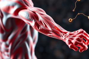
Les mouvements résultent de la contraction des muscles squelettiques striés.
Conversion d'énergie musculaire
Conversion d'énergie musculaire
Conversion de l'énergie chimique contenue dans l'ATP en énergie mécanique pour la contraction.
Phase de latence
Phase de latence
Elle correspond à la durée entre l'excitation et le début de la réponse musculaire.
Phase de contraction
Phase de contraction
Signup and view all the flashcards
Phase de relâchement
Phase de relâchement
Signup and view all the flashcards
Loi de recrutement
Loi de recrutement
Signup and view all the flashcards
Fatigue musculaire
Fatigue musculaire
Signup and view all the flashcards
Chaleur musculaire
Chaleur musculaire
Signup and view all the flashcards
Source de chaleur retardée
Source de chaleur retardée
Signup and view all the flashcards
Myofibrille
Myofibrille
Signup and view all the flashcards
Fibre musculaire
Fibre musculaire
Signup and view all the flashcards
Sarcomère
Sarcomère
Signup and view all the flashcards
Myofilaments
Myofilaments
Signup and view all the flashcards
Actine
Actine
Signup and view all the flashcards
Contraction musculaire - exigences
Contraction musculaire - exigences
Signup and view all the flashcards
rôle des ions calcium
rôle des ions calcium
Signup and view all the flashcards
Voies anaérobies immédiates
Voies anaérobies immédiates
Signup and view all the flashcards
Voies anaérobies de moyenne vitesse
Voies anaérobies de moyenne vitesse
Signup and view all the flashcards
Voies aérobies lentes
Voies aérobies lentes
Signup and view all the flashcards
Fibres de type I
Fibres de type I
Signup and view all the flashcards
Study Notes
Chapter 2: The Role of Striated Skeletal Muscle in Energy Conversion
- The body can perform multiple and varied movements resulting from the contraction of striated skeletal muscles.
- The energy required for contraction is supplied to the muscle cell by ATP molecules.
- Inside muscle cells, there is a conversion of chemical energy (ATP) into mechanical energy.
Experimental Study of Muscle Contraction
- Method of recording muscle contractions: (See document 1)
- Document 1: Recording muscle contraction in the frog
- To study the contractile activity of a muscle, the gastrocnemius muscle of a demedullated and decerebrate frog is used.
- The animal is placed on a board, ventral side against the cork, and the knee of one of the lower limbs is fixed.
- The skin of the immobilized leg is removed, and the gastrocnemius muscle and sciatic nerve are exposed.
- The lower tendon of the muscle is cut and attached to a myograph with a thread.
- Excitation electrodes are placed on the surface of the sciatic nerve or on the surface of the muscle.
- Electrical excitations are then induced, and the muscle contraction is recorded using a stylus that marks a paper fixed on a rotating cylinder with uniform and adjustable movement.
Muscle Response to Electrical Stimuli: Single Excitation
- Document 2: Response of muscle to single excitation.
- The document represents the recording obtained following the application of a single and effective excitation.
- M = Myogram; S = Excitation signal; T = Chronograph; a = amplitude. φ1 = Latency phase; φ2 = Contraction phase; φ3 = Relaxation phase.
- Describe the recorded muscular response.
- When a single, effective electrical excitation is applied directly to the muscle or its motor nerve, a brief and isolated contraction is obtained, called a muscle twitch.
- The obtained myogram consists of three phases:
- The latency phase: corresponds to the duration between the moment of excitation and the beginning of the response; this is the time required for the nerve impulse to reach the muscle.
- The contraction phase: the phase during which the length of the muscle decreases (muscle shortening).
- The relaxation phase: the phase during which the muscle returns to its initial dimensions (its duration is slightly longer than that of the contraction phase).
Muscle Response to Electrical Stimuli: Multiple Excitations at Increasing Intensity
- Document 3: Response of muscle to multiple excitations
- A frog's gastrocnemius muscle is subjected to a series of isolated excitations (i1, i2, i3, ..., i13) of increasing intensity.
- The recording cylinder is stationary, and it is turned by hand after each excitation.
- The obtained myogram is represented by the figure.
- Describe the obtained results and then establish the relationship between the intensity of the excitation and the amplitude of the response.
- Excitation (i1) does not produce a response. This excitation is therefore ineffective, as the excitation threshold has not yet been reached.
- From excitation (i2) (Excitability threshold) onwards, a response is recorded whose amplitude progressively increases. This increase in amplitude is a result of the recruitment of an increasing number of muscle units. This is the law of recruitment.
- When the excitation intensity reaches a maximum value (i12), the amplitude of the response remains constant even if the excitation intensity continues to increase, because all the units constituting the muscle are contracting.
- Therefore, there is a relationship between the intensity of the excitation and the amplitude of the response:
- A minimal muscle response is obtained when the intensity of the excitant reaches the excitation threshold, called the rheobase. From this threshold, all excitations are effective, and the amplitude of the response increases with the increase in the intensity of the excitation.
Muscle Response to Electrical Stimuli: Two Successive Close Excitations
- Document 4: Response of muscle to two close effective excitations: A muscle is subjected several times to two successive effective excitations of the same intensity (i1, i2).
- Each time, the time interval between the two excitations is reduced.
- The myograms represented by the figure are then obtained.
- Describe the recording and establish the relationship between the time interval between two successive excitations and the appearance of the muscular response.
- When the muscle is subjected to two successive effective excitations, the response differs depending on when the second excitation is applied:
- If the two excitations are sufficiently distant, two isolated muscle twitches of the same amplitude are recorded.
- If the two excitations are close together and the 2nd excitation reaches the muscle during the relaxation phase of the previous response, incomplete (partial) fusion of the two muscle twitches occurs, with an increase in the amplitude of the 2nd twitch.
- If the two excitations are very close together and the 2nd excitation reaches the muscle during the contraction phase of the previous response, complete (total) fusion of the two twitches is observed, appearing as if there is only one muscle twitch of a greater amplitude.
Muscle Response to Electrical Stimuli: Series of Successive Excitations
- Document 5: Response of muscle to a series of effective excitations: A muscle is subjected to a series of effective successive excitations of the same intensity while varying the frequency of the excitations:
- With a frequency of 12 excitations per second, the myogram in Figure 1 is obtained.
- With a frequency of 32 excitations per second, the myogram in Figure 2 is obtained.
- Compare the myograms in Figures 1 and 2 and explain the observed phenomena.
- Figure 1: When the frequency of excitations is low, the obtained myogram takes the appearance of a sinuous plateau.
- The muscle response is then called incomplete tetanus.
- This phenomenon can be explained by the incomplete fusion of muscle twitches, because each excitation reaches the muscle during the relaxation phase of the previous response.
- Figure 2: When the frequency of excitations is high, the obtained myogram takes the appearance of a straight plateau.
- The muscle response is then called perfect tetanus.
- This phenomenon can be explained by the complete fusion of muscle twitches, because each excitation reaches the muscle during the contraction phase of the previous response.
Effect of fatigue on muscle contraction
- Document 6: Muscle fatigue:
- A series of excitations of the same intensity are applied to a muscle for a very long duration.
- To obtain an overlap of the recordings, the rotation speed of the recording cylinder is adjusted in such a way that a single excitation occurs on each revolution.
- The results are represented by Figure 1 and Figure 2.
- Exploiting the data in this document, determine how muscle fatigue manifests at the level of the muscle twitch.
- It is observed from the results presented in Figure 1 and Figure 2 that there is a progressive decrease in the amplitude of muscle twitches with an increase in the duration of relaxation.
- Figure 3 shows a progressive decrease in the amplitude of the twitches until complete immobility of the muscle. Progressive fatigue of the muscle has therefore occurred.
- Muscle fatigue is therefore manifested by a decrease in the amplitude of the muscle response and by an increase in the time of relaxation.
Phenomena Accompanying Muscle Contraction
- The different aspects of muscle contraction constitute the mechanical phenomena.
- These are accompanied by thermal, chemical, and energy phenomena.
Thermal Phenomena
- a) Experimental protocol: (See document 7)
- Document 7: Thermal phenomena accompanying contraction:
- To measure the heat released during muscle contraction, Hill and Haltrée used an apparatus called a thermopile (Figure 1). The latter includes two thermoelectric needles made of two different metals (Copper-Nickel), one is introduced into the muscle, the other is maintained at a constant temperature. The temperature difference between the two needles results in a potential difference (ddp) whose value is proportional to the temperature of the contracted muscle. This ddp is translated on the oscilloscope in the form of curves (Figure 2).
- Exploiting, in parallel, the myogram and the curve of variation of the released heat, determine the different types of heat released by the muscle during muscular activity.
- Since Hill's experiment is repeated in an anaerobic environment, the release of heat 1 is observed, but heat 2 is practically zero.
Exploitation of Results
- During muscular activity, the muscle releases heat in two stages:
- Initial heat (①), which is released rapidly during the muscle twitch and of which a part is released during the contraction phase (Heat of contraction) and sustaining heat, and the other part during the relaxation phase (Relaxation heat (0)).
- Delayed heat seeps out slowly after the muscle twitch.
- The absence of delayed heat release in an anaerobic environment proves that cellular respiration is its source, whereas the origin of the initial heat is lactic fermentation.
Chemical and Energy Phenomena
- Experimental data: (See document 8)
- Document 8: Chemico-energetic phenomena accompanying muscle contraction:
- Blood analysis at muscle entry and exit both at rest and and after activity has been performed
- The results are presented in the table:
- Muscle and blood volume at rest
- O2 and CO2 volume at rest
- Glucose, protide, and lipids at rest
- Energy provided per kilogram of muscle
- O2 use per liter
- Acidic or lactic levels per gram
- During three exercises of increasing intensity the glucose consumption is measured in the leg muscles
- Glycogen levels in the lower leg
Structure and Ultrastructure of Striated Skeletal Muscle
- Structure of Striated Skeletal Muscle:
- Observations with the naked eye: (See document 9)
- Document 9: Structure of Striated Skeletal Muscle
- Annotate the diagram and identify the essential constituents of striated skeletal muscle and determine some characteristics of these constituents.
- Skeletal muscles are composed of muscle tissue, as well as nerves, blood vessels, and connective tissues. Connective tissue sheaths permeate the muscle:
- Epimysium: Surrounds the entire muscle.
- Perimysium: Surrounds bundles of muscle fibers (fascicles).
- Endomysium: Surrounds each individual muscle fiber.
- Muscles attach to bones via tendons.
- Muscles are fixed on bones
Microscopic Observations
- Document 10: Structure of skeletal striated muscle.
- The figure shows a microscopic observation of a longitudinal section of a part of a striated skeletal muscle.
- Describe the structure of the muscle fiber and justify the expression "striated muscle".
- Give a labeled explanatory drawing of the structure of the muscle fiber, of the striated skeletal muscle
- Microscopic observation shows that the muscle is made up of thousands of muscle fibers, which are elongated and multinucleated cells.
- The muscle fiber with a diameter of 10 to 100µm and a length that can reach several centimeters, is made up of a sarcoplasm containing several nuclei distributed in the periphery.
- From microscopic observation, it can be seen that the muscle tissue shows transverse striations in addition to longitudinal striations.
Ultrastructure of Skeletal Striated Muscle
- To define the ultrastructure of striated skeletal muscle, the electron microscope is used, which makes it possible to observe in detail the various organelles that make it up.
- Electron microscope observations:
- Document 11: Ultrastructure of striated skeletal muscle.
- Exploit data from the document to explain the striated appearance of muscles under microscopic observation.
- From the results of Figure 2, it can be deduced that:
- Myofibrils are made up of two types of myofilaments: Thick myofilaments made of myosin (diameter 16 nm); Thin filament of actin are 5 nanometers in diameter
- Light bands are made of actin myofilaments while dark bands are made of actin and myosin myofilaments. Myosin
Molecular Structure of Myofilaments
- The thin myofilaments consist of three types of protein:
- Actin: This is the essential constituent, it is a globular protein with a binding site for the myosin molecule on its surface.
- Troponin: Globular proteins.
- Tropomyosin: Fibrous proteins.
- Thick myofilaments are bundles of about 200 molecules of myosin. Each molecule consists of a stem and two globular heads.
Mechanism of Muscle Contraction
Microscopic Observations
- Document 13: Mechanism of Muscle Contraction
- Compare the appearance of sarcomeres at rest and in a state of contraction. Determine which changes have affected the myofibrils during contraction. What can you deduce from this comparison?
- Contraction of a sarcomere is manifested by a shortening of the sarcomeres.
- Shortening of the clear bands and H bands
- Invariant dark or sombre bands
Requirements of Muscle Contraction
- To clarify the conditions of muscle contraction, the following experiment is carried out: Isolated myofilaments are placed in a medium rich in ATP and Ca2+. Salyrgan (or a substance that blocks the hydrolysis of ATP) and a chelator (a substance that binds Ca++ ions) are added to this medium, and the myofibril tension is measured.
- Figure 2: In the presence of ATP and Ca2+ ions, an increase in myofibril tension is observed when salyrgan is added. Myofibril tension diminishes rapidly, stopping contraction. This stop in the contraction is explained with the absence of ATP hydrolysis.
- Figure 3:After addition of the chelator, the myofibril tension declines rapidly, stopping contraction, even in the presence of ATP. The contraction stop action is explained by the suppression of Ca2+ ions which interferes with operation of the same.
Regeneration of ATP During Muscle Contraction
- Evidence of ATP renewal
- Document 17: Evidence of ATP renewal
- An average ATP store is 4 to 6 mmol/kilograms.
- Exploiting document data the constant rate of ATP is compared to the energy output of muscles. These low levers suggest that ATP is generated rapidly or renewed
- Two pathways of ATP renewal:
- Experimental data
Immediate Anaerobic Pathways
- During the first minutes of muscle effort, the ATP regenerative capacities rely on anaerobic pathways without the presence of lactate (alactic)
- Transfer of a phosphate group with ADP creatine kinase
VO pathway for ADP
- With assistance of myokinase enzyme
Anaerobic Pathways of Medium Velocity
- When ATP demand rises a fermentation processes begins which is a lactate production pathway
Slow Aerobic Pathway
- During protracted and prolonged muscle contraction the body needs more O2 for which cardiac action increases and the respiratory function intervenes.
Fiber Types
- The document discusses two types of muscle fibers: type I and type II.
- Athletes involved in endurance events have muscles with more type I fibers.
- Athletes involved in brief, maximal effort have muscles with more type II fibers.
Studying That Suits You
Use AI to generate personalized quizzes and flashcards to suit your learning preferences.