Podcast
Questions and Answers
Which of the following is the MOST common cause of vision loss associated with retinal vein occlusion (RVO)?
Which of the following is the MOST common cause of vision loss associated with retinal vein occlusion (RVO)?
- Angle closure glaucoma
- Macular ischemia or edema (correct)
- Retinal detachment
- Optic nerve atrophy
What is the MOST significant risk factor associated with branch retinal vein occlusion (BRVO)?
What is the MOST significant risk factor associated with branch retinal vein occlusion (BRVO)?
- History of uveitis
- Increased cup-to-disc ratio
- Myopia
- Arterial-venous crossing changes (correct)
A patient presents with sudden, unilateral painless vision loss. Which diagnostic test is MOST crucial in differentiating between ischemic and non-ischemic RVO?
A patient presents with sudden, unilateral painless vision loss. Which diagnostic test is MOST crucial in differentiating between ischemic and non-ischemic RVO?
- Fluorescein angiography (FA) (correct)
- Visual field testing
- Amsler grid
- Optical coherence tomography (OCT)
Which of the following systemic conditions is NOT typically associated with an increased risk of RVO?
Which of the following systemic conditions is NOT typically associated with an increased risk of RVO?
In the evaluation of a young patient presenting with RVO, which of the following is the MOST important consideration regarding potential underlying etiologies?
In the evaluation of a young patient presenting with RVO, which of the following is the MOST important consideration regarding potential underlying etiologies?
Which of the following is NOT typically included in the initial lab workup for a patient diagnosed with RVO?
Which of the following is NOT typically included in the initial lab workup for a patient diagnosed with RVO?
What is the MOST appropriate initial treatment strategy for macular edema secondary to CRVO?
What is the MOST appropriate initial treatment strategy for macular edema secondary to CRVO?
In cases of RVO with neovascularization of the iris or angle, what is the MOST appropriate treatment approach?
In cases of RVO with neovascularization of the iris or angle, what is the MOST appropriate treatment approach?
When is prophylactic PRP recommended in ischemic CRVO?
When is prophylactic PRP recommended in ischemic CRVO?
What is the PRIMARY mechanism behind arteriovenous compression in central retinal vein occlusion (CRVO)?
What is the PRIMARY mechanism behind arteriovenous compression in central retinal vein occlusion (CRVO)?
Which finding is MOST indicative of ischemic CRVO compared to non-ischemic CRVO?
Which finding is MOST indicative of ischemic CRVO compared to non-ischemic CRVO?
A patient presents with mild blurring that is worse upon awakening and improves during the day. Examination reveals mild dilation and tortuosity of veins with scattered flame-shaped hemorrhages. Which condition is MOST likely?
A patient presents with mild blurring that is worse upon awakening and improves during the day. Examination reveals mild dilation and tortuosity of veins with scattered flame-shaped hemorrhages. Which condition is MOST likely?
In hemiretinal vein occlusion (HRVO), which area of the retina is typically affected?
In hemiretinal vein occlusion (HRVO), which area of the retina is typically affected?
What is the PRIMARY mechanism behind the development of branch retinal vein occlusion (BRVO) at an arteriovenous crossing point?
What is the PRIMARY mechanism behind the development of branch retinal vein occlusion (BRVO) at an arteriovenous crossing point?
A patient with BRVO presents with chronic macular edema and a visual acuity of 20/50. What is the MOST appropriate treatment if macular perfusion is present?
A patient with BRVO presents with chronic macular edema and a visual acuity of 20/50. What is the MOST appropriate treatment if macular perfusion is present?
What is the INITIAL step in managing a patient presenting with acute central retinal artery occlusion (CRAO)?
What is the INITIAL step in managing a patient presenting with acute central retinal artery occlusion (CRAO)?
Which of the following emboli is MOST likely to originate from the carotid arteries?
Which of the following emboli is MOST likely to originate from the carotid arteries?
A patient presents with a history of transient monocular vision loss described as a "curtain coming down" in one eye. This symptom is MOST indicative of:
A patient presents with a history of transient monocular vision loss described as a "curtain coming down" in one eye. This symptom is MOST indicative of:
Following a CRAO, which of the following changes is typically observed in the retina during the acute phase?
Following a CRAO, which of the following changes is typically observed in the retina during the acute phase?
What finding on ERG is MOST consistent with a central retinal artery occlusion (CRAO)?
What finding on ERG is MOST consistent with a central retinal artery occlusion (CRAO)?
In cases of recurrent branch retinal artery occlusion (BRAO), which of the following conditions MUST be considered and ruled out?
In cases of recurrent branch retinal artery occlusion (BRAO), which of the following conditions MUST be considered and ruled out?
What is the MOST appropriate management strategy for iris neovascularization secondary to CRAO?
What is the MOST appropriate management strategy for iris neovascularization secondary to CRAO?
A patient with ophthalmic artery occlusion (OAO) is MOST likely to present with which of the following?
A patient with ophthalmic artery occlusion (OAO) is MOST likely to present with which of the following?
Which of the following best characterizes Susac syndrome?
Which of the following best characterizes Susac syndrome?
Which symptom is MOST commonly associated with ocular ischemic syndrome?
Which symptom is MOST commonly associated with ocular ischemic syndrome?
Which of the following is a less typical fundus finding in ocular ischemic syndrome?
Which of the following is a less typical fundus finding in ocular ischemic syndrome?
What is the MOST appropriate initial diagnostic test to evaluate for the underlying systemic cause of ocular ischemic syndrome?
What is the MOST appropriate initial diagnostic test to evaluate for the underlying systemic cause of ocular ischemic syndrome?
How does retinopathy associated with ocular ischemic syndrome differ from central retinal vein occlusion (CRVO)?
How does retinopathy associated with ocular ischemic syndrome differ from central retinal vein occlusion (CRVO)?
The BRAVO trial demonstrated the efficacy of which treatment for macular edema secondary to BRVO?
The BRAVO trial demonstrated the efficacy of which treatment for macular edema secondary to BRVO?
The SCORE-CRVO study showed that patients treated with which of the following were more likely to have a substantial visual gain at 1 year?
The SCORE-CRVO study showed that patients treated with which of the following were more likely to have a substantial visual gain at 1 year?
What is the PRIMARY advantage of using optical coherence tomography angiography (OCTA) over traditional fluorescein angiography (FA) in evaluating RVO?
What is the PRIMARY advantage of using optical coherence tomography angiography (OCTA) over traditional fluorescein angiography (FA) in evaluating RVO?
Which of the following is NOT a typical component of the proposed interventions for acute central retinal artery occlusion (CRAO)?
Which of the following is NOT a typical component of the proposed interventions for acute central retinal artery occlusion (CRAO)?
A patient presents with a history of hypertension, hyperlipidemia, and diabetes. Which of these conditions is LEAST likely to be a direct risk factor for central retinal artery occlusion (CRAO)?
A patient presents with a history of hypertension, hyperlipidemia, and diabetes. Which of these conditions is LEAST likely to be a direct risk factor for central retinal artery occlusion (CRAO)?
What class of medications may RVO patients stop taking?
What class of medications may RVO patients stop taking?
Which of these are a sign/symptom of impending CRVO?
Which of these are a sign/symptom of impending CRVO?
Aside from visual field testing and pupil dilation, what is another additional exam that is crucial to perform at the follow-ups in RVO?
Aside from visual field testing and pupil dilation, what is another additional exam that is crucial to perform at the follow-ups in RVO?
The VIBRANT trial demonstrated that what is effective for macular edema in BRVO?
The VIBRANT trial demonstrated that what is effective for macular edema in BRVO?
What is the role of digital ocular massage and breathing into a paper bag for retinal arterial occlusion and why?
What is the role of digital ocular massage and breathing into a paper bag for retinal arterial occlusion and why?
Flashcards
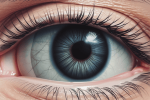
Retinal Venous Occlusion (RVO)
Retinal Venous Occlusion (RVO)
Decrease in venous outflow in the retinal circulation, leading to retinal vascular leakage, macular edema, and intraretinal hemorrhages.
RVO Main Risk Factors
RVO Main Risk Factors
Older age (>65 y/o is 50% of cases). Systemic vascular diseases and prior RVO.
BRVO Main Risk Factors
BRVO Main Risk Factors
Arterial-venous crossing changes (AR retinopathy) and high IOP.
RVO Signs and Symptoms
RVO Signs and Symptoms
Signup and view all the flashcards
RVO Testing Methods
RVO Testing Methods
Signup and view all the flashcards
RVO Required Lab Work
RVO Required Lab Work
Signup and view all the flashcards
Retinal Imaging for RVO
Retinal Imaging for RVO
Signup and view all the flashcards
RVO Differentials
RVO Differentials
Signup and view all the flashcards
RVO Treatment
RVO Treatment
Signup and view all the flashcards
Anti-VEGF for BRVO
Anti-VEGF for BRVO
Signup and view all the flashcards
Vabysmo Function
Vabysmo Function
Signup and view all the flashcards
Macular Edema Treatment
Macular Edema Treatment
Signup and view all the flashcards
Treatment for NVI/NVA
Treatment for NVI/NVA
Signup and view all the flashcards
RVO Education
RVO Education
Signup and view all the flashcards
Central Retinal Vein Occlusion (CRVO)
Central Retinal Vein Occlusion (CRVO)
Signup and view all the flashcards
CRVO Etiology
CRVO Etiology
Signup and view all the flashcards
Non-Ischemic CRVO Signs
Non-Ischemic CRVO Signs
Signup and view all the flashcards
Ischemic CRVO Signs
Ischemic CRVO Signs
Signup and view all the flashcards
Ischemic CRVO Neovascularization
Ischemic CRVO Neovascularization
Signup and view all the flashcards
Impending CRVO Signs
Impending CRVO Signs
Signup and view all the flashcards
Hemiretinal Vein Occlusion (HRVO)
Hemiretinal Vein Occlusion (HRVO)
Signup and view all the flashcards
BRVO Risk Factors
BRVO Risk Factors
Signup and view all the flashcards
BRVO Signs
BRVO Signs
Signup and view all the flashcards
Late BRVO Signs
Late BRVO Signs
Signup and view all the flashcards
BRVO Differentials
BRVO Differentials
Signup and view all the flashcards
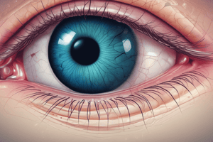
Retinal Arterial Occlusions (RAO)
Retinal Arterial Occlusions (RAO)
Signup and view all the flashcards
RAO Cause
RAO Cause
Signup and view all the flashcards
Emboli Types
Emboli Types
Signup and view all the flashcards
Rule out for RAO
Rule out for RAO
Signup and view all the flashcards
CRAO Symptoms
CRAO Symptoms
Signup and view all the flashcards
Amaurosis Fugax
Amaurosis Fugax
Signup and view all the flashcards
CRAO Signs
CRAO Signs
Signup and view all the flashcards
OCT-CRAO
OCT-CRAO
Signup and view all the flashcards
OCTA findings in CRAO
OCTA findings in CRAO
Signup and view all the flashcards
ERG
ERG
Signup and view all the flashcards
Susac Syndrome
Susac Syndrome
Signup and view all the flashcards
Acute RAO Management
Acute RAO Management
Signup and view all the flashcards
Treat RAO
Treat RAO
Signup and view all the flashcards
Proposed RAO Interventions
Proposed RAO Interventions
Signup and view all the flashcards
Susac Syndrome Workup
Susac Syndrome Workup
Signup and view all the flashcards
Study Notes
- Retinal Venous Occlusion (RVO) involves decreased venous outflow, leading to retinal vascular leakage, macular edema, and intraretinal hemorrhages.
- Vision loss in RVO is associated with macular ischemia/edema, retinal hemorrhages, vitreous hemorrhage, epiretinal membrane formation, rubeosis iridis, and neovascular glaucoma.
- Additional findings include retinal arterial macroaneurysm formation and cilioretinal artery occlusions.
RVO Risk Factors
- Older age is the main risk factor for both CRVO and BRVO, with 50% of patients being over 65 years old.
- A prior RVO in one eye increases the risk of RVO in the fellow eye.
- Patients with a BRVO in one eye have a 10% risk of developing an RVO in the fellow eye over 3 years.
- Arteriovenous crossing changes and high IOP are the primary risks for BRVO.
- Systemic conditions like HBP, hyperlipidemia, diabetes, and coronary artery disease are also risk factors for BRVO.
Classification
- RVO can be classified by the extent of retinal ischemia.
Signs and Symptoms
- Sudden onset unilateral painless vision loss occurs, with the degree of loss depending on whether the RVO is ischemic or non-ischemic.
- Vision worse than 20/200 is associated with non-perfusion (ischemic RVO).
Testing
- FA helps evaluate the degree of ischemia, defined as eyes with >10 disc areas of capillary nonperfusion and extensive retinal hemorrhages.
- OCT is used for detecting macular edema.
- OCTA accurately evaluates changes in microvasculature, differentiating between ischemic and non-ischemic RVO.
- VF testing is part of testing.
Evaluation
- Complete medical and ocular history, retinal imaging, and lab work are needed.
- Lab work consists of CBC, glucose tolerance test, lipid profile, serum protein electrophoresis, chemistry profile, hematologic tests, syphilis serology, and thrombophilic screening.
Retinal Imaging
- FA is essential to detect non-perfused capillary areas and the extent of macular ischemia.
- OCT quantifies the presence and extent of macular edema.
- OCTA accurately evaluates changes in microvasculature.
Differentials
- Ocular Ischemic Syndrome & retinopathy of carotid occlusive disease
- Diabetic Retinopathy DOES NOT respect horizontal
- Papilledema, papillophlebitis
- Radiation therapy
RVO Treatment
- Treat underlying medical conditions, discontinue oral contraceptives, ensure hydration, and consider alternative diuretics.
- Reduce IOP, if elevated.
- Anti-VEGF injections treat macular edema and neovascularization.
- PRP is indicated if NVD or retinal neovascularization is present.
- Aspirin is often recommended, though its efficacy hasn't been proven.
Anti-VEGF
- Vabysmo is a bispecific antibody approved for ME following RVO, with inhibitory effects on both VEGF-A and Ang-2.
Steroids
- Steroids can treat macular edema associated with RVO, but carry risks of cataracts and glaucoma.
Macular Edema Treatment
- First-line treatment involves anti-VEGF therapy with intravitreal bevacizumab, ranibizumab, aflibercept, or faricimab-svoa.
- Second-line treatments include intravitreal triamcinolone and dexamethasone implant.
Neovascularization Treatment
- Peripheral PRP is recommended for patients with NVI and NVA angle.
Laser Photocoagulation
- BVOS demonstrated the efficacy of grid laser photocoagulation surgery for macular edema due to BRVO.
- CVOS did not show any value of focal photocoagulation for macular edema in patients with CRVO.
Follow Up
- Educate patients on the 8-10% risk of an RVO in the other eye.
- Follow-up intervals vary based on VA, with frequent monitoring for neovascularization.
Central Retinal Vein Occlusion (CRVO)
- CRVO: occlusion of the central retinal vein at or posterior to the lamina cribosa.
- Most patients with CRVO are over 50 and men get this slightly more than women.
- Younger patients often have an inflammatory cause and a more benign course.
- Systemic HTN, DM, and open-angle glaucoma are risk factors.
Etiology
- A/V mechanical compression occurs.
- Occlusion at the lamina cribosa occurs.
- Blood disorders & inflammation are causes.
Non-Ischemic CRVO
- VA is better than 20/400.
- Has no/mild RAPD.
- Signs: tortuous and dilated veins, dot & blot & flame-shaped hemorrhages, some cotton wool spots, disc edema, macular edema.
- Often converts to ischemic form.
Ischemic CRVO
- VA is worse than 20/400.
- RAPD is present.
- ERG shows decreased b wave amplitude.
- VF shows constriction.
- Signs: retinal hemorrhages (4 quadrants), dilated & tortuous retinal veins, superficial hemorrhages, CWS, and macular edema.
- Neovascularization (anterior > posterior) occurs.
- Can have a vitreous hemorrhage.
- Neovascular glaucoma occurs in 60% of patients.
Impending CRVO
- Mild blurring, worse on awakening and improves during the day are symptoms.
- Mild dilation and tortuosity of veins and scattered flame-shaped hemorrhages are signs.
Hemiretinal Vein Occlusion (HRVO)
- HRVO is an occlusion at the disc involving half of the retinal venous drainage.
- Vascular tortuosity and dilation, retinal and macular edema & retinal hemorrhages noted.
- Late findings: collaterals, disc edema, neovascularization, vitreous hemorrhage, neovascular glaucoma.
Branch Retinal Vein Occlusions (BRVO)
- BRVOs occur at arteriovenous crossing points.
- Vein compression causes endothelial cell loss, thrombus formation and potential occlusion.
- Complete or partial obstruction at a branch of the CRV.
- Have sudden, onset, unilateral painless decreased vision loss w/ a VF defect.
Signs of BRVO
- Vein dilated & tortuous
- Occlusion site at AV cross point
- Flame shape, dot/blot heme
- Retinal edema
- May have CWS in area
BRVO Late Clinical Findings:
- Collaterals
- Sclerosed vessels, exudate
- Neovascularization: posterior segment > anterior segment that may lead to recurrent vitreous heme or pre-retinal heme, and occasionally tractional retinal detachment.
- Macular edema; chronic macular edema most common cause of persistent poor VA.
Retinal Arterial Occlusions (RAO)
- RAO is a blockage of one or more arteries of the retina.
- Treated as a brain stroke.
- GCA should be suspected in patients over 50 years of age, consider urgent systemic corticosteroid therapy when GCA is diagnosed.
- Partial or complete obstruction of any of the branch tributaries of the CRA.
Pathophysiology
- Emboli are the MOST COMMON CAUSE!
- Non-embolic: Thrombosis, Atherosclerosis, Vasculitis
Risk Factors
- Common ones: HTN, Hyperlipidemia, Carotid occlusive disease, Cardiac disease, Diabetes
Central Retinal Artery Occlusion
- Impending stroke = patient needs to be sent to hospital immediately
- Causes sudden, unilateral painless decrease in vision and field of vision.
- Preceded by amaurosis fugax.
- Early signs: RAPD, emboli, whitening of the posterior pole of retina, cherry red spot in the macula & box-carring or segmentation of arterioles.
- OCt shows thickening & increased inner retinal reflectivity may be present, followed by inner retinal thinning in chronic RAO
- FA shows a delayed arm to retina time period.
Branch Retinal Artery Occlusion
- Causes Unilateral, painless, sudden partial VF loss
Management
- Acute, symptomatic artery occlusions with visible emboli should prompt an immediate referral to the nearest stroke referral center!
- Occlusion beyond the 240 minutes results in catastrophic, irreversible retinal damage.
- Digital ocular massage as well as topical pressure lowering therapies is proposed as an intervention.
Ophthalmic Artery Occlusion
- Causes a Diffusely pale retina with No cherry red spot and LP/NLP in vision.
- GCA is likely!
Susac Syndrome
- Usually affects young women and involves BRAO, hearing loss & headaches with focal neurologic signs. AWH = Arterial Wall Hyperflourescence and Gass plaques can be seen
Ocular Ischemic Syndrome
- Severe blood restriction to the anterior and posterior ocular segments.
- Usually monocular in 80% of cases.
- Causes vision loss and ocular angina.
- Retinal arteries narrowing and dilated veins with no signs of tortuosity.
- Retinopathy of occlusion, Hypoperfusion retinopathy/venous stasis retinopathy veins are dilated and not tortuous & in the mid-periphery
Studying That Suits You
Use AI to generate personalized quizzes and flashcards to suit your learning preferences.