Podcast
Questions and Answers
What is the primary shape of the external nose?
What is the primary shape of the external nose?
- Cylindrical
- Spherical
- Pyramidal (correct)
- Cuboidal
Which structure forms the medial wall of the nasal cavity?
Which structure forms the medial wall of the nasal cavity?
- Ala of the nose
- Dorsum of the nose
- Nasal septum (correct)
- Piriform aperture
What is the primary blood supply to the alar and septal regions of the nose?
What is the primary blood supply to the alar and septal regions of the nose?
- Facial artery (correct)
- Dorsal nasal branch
- Ophthalmic artery
- Maxillary artery
Which nerve provides sensory innervation to the external nose?
Which nerve provides sensory innervation to the external nose?
What type of tissue lines the vestibule of the nasal cavity?
What type of tissue lines the vestibule of the nasal cavity?
What is the approximate height of each half of the nasal cavity?
What is the approximate height of each half of the nasal cavity?
Which structure forms the roof of the nasal cavity?
Which structure forms the roof of the nasal cavity?
Which of the following structures contributes to the formation of the nasal septum?
Which of the following structures contributes to the formation of the nasal septum?
What type of cartilage is found in the nasal septum?
What type of cartilage is found in the nasal septum?
In the context of the nasal septum, what is the significance of Kiesselbach's area?
In the context of the nasal septum, what is the significance of Kiesselbach's area?
Through which vein does the mobile part of the septum drain?
Through which vein does the mobile part of the septum drain?
How is the olfactory zone innervated in the nasal septum?
How is the olfactory zone innervated in the nasal septum?
What are the three parts into which the lateral wall of the nasal cavity can be subdivided?
What are the three parts into which the lateral wall of the nasal cavity can be subdivided?
What is the primary function of the hairs (Vibrissae) found in the vestibule of the lateral nasal wall?
What is the primary function of the hairs (Vibrissae) found in the vestibule of the lateral nasal wall?
Which structure is located above the superior concha?
Which structure is located above the superior concha?
In which nasal meatus does the nasolacrimal duct terminate?
In which nasal meatus does the nasolacrimal duct terminate?
Which sinus(es) opens directly into the middle meatus?
Which sinus(es) opens directly into the middle meatus?
The olfactory nerve is responsible for special sensory innervation of which part of the lateral nasal wall?
The olfactory nerve is responsible for special sensory innervation of which part of the lateral nasal wall?
What anatomical structure is sometimes affected by infection spreading from the nasal cavity through the cribriform plate?
What anatomical structure is sometimes affected by infection spreading from the nasal cavity through the cribriform plate?
What condition results from any side of septal deviation, causing congestion and obstruction?
What condition results from any side of septal deviation, causing congestion and obstruction?
Which area is described as where four arteries anastomose to form a vascular plexus, and is a common site for nosebleeds?
Which area is described as where four arteries anastomose to form a vascular plexus, and is a common site for nosebleeds?
Which of the following conditions might be associated with hypertrophy of mucosa over the inferior nasal concha?
Which of the following conditions might be associated with hypertrophy of mucosa over the inferior nasal concha?
What is the typical length of the pharynx?
What is the typical length of the pharynx?
The pharynx extends from the cranial base to which of the following anatomical landmarks?
The pharynx extends from the cranial base to which of the following anatomical landmarks?
What structure marks the communication point between the oral cavity and the pharynx?
What structure marks the communication point between the oral cavity and the pharynx?
Which feature is associated with the nasopharynx?
Which feature is associated with the nasopharynx?
In relation to the vertebral column, where does the oropharynx lie?
In relation to the vertebral column, where does the oropharynx lie?
The buccopharyngeal fascia coats which part of the pharynx?
The buccopharyngeal fascia coats which part of the pharynx?
An anatomical weak spot in the pharyngeal wall between the thyropharyngeus and cricopharyngeus muscles is known as:
An anatomical weak spot in the pharyngeal wall between the thyropharyngeus and cricopharyngeus muscles is known as:
What is the main function of constrictor muscles of pharynx?
What is the main function of constrictor muscles of pharynx?
By which nerve are all constrictors of the pharynx supplied?
By which nerve are all constrictors of the pharynx supplied?
What kind of spaces are paranasal sinuses?
What kind of spaces are paranasal sinuses?
How many groups of sinuses are there?
How many groups of sinuses are there?
When do Frontal and Sphenoidal sinuses typically develop?
When do Frontal and Sphenoidal sinuses typically develop?
Paranasal sinuses are lined with:
Paranasal sinuses are lined with:
Which bone does NOT contain paranasal sinuses?
Which bone does NOT contain paranasal sinuses?
Which of the following statements best describes the function of paranasal sinuses?
Which of the following statements best describes the function of paranasal sinuses?
Why is it that people may have discomfort in talking when they are sick?
Why is it that people may have discomfort in talking when they are sick?
What is the difference between acute and chronic sinusitis?
What is the difference between acute and chronic sinusitis?
If a patient has a deviated nasal septum that is causing significant nasal congestion, which of the following interventions would be the most appropriate initial step?
If a patient has a deviated nasal septum that is causing significant nasal congestion, which of the following interventions would be the most appropriate initial step?
A patient complains of frequent nosebleeds originating from the anterior part of the nasal septum. Damage to which area is most likely the cause?
A patient complains of frequent nosebleeds originating from the anterior part of the nasal septum. Damage to which area is most likely the cause?
A patient reports a loss of smell following a head trauma. Which of the following structures is most likely damaged?
A patient reports a loss of smell following a head trauma. Which of the following structures is most likely damaged?
A patient with a severe sinus infection experiences vision changes and is suspected of having an intracranial spread of infection. Which anatomical structure is the most likely route for the spread of infection?
A patient with a severe sinus infection experiences vision changes and is suspected of having an intracranial spread of infection. Which anatomical structure is the most likely route for the spread of infection?
During a physical examination, the doctor notes inflammation of the nasal mucosa, particularly around the openings into the paranasal sinuses. Where is the most likely location of this inflammation?
During a physical examination, the doctor notes inflammation of the nasal mucosa, particularly around the openings into the paranasal sinuses. Where is the most likely location of this inflammation?
A surgeon is planning a procedure involving the lateral nasal wall. Which structure must the surgeon be cautious of damaging to prevent the entry of tears in the nasal cavity?
A surgeon is planning a procedure involving the lateral nasal wall. Which structure must the surgeon be cautious of damaging to prevent the entry of tears in the nasal cavity?
Following a motor vehicle accident, a patient presents with cerebrospinal fluid (CSF) rhinorrhea. Which anatomical structure has most likely been damaged?
Following a motor vehicle accident, a patient presents with cerebrospinal fluid (CSF) rhinorrhea. Which anatomical structure has most likely been damaged?
A patient is diagnosed with a condition affecting the pharyngeal branch of the vagus nerve. Which of the following muscles would be most directly affected by this condition?
A patient is diagnosed with a condition affecting the pharyngeal branch of the vagus nerve. Which of the following muscles would be most directly affected by this condition?
A patient who has difficulty swallowing is found to have a weakened area in the posterior pharyngeal wall. Which anatomical area is most likely compromised?
A patient who has difficulty swallowing is found to have a weakened area in the posterior pharyngeal wall. Which anatomical area is most likely compromised?
During an endoscopic examination of the nasal cavity, a doctor identifies the sphenoethmoidal recess. Where should the endoscope be directed to visualize this recess?
During an endoscopic examination of the nasal cavity, a doctor identifies the sphenoethmoidal recess. Where should the endoscope be directed to visualize this recess?
Which aspect of the nasal cavity does the Palatine bone contribute to?
Which aspect of the nasal cavity does the Palatine bone contribute to?
What would be the result of damage of the olfactory nerve?
What would be the result of damage of the olfactory nerve?
Which of the options best describes the location of the Oropharynx?
Which of the options best describes the location of the Oropharynx?
What is the normal length of the pharynx?
What is the normal length of the pharynx?
Flashcards
Learning Outcomes
Learning Outcomes
Provides introduction and functions of the respiratory tract, structural and functional divisions, innervation, and the structure and functions of related organs.
External Nose
External Nose
The visible part of the nose, shaped like a pyramid.
Nostrils (Nares)
Nostrils (Nares)
Pair of openings in the external nose.
Median Nasal Septum
Median Nasal Septum
Signup and view all the flashcards
Vestibule of Nose
Vestibule of Nose
Signup and view all the flashcards
Nasal Cavity Roof
Nasal Cavity Roof
Signup and view all the flashcards
Nasal Cavity Floor
Nasal Cavity Floor
Signup and view all the flashcards
Nasal Septum Function
Nasal Septum Function
Signup and view all the flashcards
Nasal Septum Bony Part
Nasal Septum Bony Part
Signup and view all the flashcards
Nasal Septum Cartilaginous Part
Nasal Septum Cartilaginous Part
Signup and view all the flashcards
Kiesselbach's Area
Kiesselbach's Area
Signup and view all the flashcards
Venous Drainage
Venous Drainage
Signup and view all the flashcards
Nasal Meatuses and Conchae
Nasal Meatuses and Conchae
Signup and view all the flashcards
Nasal Conchae (Turbinate Bones)
Nasal Conchae (Turbinate Bones)
Signup and view all the flashcards
inferior meatus
inferior meatus
Signup and view all the flashcards
Superior Meatus
Superior Meatus
Signup and view all the flashcards
Superior Meatus drainage
Superior Meatus drainage
Signup and view all the flashcards
Sinusitis
Sinusitis
Signup and view all the flashcards
Paranasal Air Sinuses
Paranasal Air Sinuses
Signup and view all the flashcards
Functions of Paranasal Sinuses
Functions of Paranasal Sinuses
Signup and view all the flashcards
Pharynx Description
Pharynx Description
Signup and view all the flashcards
Pharynx Extent
Pharynx Extent
Signup and view all the flashcards
Features of Nasopharynx
Features of Nasopharynx
Signup and view all the flashcards
Features of oropharynx
Features of oropharynx
Signup and view all the flashcards
How wall of pharynx are formed
How wall of pharynx are formed
Signup and view all the flashcards
what are the arteries to the pharynx?
what are the arteries to the pharynx?
Signup and view all the flashcards
Bleeding from Nose (Epistaxis)
Bleeding from Nose (Epistaxis)
Signup and view all the flashcards
Killian's Dehiscence
Killian's Dehiscence
Signup and view all the flashcards
Trachea and Bronchial Tree
Trachea and Bronchial Tree
Signup and view all the flashcards
Study Notes
- The lecture is about the anatomy of the respiratory tract
Objectives of the Lecture
- Introduction and functions of the respiratory tract
- Structural and functional divisions of the respiratory tract
- Innervation of the respiratory tract
- Structure and functions of the nose, pharynx, larynx, trachea, and their divisions
Parts of Upper Respiratory Tract
- Frontal sinus
- Superior, middle, inferior nasal conchae
- Sphenoidal sinus
- Nasopharynx
- Oropharynx
- Laryngopharynx
- Tongue
- Epiglottis
- Trachea
- Esophagus
Nose - Parts
- External Nose: Pyramidal in shape with parts including the apex (tip), root, and dorsum
- Nostrils/Nares: Pair of piriform apertures
- Nasal Cavity:
- Medial wall of the nasal cavity is made by the nasal septum
- Medially, there is the nasal septum
- Laterally, there is the ala of the nose
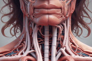
Blood Supply and Innervation of the Nose
- Arteries: Dorsal nasal branch (ophthalmic), infraorbital (maxillary), alar, and septal (facial)
- Sensory Innervation: External nasal, infratrochlear (opthalmic), and infraorbital (maxillary)
- Motor Innervation: Facial nerve branches
Nasal Cavity
- Divided into 2 halves by the median nasal septum
- Asymmetrical, with elevations and depressions representing the lateral wall
- Extends from the nostrils to the choanae
- Vestibule: The part of the nasal cavity close to the nostrils
- Lined by skin with hairs and sebaceous glands
Nasal Cavity - Extent and Division
- External nares are anterior and the posterior nasal aperture (choanae) is posterior
- Nasal septum consists of both bony and cartilaginous parts
- Anteriorly, the nasal septum is made up of nasal cartilage
- Posteriorly, the nasal septum is bone which is vomer
Nasal Cavity - Halves
- Subdivided into two halves
- Each half has a roof, floor, medial wall, and lateral wall
- The roof is made of a hard pallet and the ethmoid bone
- The floor is formed by the hard pallet (anteriorly) and the soft pellet (posteriorly)
- The medial wall is the nasal septum
- Each half measures:
- 5cm in height
- 5-7cm in length from slope to slope
- 1.5cm width near the floor
- 1-2mm near the roof
Nasal Cavity - Roof
- Anterior slope consists of the nasal bone and nasal catilage
- Posterior consists of the inferior surface of the sphenoid bone
- Ethmoid : cribriform plate
Nasal Cavity - Roof
- 7cm long
- 2mm wide
- Slopes downwards both in front and behind
- Middle horizontal part: cribriform plate of ethmoid
- Anterior slope forms from the nasal part of frontal bone, nasal bone, and nasal cartilages
- Posterior slope forms from the inferior surface of the body of sphenoid
Nasal Cavity - Floor
- 5cm long and 1.5cm wide
- Floor is made of anterior hard pallat and posterior hard packet
- Formed by the palatine process of the maxilla and the horizontal plate of the palatine bone
- Concave from side to side
Nasal Septum (Medial Wall)
- Median osseocartilaginous partition between the two halves of the nasal cavity
- Covered by mucous membrane on each side and forms the medial wall of both nasal cavities
- Consists of bony and cartilaginous parts
Bony Nasal Septum
- Formation includes the perpendicular plate of the ethmoid and the vomer
Cartilaginous Nasal Septum
- Includes the septal cartilage and inferior nasal cartilage (which one can touch)
Cuticular Nasal Septum
- Includes the fibro-fatty tissue and skin lining the anterior part
- The columella, lined by skin, contains sebaceous glands and the vomero-nasal organ of Jacobson
Nasal Septum - Arterial Supply
- The blood drains into the medial wall
- Bleeds Heavily: because 80 many arteries and anastomosing
- Includes the Kiesselbach’s plexus
Venous Drainage
- Accompany by olfactory nerves into inferior cerebral veins
- Superior: Superior ophthalmic vein
- Mobile part of the septum : Facial vein to IJV (dangerous area of face), dental ligures can spread from tooth
- Postero- inferior: Pterygoid venous plexus (surrounds mandible) one of the peripusal
Nasal Septum: Venous Drainage
- Includes the Kiesselbach's plexus
- This drains into the Facial vein which drains into the sphenopalatine vein which drains into the pterygoid venous plexus
Nasal Septum - Nerve Supply
- Only supplied by Olfactory nerves
- Long Sphenopalatine
- Short Sphenopalatine and Greater palatine N. (Pterygo palatine ganglion & Maxillary N)
Nasal Septum: Sensory Nerve Supply
- Involves special sensory and general sensory
Lateral Wall
- Subdivided into 3 parts: vestibule, atrium, and concha
- A small depressed area in the anterior part called the vestibule where hair folicules are located
- Vestibule is lined by modified skin containing short, stiff, curved hairs called vibrissae and contain sweat and sebaceous glands
- Middle part is the atrium of middle meatus
- Posterior part contains the concha: elevations
- Spaces separating the concha called Meatuses: depressions
Lateral Wall of Nose
- Agger nasi: mucous ridge
- Atrium
- Of middle meatus
- Limen nasi: mucocutaneous ridge
- Vestibule
Nasal Conchae (Turbinate Bones)
- Inferior Concha is a separate bone that extends horizontally backwards
- Ends 1.25cm in front of the pharyngeal opening of the auditory tube
- Anteriorly continuous as the vestibule
- Space under the inferior concha is the inferior meatus
- This is the termination of the naso lacrimal duct
- Sinos open into inferior meatus
Middle Concha
- Middle meatus is under middle concha
- This is a bony bulging of the middle meatus , receives opening of middle ethamoidal air sinuses
- Anterior ethmoidal + frontal sinus sinuses open here
- A short passage at anterior end of the hiatus
Superior Concha
- Shortest concha
- Superior meatus- Posterior ethamoidal sinus opens
- Superior meatus is b/w superior concha & roof of nasal cavity
- Sphenoethamoidal recess is the depression above &behind the superior concha
- Sphenoidal air sinuses opens
Nasal Passage
- Inferior meatus drains the nasolacrimal duct
- Middle meatus drains the frontal sinus, maxillary sinus, anterior ethmoidal cells, middle ethmoidal cells
- Superior meatus drains posterior ethmoidal cells
- Sphenoethmoidal Recess drains Sphenoidal sinus
Lateral Wall of Nose - Arterial Supply
- Involves the Anterior ethmoidal, Posterior ethmoidal, Facial, and sphenopalatine
Lateral Wall of Nose - Nerve Supply
- Anterior ethmoidal : ophthalmic
- Anterior superior alveolar: maxillary
- Posterior superior lateral nasal: Pterygopalatine ganglion (maxillary nerve)
- Anterior palatine: Pterygopalatine ganglion (maxillary nerve)
Olfactory Nerve Supply
- Olfactory nerve is the special sensory nerve for the lateral wall of the nose
Applied Anatomy - Spread of Infection
- Middle cranial fossa via the cribriform plate (infection can go from uge skull)
- Lacrimal apparatus causes Nasolacrimal duct
Applied Anatomy
- Septal deviation of septum any side causes congestion
- Septal deviations corrected by sub mucosal resection of the nasal septum
- Epistaxis - bleeding from the nose, can lead to increase of intracranial pressure
- In older people it is important to go to the hospital for epistaxis
- Dangerous area of the nose infection to the cranial cavity via the olfactory nerves
- Broken nose - nasal fractures that typically occur over the bridge of the nose or in the septum, which is the area that divides your nostrils.
Applied Anatomy - Pathologies of the Nose
- DNS- Deviated Nasal Septum is an abnormal shift in the septum Nasal Polyp - Touched mucous membrane becomes inflamed + enlarged which can be surgical
Little's Area
- Kiesselbach's plexus is in the Kiesselbach's area/triangle, or Little's area
- Region in the anteroinferior part of the nasal septum where four arteries anastomose to form a vascular plexus.
- Common site of bleeding from nose (Epistaxis).
- Involves 4 Arteries:
- Anterior + posterior ethmoidal
- Sphenopalatine
- Greater palatine artery
Applied Anatomy - Allergic Rhinitis
- Hypertrophy of mucosa over inferior nasal concha causing constant sneezing
Pharynx
- The pharynx is a 12-14 cm long
- The pharynx lies behind, and communicates with, the nasal, oral and laryngeal cavities via the nasopharynx, oropharynx and laryngopharynx, respectively.
- It is a musculomembranous tube shaped like an inverted cone
- Extends from the cranial base to the lower border of the cricoid cartilage (at the level of the sixth cervical vertebra), where it becomes continuous with the esophagus.
- Communication between oral cavity & pharynx is oropharyngeal isthmus
- Divisible into nasopharynx, oropharynx,& laryngopharynx.
Features on Nasopharynx:
- Includes opening of auditory tube
- Includes tubal elevation and salpingopharyngeal & salpingopalatine fold
- Communication between naso & oropharynx is pharyngeal isthmus Tubal tonsil & pharyngeal tonsil (adenoids) are present
Features on Oropharynx:
- Lies in front of C2 & upper part of C3 vertebra
- Palatopharyngeal folds/arches
- Palatine tonsil & tonsillar sinus
Pharynx Anatomy
- Isthmus: b/w naso toro pharnyx
- Includes the Nasal Cavity, tongues, lingual tonsils, nasopharynx, oropharynx, laryngopharynx etc
Features on Laryngopharynx:
- Lies in front of C3-C6 vertebra- entry point in blarynx (larynx)
- Upper part formed by inlet of larynx
- Below and posterior surfaces by arytenoid & cricoid cartilages
Wall of Pharynx:
- Formed by skeletal muscles and fascia
- Buccopharyngeal fascia coats outside of muscle wall
- Pharyngobasilar fascia lines inside of the muscle wall
Constrictors Muscles of Pharynx:
- Includes the superior middle & inferior constrictors
- All originate posterior to nasal, oral & laryngeal cavities
- Fibers pass to posterior & lateral walls of pharynx meeting to form midline raphe.
- Longitudinal muscles also present
Longitudinal Muscles of Pharynx:
- Stylopharyngeus, palatopharyngeus & salpingopharyngeus.
Nerve Supply of Muscles of the Pharynx:
- All constrictors & salpingopharyngeus by pharyngeal branch of vagus
- Stylopharyngeus by glossopharyngeal
- Palatopharyngeus by cranial part of accessory
Blood Vessels & Lymphatics of Pharynx:
- Ascending pharyngeal, lingual, facial, maxillary arteries
- Venous plexus surrounding pharynx drains into internal jugular & facial veins
- Lymph vessels drain into deep cervical lymph nodes.
Killian's Dehiscence
- Known as Killian's triangle, which is a triangular area in the wall of the pharynx between the thyropharyngeal and cricopharyngeus of the inferior constrictor of the pharynx.
- Tonsils are arranged in circular arrangement
- Represents a potentially weak spot where a pharyngoesophageal diverticulum (Zenker's diverticulum) is more likely to occur
Paranasal Air Sinus
- Air filled spaces in bones that surround the nasal cavity
- Named according to the bones in which they're located
- There are four types - maxillary, ethmoid, frontal, and sphenoid
Paranasal Air Sinus
- Located in the interior of the maxilla, frontal, sphenoid, and ethmoid bones
- Lined with mucoperiosteum and filled with air and communicate with the nasal cavity
- Lined by pseudostratified ciliated columnar epithelium.
- Maxillary sinuses are aerated at birth
- Frontal and sphenoidal sinuses develop at :age 6-7 years
- Ethmoid develops during and shortly after puberty
Paranasal Sinus
- Sphenoid bone
- Ethmoid bone
- Inferior Nasal Concha
- Vomer
- Palatine are all apart of this sinus
Paranasal Air Sinus - Ethmoid
- Contains the perpendicular plate, ethmoidal sinuses, crista galli, orbital plate, and cribriform
Function of Paranasal Air Sinus
- Decrease skull bone weight
- Warm, moisten and filter incoming air
- Serve as a resonating chamber for voice (which accounts for discomfort in talking when sick)
- Act as shock absorbers in trauma (like air bags in a car)
- Possibly help control Immune system
- The voice quality would markedly changed if the apertures of the sinuses are blocked or they become filled with fluid
Applied Anatomy of Paranasal Air Sinus
- Sinusitis is caused by inflammation or swelling of the tissue lining the sinus, which can block the flow of air
- Healthy sinuses are filled with air, but diseased sinuses are filled with fluid and cause infections
- Sinusitis has a variety of classifications including: acute, subacute, chronic, and recurrent
- Sinusitis also caused by: common cold, allergic rhinitis, nasal polyp and deviated nasal septum
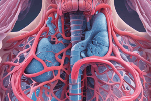
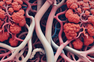
Trachea and Bronchial Tree
- After the larynx
- Involves various bronchioles and aveoli
- Gas exchange takes place from air sacs to tissues Note: Look at figure 9 for gas exchange and aveolar region
Studying That Suits You
Use AI to generate personalized quizzes and flashcards to suit your learning preferences.