Podcast
Questions and Answers
Which of the following is a component of the lower repiratory tract?
Which of the following is a component of the lower repiratory tract?
- External Nose
- Nasal Cavity
- Larynx (correct)
- Pharynx
What is the main function of the conducting zone within the respiratory system?
What is the main function of the conducting zone within the respiratory system?
- To filter, warm, and moisten air (correct)
- To facilitate gas exchange between air and blood
- To regulate blood flow to the lungs
- To produce respiratory secretions
In which structure does the conducting portion of the respiratory system terminate?
In which structure does the conducting portion of the respiratory system terminate?
- Alveolar Sacs
- Terminal Bronchioles (correct)
- Alveoli
- Respiratory Bronchioles
Which of the following structures is part of the respiratory portion of the respiratory system?
Which of the following structures is part of the respiratory portion of the respiratory system?
What is the approximate length of the trachea?
What is the approximate length of the trachea?
At which vertebral level does the trachea begin?
At which vertebral level does the trachea begin?
What anatomical structure marks the end of the trachea and the beginning of the primary bronchi?
What anatomical structure marks the end of the trachea and the beginning of the primary bronchi?
What is the role of the trachealis muscle, located on the posterior part of the trachea?
What is the role of the trachealis muscle, located on the posterior part of the trachea?
Which structure is located posterior to the trachea in the neck?
Which structure is located posterior to the trachea in the neck?
The trachea bifurcates into the primary bronchi at the level of the sternal angle, corresponding to which vertebral level?
The trachea bifurcates into the primary bronchi at the level of the sternal angle, corresponding to which vertebral level?
Which statement accurately describes the carina?
Which statement accurately describes the carina?
How many incomplete cartilaginous rings typically support the trachea, ensuring it remains open during respiration?
How many incomplete cartilaginous rings typically support the trachea, ensuring it remains open during respiration?
Which of the following relations is found anterior to the trachea in the superior mediastinum?
Which of the following relations is found anterior to the trachea in the superior mediastinum?
Which of the following best describes the right primary bronchus compared to the left?
Which of the following best describes the right primary bronchus compared to the left?
Upon entering the hilum, into which lobes does the left primary bronchus divide?
Upon entering the hilum, into which lobes does the left primary bronchus divide?
What structural change occurs in the bronchial tree as branching progresses from bronchi to bronchioles?
What structural change occurs in the bronchial tree as branching progresses from bronchi to bronchioles?
Why are foreign particles more likely to lodge in the right primary bronchus compared to the left?
Why are foreign particles more likely to lodge in the right primary bronchus compared to the left?
The system of air-conducting passages that originate from the primary bronchi are collectively referred to as what?
The system of air-conducting passages that originate from the primary bronchi are collectively referred to as what?
Each lung is divided by fissures into lobes. How many lobes are typically found in the right lung?
Each lung is divided by fissures into lobes. How many lobes are typically found in the right lung?
Which structures are related to the base (diaphragmatic surface) of the right lung?
Which structures are related to the base (diaphragmatic surface) of the right lung?
Which of the following is a characteristic of the apex of each lung?
Which of the following is a characteristic of the apex of each lung?
Which anatomical feature is present on the anterior border of the left lung?
Which anatomical feature is present on the anterior border of the left lung?
Which surface of the lung is in direct contact with the ribs and intercostal muscles?
Which surface of the lung is in direct contact with the ribs and intercostal muscles?
What is the hilum of the lung?
What is the hilum of the lung?
Identify the structure that passes above the hilum of the right lung.
Identify the structure that passes above the hilum of the right lung.
What comprises a bronchopulmonary segment?
What comprises a bronchopulmonary segment?
Veins carrying oxygenated blood are located in which area relative to the bronchopulmonary segments??
Veins carrying oxygenated blood are located in which area relative to the bronchopulmonary segments??
Which of the following outlines the segments of the left upper lobe?
Which of the following outlines the segments of the left upper lobe?
What are the fissures generally composed of?
What are the fissures generally composed of?
Which of the following is NOT a segment of the right lung?
Which of the following is NOT a segment of the right lung?
Which statement best describes the anatomical relationship of the esophagus to the left lung?
Which statement best describes the anatomical relationship of the esophagus to the left lung?
The mediastinal pleura covers which aspect of the mediastinum?
The mediastinal pleura covers which aspect of the mediastinum?
What is the function of the fluid found in the plueral space?
What is the function of the fluid found in the plueral space?
The visceral pleura directly covers what structure?
The visceral pleura directly covers what structure?
The cervical pleura directly lines which part of the body?
The cervical pleura directly lines which part of the body?
Flashcards
Respiratory System Classification
Respiratory System Classification
The respiratory system is classified into upper and lower tracts based on structure and function.
Upper Respiratory Tract
Upper Respiratory Tract
Includes the external nose, nasal cavity & paranasal sinuses, and pharynx.
Lower Respiratory Tract
Lower Respiratory Tract
Includes the larynx, trachea (windpipe), bronchi, and lungs.
Conducting Zone
Conducting Zone
Signup and view all the flashcards
Respiratory Zone
Respiratory Zone
Signup and view all the flashcards
Conducting Portion structures
Conducting Portion structures
Signup and view all the flashcards
Respiratory Portion Structures
Respiratory Portion Structures
Signup and view all the flashcards
Trachea
Trachea
Signup and view all the flashcards
Trachea Beginning
Trachea Beginning
Signup and view all the flashcards
Trachea Ending
Trachea Ending
Signup and view all the flashcards
Carina
Carina
Signup and view all the flashcards
Tracheal cartilage
Tracheal cartilage
Signup and view all the flashcards
Carina
Carina
Signup and view all the flashcards
Tracheal Rings
Tracheal Rings
Signup and view all the flashcards
Posterior Part of Trachea
Posterior Part of Trachea
Signup and view all the flashcards
Trachea Relations In the Neck
Trachea Relations In the Neck
Signup and view all the flashcards
Anterior Relations of Trachea
Anterior Relations of Trachea
Signup and view all the flashcards
Left Side Relations of Trachea
Left Side Relations of Trachea
Signup and view all the flashcards
Right Side Relations of Trachea
Right Side Relations of Trachea
Signup and view all the flashcards
Primary Bronchi Function
Primary Bronchi Function
Signup and view all the flashcards
Right Principal Bronchus
Right Principal Bronchus
Signup and view all the flashcards
Left Principal Bronchus
Left Principal Bronchus
Signup and view all the flashcards
Conduction Zone Branches
Conduction Zone Branches
Signup and view all the flashcards
Respiratory Zone Branches
Respiratory Zone Branches
Signup and view all the flashcards
Primary Bronchi Orientation
Primary Bronchi Orientation
Signup and view all the flashcards
Foreign Particles in Bronchi
Foreign Particles in Bronchi
Signup and view all the flashcards
Bronchial Tree
Bronchial Tree
Signup and view all the flashcards
Bronchial Tree Wall Changes
Bronchial Tree Wall Changes
Signup and view all the flashcards
Epithelium Changes in Bronchi
Epithelium Changes in Bronchi
Signup and view all the flashcards
Lungs
Lungs
Signup and view all the flashcards
Lungs: General Features
Lungs: General Features
Signup and view all the flashcards
Lung Apex
Lung Apex
Signup and view all the flashcards
Lung Base
Lung Base
Signup and view all the flashcards
Lungs Anterior Border
Lungs Anterior Border
Signup and view all the flashcards
Lung Posterior Border
Lung Posterior Border
Signup and view all the flashcards
Lung Inferior Border
Lung Inferior Border
Signup and view all the flashcards
Left Lung Anterior Border
Left Lung Anterior Border
Signup and view all the flashcards
Lung Costal Surface
Lung Costal Surface
Signup and view all the flashcards
Lung Mediastinal Surface
Lung Mediastinal Surface
Signup and view all the flashcards
Lung Vertebral Surface
Lung Vertebral Surface
Signup and view all the flashcards
Study Notes
Respiratory System Classification
- The classification is based on structure and function.
- The upper respiratory tract includes the external nose, nasal cavity, paranasal sinuses, and pharynx.
- The lower respiratory tract includes the larynx, trachea, bronchi, bronchioli, and lungs.
Respiratory System Functional Zones
- The conducting zone includes the nose, nasal cavity, pharynx, larynx, trachea, bronchi, and terminal bronchioles.
- It filters, warms, and moistens air, then conducts it into the lungs.
- The respiratory zone includes the respiratory bronchiole, alveolar ducts, alveolar sac, and alveoli.
- It serves as the major location for gas exchange between air and blood.
Conducting Portion
- It is responsible for transporting air.
Included are:
- Nasal cavity
- Pharynx
- Larynx
- Trachea
- Main bronchus
- Lobar bronchus
- Segmental bronchus
- Terminal bronchioles
Respiratory Portion
- This supports gas exchange; the parts include:
- Respiratory Bronchioles
- Alveolar Ducts
- Alveoli
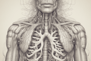
Trachea (Windpipe)
- It's a mobile fibrocartilaginous tube, 5 inches long and 1 inch in diameter.
- Superiorly, it begins in the neck below the cricoid cartilage of the larynx, at the level of the C6 vertebra.
- Inferiorly, it ends below in the thorax at the level of the sternal angle (lower border of T4), by dividing into the right and left primary bronchi.
- The carina is a ridge found at the bifurcation; it is the most sensitive part of the tract and enables the cough reflex.
- The trachea's structure includes tracheal cartilage resembling a horseshoe.
- Annular ligaments connect the tracheal cartilages.
- A fibromuscular membrane connects the tracheal cartilages called the membranous part of the trachea.
- It extends through the lower neck and upper mediastinum, occupying from C6-T4 vertebrae.
- At the level of the sternal angle it bifurcates into right and left primary bronchi.
- Known as the carina, the angle between right and left main bronchi forms through a hook from the last tracheal ring.
- 15-20 cartilaginous rings keep the trachea open during all respiratory phases, and the posterior part contains the trachealis muscle.
- This part allows the esophagus to expand during swallowing.
Trachea Important Relations
- It's partially covered by the sternocleidomastoid muscle and the isthmus and lobes of the thyroid gland, with an anterior arch of the aorta.
- Posteriorly in the neck in relation to the esophagus.
Relations in the Superior Mediastinum
- Anteriorly the trachea is near to the sternum, thymus, left brachiocephalic vein, brachiocephalic artery, and left common carotid.
- Posteriorly it is related to the esophagus and the left recurrent laryngeal nerve.
- Relations on the left side consists of the arch of the aorta, left common carotid artery, left subclavian artery, left vagus, and phrenic nerves, as well as pleural tissue.
- Relations on the right include azygos vein, right vagus nerve, and pleura.
Primary Bronchi
- They project laterally toward their respective lung.
- They enter the hilus of each lung along with the pulmonary vessels, lymphatic vessels, and nerves.
- They branch into several secondary (lobar) bronchi.
- The right principal bronchus is shorter (one inch), wider, shorter and more vertical than the left.
- It gives off the superior lobar bronchus before entering the hilum of the right lung.
- It divides into the middle and inferior lobar bronchi upon entering the hilum.
- The left principal bronchus is about two inches in length has a narrower, longer and more horizontal structure than the right.
- It passes to the left below the aortic arch and in front of the esophagus.
- It divides into the superior and inferior lobar bronchi once entering the hilum of the left lung.
Bronchial Divisions
- Within the lung, each bronchus divides and redivides into numerous branches, which can be split into two groups.
- Conduction Zone Branches include the Primary/main, Secondary/lobar, Tertiary/segmental bronchi, Smaller bronchi, Bronchioles, and Terminal bronchioles.
- Respiratory Zone Branches include the Respiratory bronchioles, Alveolar ducts, Alveolar sacs, and Alveoli.
- Right main (primary) bronchus is shorter, wider, and more vertically oriented than the left.
- Foreign particles are more likely to lodge in right primary bronchus.
- The Bronchial tree systems air-conducting passages from left and right primary bronchi, that progressively branch into narrower tubes spreading throughout the lungs before ending as terminal bronchioles.
- The Bronchial tree wall structure, with successive branching amount of cartilage decreases and smooth muscle amounts increase, for variations in airway diameter.
- And the Epithelium gradually changes from ciliated pseudostratified columnar epithelium to simple cuboidal epithelium in terminal bronchioles.
Lungs
- The lungs are half conical spongy organs lying in the lateral aspects of the thoracic cage enclosed within the pleural sacs.
- Each lung features an apex, a base, 3 borders, and 2 surfaces, and is divided by fissures into 2 or 3 lobes.
- The Apex is blunt, extending into the neck, an inch above the medial 1/3rd of the clavicle, it is cervical pleura and a suprapleural membrane.
- Base (Diaphragmatic surface) is semilunar and concave, related to the dome of the diaphragm.
- The liver separates the right side, while the stomach and spleen separate from the left.
- The lung has three borders: anterior, posterior, and inferior.
- Anterior border is sharp and shorter than the posterior border.
- Posterior border is thick and rounded.
- Inferior border is semilunar in shape.
- The left side presents a concave cardiac notch, and the tongue-shaped projection below the cardiac notch is the Lingula.
- The lung has three surfaces: costal, mediastinal, and vertebral. The costal surface is convex, related to the intercostal muscles and ribs.
- The medial surface divides into 2: Vertebral and Mediastinal.
- The vertebral surfaces occupies the posterior third of the medial surface relating to vertebral bodies and intervertebral discs, the mediastinal to the anterior two thirds relating to mediastinal structure.
- The lung's hilum is a depression in the mediastinal part where bronchi, vessels, and nerves enter/leave.
The Right Lung vs Left Lung
- Anterior to the hilum rests pericardial impression for pericardium, the inferior vena cava, right brachiocephalic vein and superior vena cava, ascending aorta, remains of the thymus gland, and right phrenic nerve.
- Posterior to the hilum resides to Esophagus and the Azygos vein.
- Situated above the Hilum rests arch of the azygos vein, trachea, and right vagus, esophagus.
- The Left Lung is divided 2 lobes.
- The Anterior to the hilum contains pericardial impression in order of the for pericardium and pulmonary trunk with remnants of thymus gland.
- The Posterior part of the descending aorta, short groove, and esophagus.
- Above sits Aorta , left common carotid and left subclavian arteries, Esophagus thoracic duct, and left recurrent laryngeal nerve.
- Fissures are lined by visceral pleura to enable expansion, the right lung has 3 lobes, has usually 2 fissures: Oblique and transverse/horizontal.
- The left lobe has 2 lobes, and 1 fissure: Oblique.
- Oblique Fissures separate the superior lobe from lower lobes in both lungs.
- Horizontal separates the right upper lobe from the right middle lobe.
- Lungs are anatomically, functionally, and surgically bronchopulmonary segments.
- They are pyramid shaped, with their apex toward the lung root and their base towards the surface of the lung.
- Each has a segmental bronchus, a segmental artery from the pulmonary artery, lymph vessel, and autonomic nerves, surrounded by connective tissue
Lung Lobes
- The left lung has 2 lobes:
- Left upper lobe, including the apicoposterior, anterior, superior lingular, and inferior lingular segments.
- The left lower lobe containing: superior, anteromedial, lateral and posterior segments
- The right lung is divided into 3 lobes:
- Right upper lobe, including the apical segment, posterior segment, and anterior segment.
- Right middle lobe, including the lateral segment, and medial segment.
- Right lower lobe consists of: superior segment, medial segment, anterior segment, lateral segment, and posterior segment.
Parietal Pleura
- Covering: The pleura includes two thin layers of tissue.
- Location: Parietal pleura exterior lung portion extending to the interlobar fissures .
- Structure: Visceral pleura internal part of the thoracic cavity to its point of contact.
- The Mediastinal pleura covers the lateral aspect of the mediastinum .
- Cervical pleura lines the pleural cavity in the neck, Costal is the inner aspect of the ribs.
- Lastly, the Diaphragmatic pleura covers the diaphragm surface, and the parietal & visceral pleura are separated by a cavity.
- With a fluid lubricating to slide those layers, called pleural.
Studying That Suits You
Use AI to generate personalized quizzes and flashcards to suit your learning preferences.