Podcast
Questions and Answers
What is the primary function of the tracheal cartilages?
What is the primary function of the tracheal cartilages?
- To assist in gas exchange
- To provide rigidity and maintain airway patency (correct)
- To support the lungs during inhalation
- To produce mucus for airway lubrication
At what anatomical level does the trachea bifurcate into the primary bronchi?
At what anatomical level does the trachea bifurcate into the primary bronchi?
- T4 (correct)
- T6
- C4
- T2
Which structure lies posterior to the trachea?
Which structure lies posterior to the trachea?
- Larynx
- Esophagus (correct)
- Carina
- Bronchi
What type of muscle closes the posterior parts of the tracheal rings?
What type of muscle closes the posterior parts of the tracheal rings?
What do the bronchi branch into after leaving the trachea?
What do the bronchi branch into after leaving the trachea?
Which of the following accurately describes the carina?
Which of the following accurately describes the carina?
Which statement about the bronchial tree is correct?
Which statement about the bronchial tree is correct?
What are the elastic sheets connecting the tracheal cartilage rings called?
What are the elastic sheets connecting the tracheal cartilage rings called?
Which structure is NOT a part of the bronchi system?
Which structure is NOT a part of the bronchi system?
How many C-shaped cartilages typically form the trachea?
How many C-shaped cartilages typically form the trachea?
What is a primary function of the hyaline cartilage in the bronchial tree?
What is a primary function of the hyaline cartilage in the bronchial tree?
Which statement accurately describes the right primary bronchus?
Which statement accurately describes the right primary bronchus?
How many secondary bronchi does the left lung have?
How many secondary bronchi does the left lung have?
What are tertiary bronchi also known as?
What are tertiary bronchi also known as?
What causes inhaled foreign bodies to more likely lodge in the right primary bronchus?
What causes inhaled foreign bodies to more likely lodge in the right primary bronchus?
How many tertiary bronchi does the right lung typically have?
How many tertiary bronchi does the right lung typically have?
Which bronchi is responsible for supplying a specific lobe of the lung?
Which bronchi is responsible for supplying a specific lobe of the lung?
What is the role of terminal bronchioles in the bronchial tree?
What is the role of terminal bronchioles in the bronchial tree?
Which structure follows the tertiary bronchi in the branching of the bronchial tree?
Which structure follows the tertiary bronchi in the branching of the bronchial tree?
What anatomical feature serves as the entry point for bronchi into the lungs?
What anatomical feature serves as the entry point for bronchi into the lungs?
Flashcards
What is the trachea?
What is the trachea?
The trachea, also known as the windpipe, is a flexible tube that carries air from the larynx to the lungs.
Where is the trachea located?
Where is the trachea located?
The trachea is located in the mediastinum, a space in the chest containing the heart and major blood vessels.
What keeps the trachea open?
What keeps the trachea open?
The trachea is reinforced by C-shaped cartilage rings that prevent it from collapsing.
What happens to the trachea at the sternal angle?
What happens to the trachea at the sternal angle?
Signup and view all the flashcards
What is the carina?
What is the carina?
Signup and view all the flashcards
What happens if the carina is irritated?
What happens if the carina is irritated?
Signup and view all the flashcards
What is the length of the trachea?
What is the length of the trachea?
Signup and view all the flashcards
What completes the tracheal rings posteriorly?
What completes the tracheal rings posteriorly?
Signup and view all the flashcards
What are the sections of the trachea?
What are the sections of the trachea?
Signup and view all the flashcards
What are the bronchi?
What are the bronchi?
Signup and view all the flashcards
What are primary bronchi?
What are primary bronchi?
Signup and view all the flashcards
How does the right primary bronchus differ from the left?
How does the right primary bronchus differ from the left?
Signup and view all the flashcards
Describe the basic branching pattern of the bronchial tree.
Describe the basic branching pattern of the bronchial tree.
Signup and view all the flashcards
How many lobes does each lung have and how many secondary bronchi supply them?
How many lobes does each lung have and how many secondary bronchi supply them?
Signup and view all the flashcards
What are tertiary bronchi and what do they supply?
What are tertiary bronchi and what do they supply?
Signup and view all the flashcards
What is a bronchopulmonary segment and why is it important?
What is a bronchopulmonary segment and why is it important?
Signup and view all the flashcards
How many tertiary bronchi are found in each lung?
How many tertiary bronchi are found in each lung?
Signup and view all the flashcards
What are bronchioles and what is their role?
What are bronchioles and what is their role?
Signup and view all the flashcards
What is the bronchial tree and what is its importance?
What is the bronchial tree and what is its importance?
Signup and view all the flashcards
Describe the hierarchy of the bronchial tree branch system.
Describe the hierarchy of the bronchial tree branch system.
Signup and view all the flashcards
Study Notes
Respiratory System Anatomy
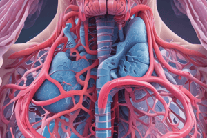
- The trachea is a flexible, slightly rigid tubular organ, often called the windpipe.
- It extends through the mediastinum, anterior to the esophagus and inferior to the larynx, superior to the primary bronchi of the lungs.
- The trachea is supported by 15-20 C-shaped cartilages, which provide rigidity to ensure it remains open.
- Cartilage rings are connected by elastic sheets called anular ligaments.
Trachea
- At the level of the sternal angle, the trachea bifurcates into two smaller tubes called the right and left primary bronchi.
- Each primary bronchus projects laterally toward its corresponding lung.
- The most inferior tracheal cartilage separates the primary bronchi at their origin and forms an internal ridge called the carina.
- The carina is highly sensitive to irritation, triggering the cough reflex.
Bronchi
- Tubes that branch off the trachea and enter the lungs are called bronchi.
- Branches in the bronchi tree are: primary bronchi, secondary bronchi (lobar bronchi), tertiary bronchi (segmental bronchi), bronchioles, terminal bronchioles, respiratory bronchioles, alveolar ducts, alveolar sacs, and alveoli.
- Primary bronchus: Incomplete rings of hyaline cartilage ensure that they stay open. The right primary bronchus is shorter, wider, and more vertically oriented than the left, increasing the likelihood of foreign particles lodging in the right.
Lungs
- The lungs are cone-shaped, with a wide concave base resting on the diaphragm and a relatively blunt superior region (apex).
- They are bordered by the thoracic wall anteriorly, laterally, and posteriorly, and supported by the rib cage.
- They are separated from one another in the midline by the mediastinum.
- Each lung is divided into lobes by fissures.
- The left lung has two lobes, separated by an oblique fissure.
- The right lung has three lobes divided by oblique and horizontal fissures.
- Lungs have structures like cardiac notches and lingula.
Bronchial Tree
- A highly branched system of air-conducting passages originating from the right and left primary bronchi.
- The bronchi progressively branch into narrower tubes, diverging throughout the lungs before terminating in terminal bronchioles.
Respiratory Bronchioles, Alveolar Ducts, and Alveoli
- Contain small saccular outpocketings called alveoli.
- An average alveolus is about 0.25 to 0.5 millimeters in diameter.
- Alveoli have thin walls for efficient gas exchange between the alveolus and the blood in the pulmonary capillaries.
- The lungs contain approximately 300-400 million alveoli contributing to its spongy texture due to their packing.
Bronchopulmonary Segments
- The anatomical divisions of each lung are called bronchopulmonary segments.
- Each segment is served by a tertiary bronchus. The left lung has 8-10 segments, and the right lung has 10 segments.
Pleura and Pleural Cavities
- The outer surface of each lung and adjacent internal thoracic wall is lined by a serous membrane called pleura, formed from simple squamous epithelium.
- The surface of each lung is covered by visceral pleura, while the internal thoracic walls are lined by parietal pleura.
- The parietal and visceral layers connect at the hilum of each lung. There is a potential space between these layers, the pleural cavity, containing serous fluid for reduced friction during breathing.
Breathing Mechanism
- During inhalation, muscles like the diaphragm and external intercostals contract increasing the size of the thoracic cavity and lowering internal pressure.
- Oppositely, during exhalation, muscles like the internal intercostals and abdominal muscles relax, decreasing the size of the thoracic cavity and raising internal pressure.
- The pleura and the pleural cavities play an important role in the breathing mechanics of the lungs.
Arteries and Veins
- Bronchial arteries supply blood to lung tissue.
- Pulmonary arteries carry deoxygenated blood from the heart to the lungs for oxygenation.
- Pulmonary veins return oxygenated blood from the lungs to the heart.
- Specific bronchial veins and lymphatic drainage systems are found surrounding the lungs.
Auscultation of the lungs
- Listening to the sounds of air movement in the lungs. These sounds are used as diagnostics.
Thoracic Wall Dimensional Changes
- The thoracic wall expands and contracts during breathing. This is driven by muscle contractions in the ribs, diaphragm, and additional accessory muscles to expand the thoracic cage laterally, vertically and anterior-posteriorly.
Studying That Suits You
Use AI to generate personalized quizzes and flashcards to suit your learning preferences.


