Podcast
Questions and Answers
What type of epithelium lines the mucous membrane of the trachea?
What type of epithelium lines the mucous membrane of the trachea?
Pseudostratified ciliated columnar epithelium with goblet cells.
What is found deep to the basement membrane in the trachea's mucous membrane?
What is found deep to the basement membrane in the trachea's mucous membrane?
The lamina propria, which is made up of areolar tissue with elastic fibers and lymphocytes.
Describe the submucosa layer of the trachea.
Describe the submucosa layer of the trachea.
It is composed of loose connective tissue containing large blood vessels and nerves, along with mucous, serous, and mixed glands.
What distinguishes the posterior wall of the trachea?
What distinguishes the posterior wall of the trachea?
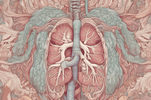
Which structures are typically included in a section through the lung?
Which structures are typically included in a section through the lung?
What type of cells line the alveoli?
What type of cells line the alveoli?
What is the role of type 2 cells in the alveoli?
What is the role of type 2 cells in the alveoli?
What type of cartilage is found in the bronchi and how is it characterized?
What type of cartilage is found in the bronchi and how is it characterized?
How does the structure of bronchioles differ from bronchi?
How does the structure of bronchioles differ from bronchi?
What components are found between the alveoli?
What components are found between the alveoli?
Flashcards are hidden until you start studying
Study Notes
Trachea
- Lined by pseudostratified ciliated columnar epithelium with goblet cells and a basement membrane.
- Lamina propria (beneath basement membrane) contains areolar tissue, elastic fibers, and lymphocytes.
- Submucosa has loose connective tissue, large blood vessels, nerves, and mucous, serous, and mixed glands. Gland ducts open onto the epithelium.
- Hyaline cartilage is external to the submucosa, covered by perichondrium. Cartilage pieces are interconnected by an elastic fibrous membrane.
- The posterior tracheal wall lacks cartilage; it contains the trachealis muscle (inner circular and outer longitudinal smooth muscle layers).
- Adventitia (outermost layer) contains connective tissue, blood vessels, and nerves.
Lung
- Lung sections reveal bronchi, bronchioles, alveoli, pulmonary artery branches, and pulmonary vein tributaries.
Bronchi
- Mucosa: Pseudostratified ciliated columnar epithelium; highly folded due to underlying smooth muscle.
- Lamina propria: Connective tissue, smooth muscle fibers, mucous and serous glands.
- Hyaline cartilage: Present in small pieces outside the lamina propria.
Bronchioles
- Mucosa: Simple columnar epithelium with folds.
- Lamina propria: Connective tissue and smooth muscle fibers (causing mucous membrane folds); lacks glands and cartilage.
Alveoli
- Lined by simple squamous epithelium.
- Interalveolar septa (between alveoli) are thin and contain capillaries, elastic fibers, fibroblasts, mesenchymal cells, and macrophages.
- Two types of alveolar lining cells:
- Type I (squamous) cells: Simple squamous cells with nuclei protruding into the lumen.
- Type II (secretory) cells: Rounded cells projecting into the lumen.
Studying That Suits You
Use AI to generate personalized quizzes and flashcards to suit your learning preferences.