Podcast
Questions and Answers
Which pressure is typically negative and helps keep the lungs inflated?
Which pressure is typically negative and helps keep the lungs inflated?
- Transpulmonary pressure
- Intrapulmonary pressure
- Intrapleural pressure (correct)
- Atmospheric pressure
Intrapulmonary pressure is always lower than atmospheric pressure during normal breathing.
Intrapulmonary pressure is always lower than atmospheric pressure during normal breathing.
False (B)
Which muscle abducts the vocal cords to allow for inhalation?
Which muscle abducts the vocal cords to allow for inhalation?
Posterior cricoarytenoid muscle
The laryngeal muscles adjust the size of the opening between the vocal cords, called the ______.
The laryngeal muscles adjust the size of the opening between the vocal cords, called the ______.
Match the following muscles with their function in moving the vocal cords:
Match the following muscles with their function in moving the vocal cords:
What muscles are responsible for fine-tuning the tension and position of the vocal cords?
What muscles are responsible for fine-tuning the tension and position of the vocal cords?
Ventilation only refers to inhalation.
Ventilation only refers to inhalation.
Which two muscles primarily contract during inhalation?
Which two muscles primarily contract during inhalation?
During inhalation, the contraction of the diaphragm causes it to move ______.
During inhalation, the contraction of the diaphragm causes it to move ______.
Match the mechanical processes with their effect on intrapulmonary volume during normal breathing:
Match the mechanical processes with their effect on intrapulmonary volume during normal breathing:
What happens to intrapulmonary pressure during exhalation?
What happens to intrapulmonary pressure during exhalation?
Residual volume is the amount of air that can be forcefully exhaled after a normal exhalation.
Residual volume is the amount of air that can be forcefully exhaled after a normal exhalation.
Vital capacity is the sum of which three lung volumes?
Vital capacity is the sum of which three lung volumes?
The amount of air inhaled and exhaled during normal resting breathing is known as ______ volume.
The amount of air inhaled and exhaled during normal resting breathing is known as ______ volume.
Match the lung volumes with their approximate values:
Match the lung volumes with their approximate values:
Which anatomical structure is the primary site of pulmonary gas exchange?
Which anatomical structure is the primary site of pulmonary gas exchange?
Compliance refers to the ease with which the lungs can recoil during exhalation.
Compliance refers to the ease with which the lungs can recoil during exhalation.
Name two factors that affect lung compliance.
Name two factors that affect lung compliance.
A condition characterized by increased lung compliance, where the lungs lose elasticity, is ______.
A condition characterized by increased lung compliance, where the lungs lose elasticity, is ______.
Match each condition with its effect on lung compliance:
Match each condition with its effect on lung compliance:
What happens to the body's acid-base balance in respiratory acidosis?
What happens to the body's acid-base balance in respiratory acidosis?
Respiratory alkalosis is typically caused by conditions that lead to decreased ventilation.
Respiratory alkalosis is typically caused by conditions that lead to decreased ventilation.
Name three symptoms of respiratory alkalosis.
Name three symptoms of respiratory alkalosis.
Metabolic acidosis can result from excessive production of acids or a loss of ______ from the body.
Metabolic acidosis can result from excessive production of acids or a loss of ______ from the body.
Match each acid-base imbalance with a potential cause:
Match each acid-base imbalance with a potential cause:
In metabolic acidosis, the body may attempt to compensate by:
In metabolic acidosis, the body may attempt to compensate by:
Compensation always restores the pH to the normal range in acid-base imbalances.
Compensation always restores the pH to the normal range in acid-base imbalances.
What acid-base disorder is caused by a decrease in ventilation, possibly due to chronic obstructive pulmonary disease?
What acid-base disorder is caused by a decrease in ventilation, possibly due to chronic obstructive pulmonary disease?
A condition where the body attempts to restore the acid-base balance when there is an imbalance in the pH is called ______.
A condition where the body attempts to restore the acid-base balance when there is an imbalance in the pH is called ______.
Match the compensatory mechanism with its acid-base imbalance:
Match the compensatory mechanism with its acid-base imbalance:
Flashcards
Intrapleural Pressure
Intrapleural Pressure
Pressure within the pleural cavity; it's typically negative to keep lungs inflated.
Intrapulmonary Pressure
Intrapulmonary Pressure
Pressure within the air sacs of the lungs (alveoli); it generally equalizes with atmospheric pressure during normal breathing.
Atmospheric Pressure
Atmospheric Pressure
Pressure exerted by the Earth's atmosphere; at sea level, it's around 760 mmHg.
Posterior Cricoarytenoid Muscle
Posterior Cricoarytenoid Muscle
Signup and view all the flashcards
Lateral Cricoarytenoid Muscle
Lateral Cricoarytenoid Muscle
Signup and view all the flashcards
Intrinsic Laryngeal Muscles
Intrinsic Laryngeal Muscles
Signup and view all the flashcards
Extrinsic Laryngeal Muscles
Extrinsic Laryngeal Muscles
Signup and view all the flashcards
Sternothyroid Muscle
Sternothyroid Muscle
Signup and view all the flashcards
Cricothyroid Muscle
Cricothyroid Muscle
Signup and view all the flashcards
Ventilation
Ventilation
Signup and view all the flashcards
Muscles of Inhalation
Muscles of Inhalation
Signup and view all the flashcards
Tidal Volume
Tidal Volume
Signup and view all the flashcards
Inspiratory Reserve Volume (IRV)
Inspiratory Reserve Volume (IRV)
Signup and view all the flashcards
Expiratory Reserve Volume
Expiratory Reserve Volume
Signup and view all the flashcards
Residual Volume
Residual Volume
Signup and view all the flashcards
Minute Volume
Minute Volume
Signup and view all the flashcards
Vital Capacity
Vital Capacity
Signup and view all the flashcards
Inspiratory Capacity
Inspiratory Capacity
Signup and view all the flashcards
Functional Residual Capacity
Functional Residual Capacity
Signup and view all the flashcards
Forced Expiratory Vital Capacity (FEV)
Forced Expiratory Vital Capacity (FEV)
Signup and view all the flashcards
Total Lung Capacity
Total Lung Capacity
Signup and view all the flashcards
Compliance
Compliance
Signup and view all the flashcards
Factors Affecting Lung Compliance
Factors Affecting Lung Compliance
Signup and view all the flashcards
Conditions of Reduced Compliance
Conditions of Reduced Compliance
Signup and view all the flashcards
Conditions of Increased Compliance
Conditions of Increased Compliance
Signup and view all the flashcards
Obstructive Lung Disorders
Obstructive Lung Disorders
Signup and view all the flashcards
Restrictive Lung Disorders
Restrictive Lung Disorders
Signup and view all the flashcards
Acids and Bases
Acids and Bases
Signup and view all the flashcards
Bases and Acids
Bases and Acids
Signup and view all the flashcards
Study Notes
Respiratory Pressures
- Intrapleural pressure is the pressure within the pleural cavity, between the lungs and chest wall
- It is typically negative, lower than atmospheric pressure
- The negative pressure helps keep lungs inflated and prevents collapse
- Intrapulmonary pressure (alveolar pressure) is the pressure within the lung's air sacs (alveoli)
- Varies during the respiratory cycle
- It generally equalizes with atmospheric pressure during normal breathing
- Atmospheric pressure (barometric pressure) is the pressure exerted by Earth's atmosphere
- Measured in millimeters of mercury (mmHg)
- At sea level, it is typically around 760 mmHg
Laryngeal Muscles and Vocal Cord Movement
- Laryngeal muscles control the position and tension of the vocal cords within the larynx
- The larynx is located at the top of the trachea (windpipe)
- The muscles adjust the size of the opening between the vocal cords (glottis)
- This produces different sounds as air passes through
- Intrinsic and extrinsic muscles are two sets within the larynx that move the vocal cords
- Intrinsic muscles are located within the larynx, are responsible for fine-tuning vocal cord position and tension
- Extrinsic muscles attach to the larynx from the outside, helping move it
Specific Muscles
- The posterior cricoarytenoid muscle abducts (separates) the vocal cords, allowing for inhalation
- The lateral cricoarytenoid muscle adducts (brings together) the vocal cords, for sound production
- Intrinsic muscles include the thyroarytenoid
- It controls vocal cord tension along with the lateral cricoarytenoid for adduction
- Extrinsic muscles include the sternothyroid
- It lowers the larynx and lengthens vocal cords for deeper tones
- The cricothyroid muscle raises the larynx and shortens vocal cords for higher tones
- Muscle contraction adjusts the position and tension of vocal cords
- This changes the glottis' size and shape and produces different sounds
- The nervous system controls the precise movements of the laryngeal muscles
Ventilation
- Ventilation is the process of breathing, including both inhalation and exhalation
Inhalation
- The diaphragm and external intercostal muscles contract
- Intrapulmonary volume expands in all directions, including vertically
- Intrapulmonary pressure drops
- Air is drawn in as the pressure drops to reach equilibrium between intrapulmonary and atmospheric pressure
- The thoracic cavity expands, increasing its volume and expanding the lungs
- Decreased pressure inside the lungs relative to the outside
- Air flows into the lungs through the airways
Exhalation
- Internal intercostals and transverse thoracis muscles relax
- Intrapulmonary volume decreases, and pressure increases
- Air is expelled to re-establish equilibrium between intrapulmonary and atmospheric pressure
- Diaphragm and external intercostal muscles relax
- The elastic recoil of the lungs causes a decrease in volume
- This causes air to flow out of the lungs
Lung Volumes
- Four pulmonary volumes exist:
- Tidal volume: air inhaled/exhaled during normal breathing (500 mL)
- Inspiratory reserve volume: air forcefully inhaled after normal inspiration (3000 mL)
- Expiratory reserve volume (ERV): maximal air exhaled after normal exhalation (1100 mL)
- Residual volume: air left in the lung after maximal exhalation (1200 mL)
- Minute volume: normal air inspired/expired in 1 minute of normal pulmonary ventilation
Lung Capacities
- Vital capacity is the sum of inspiratory reserve volume, tidal volume, and expiratory reserve volume: 4600 mL
- Inspiratory capacity is tidal volume plus inspiratory reserve volume; the air a person can inspire maximally after normal expiration: 3500 mL
- Functional residual capacity is expiratory reserve volume plus residual volume; the air remaining in lungs after normal expiration: 2300 mL
- Forced Expiratory Vital Capacity (FEV) is the air expelled from the lungs in 1 second
- Total lung capacity is the sum of inspiratory and expiratory reserve volumes plus tidal volume and residual volume: 5800 mL
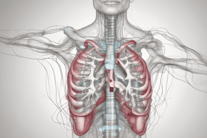
Internal Surface Area of the Lungs
- The alveoli are the site of pulmonary gas exchange that increases surface area
- They are numerous, about 300 million in each lung
- A large surface area to volume ratio exists
- The alveolar walls are very thin for efficient gas diffusion between air and blood
- The tracheobronchial tree consists of the trachea and network of air tubes in the lungs
- The trachea divides into left and right main bronchus
- Each main bronchus divides into smaller bronchi which terminate in microscopic tubes and sacs
- The bronchial tree delivers air to the alveoli where gas exchange with the bloodstream occurs
Compliance
- Compliance is the measure of how easily a structure stretches
- Defined as the change in volume per unit change in pressure
- In respiratory physiology, compliance is the ability of lungs to stretch/expand during inhalation and return during exhalation
- It is a measure of air ease moving in and out of the lungs
- Lung compliance involves elasticity of lung tissue, surface tension within the alveoli, and airway resistance
- High the compliance means the lungs expand easily with minimal effort, for efficient gas exchange
- Low compliance requires more force to expand the lungs making breathing more difficult
- Clinically measured as change in lung volume per unit change in transpulmonary pressure
Reduced Compliance
- Reduced compliance makes it harder to expand the lungs and thorax
- Potential conditions are:
- Pulmonary fibrosis: deposition of inelastic fibers in lung tissue
- Infant respiratory distress syndrome and pulmonary edema: collapse of the alveoli
- Asthma, bronchitis, and lung cancer: increased resistance to airflow
- Kyphosis and scoliosis: deformities of the thoracic wall that reduce expansion ability
Increased Compliance
- With Emphysema the lungs and thorax have lost elasticity
- It becomes easier to expand the lungs and thorax from destruction of elastic tissue
- Expiration is less efficient (COPD - barrel chest)
Lung Disorders and Compliance
- Obstructive lung disorders, (asthma and COPD) are characterized by increased resistance to airflow
- Leads to difficulty exhaling air, increased air trapping, reduced lung volume, and decreased compliance
- Compliance is decreased due to increased resistance to airflow
- Restrictive lung disorders (pulmonary fibrosis and sarcoidosis), are characterized by reduced lung volume and capacity
- The lungs become stiff and lose expansion ability, resulting in decreased lung compliance
- Compliance decreases by reducing lung volume and capacity
Blood Flow Through the Kidney
- Blood flow trace from the abdominal aorta to the inferior vena cava (IVC):
- Abdominal aorta → renal artery → segmental artery → interlobar artery → arcuate artery → afferent arteriole → glomerulus → efferent arteriole → peritubular capillaries → arcuate vein → interlobar vein → segmental vein → renal vein → IVC
Filtrate/Urine Trace
- Filtrate/urine proceeds from Bowman’s capsule to the urethra:
- Bowman’s capsule → proximal convoluted tubule → loop of Henle → distal convoluted tubule → collecting duct → minor calyx → major calyx → renal pelvis → ureter → bladder → urethra
Nephron Parts and Functions
- The nephron filters blood and produces urine.
- Renal Corpuscle:
- The glomerulus is a network of capillaries for filtration.
- Bowman's capsule surrounds the glomerulus and collects filtered fluid.
- Proximal Convoluted Tubule (PCT):
- Extends from Bowman's capsule and reabsorbs nutrients, glucose, amino acids, and electrolytes from the filtrate, returning them to the bloodstream.
- Loop of Henle:
- A U-shaped tube extending from the PCT that establishes a concentration gradient in the kidney.
- This reaction allows for reabsorption of water and electrolytes.
- Distal Convoluted Tubule (DCT):
- Connects to the Loop of Henle and regulates potassium and sodium ion concentrations in the blood.
- It secretes waste products into the urine.
- Collecting Duct:
- Receives urine from nephrons and regulates water and electrolyte concentration for fluid electrolyte balance.
Urine Formation
- Glomerular Filtration:
- The renal filtrate is plasma, without blood cells and proteins with about 99% reabsorbed
- The filtration membrane includes fenestrated endothelium, a basement membrane, and filtration slits formed by podocytes.
- Filtration pressure changes are primarily caused by glomerular capillary pressure changes.
- Tubular Reabsorption
- Filtrate is reabsorbed by passive transport, including simple and facilitated diffusion, active transport and symport
- The thin segment of the nephron loop specializes in passive transport
- The rest of the nephron and collecting ducts perform active/passive transport, and symport
- Active transport moves mainly Na+ across the wall of nephron
- Other ions and molecules move primarily with symport
- Passive transport moves water, urea, and lipid-soluble, nonpolar compounds
- Tubular Secretion
- Substances enter the proximal or distal convoluted tubules and collecting ducts
- Hydrogen ions, K+, and some substances not produced in the body/secreted by antiport mechanisms
- Active transport spoons returns good (glucose or amino acids) substances to the blood
- Salt can be removed with salt transportprotein and regulate excess to urine
Urinalysis Results
- Increased salt (NaCl) concentration in the urine indicates a high salt diet
- it is possible a salt transport protein problem
- Protein in the urine indicates a high protein diet
- It is possible kidney damage(Maybe Glomerulus capillary damage)
- Amino acids in the urine indicates a high protein diet
- It is possible an amino acid transport protein problem
- Blood cells in the urine may indicate kidney damage
- This could be ureter or bladder damage or a kidney stone
- Glucose is found in the urine for recent consumption of a large meal or diabetes
Acids, Bases, and Buffers
- An acid is a proton donor that releases hydrogen ions (H+)
- Properties include a pH value less than 7
- Strong acids have a higher concentration of H+ ions, and weak acids have a lower concentration
- A base is a proton acceptor that binds to (accepts) H+
- Properties include a pH value greater than 7
- Strong bases have a higher concentration of OH- ions and weak bases have a lower concentration.
- Acids and bases neutralize each other, forming a salt and water in neutralization
- The resulting solution's pH is 7 (neutral)
- Buffers resist pH changes when acids or bases are added
- The bicarbonate buffering system is an important mechanism for regulating the pH of bodily fluids
- Bicarbonate ions (HCO3-) act as a base
- Hydrogen ions (H+) act as an acid
Bicarbonate Buffering
- When excess acid (H+) is introduced, it reacts with bicarbonate ions (HCO3-)
- This reaction produces carbonic acid (H2CO3), which breaks down into carbon dioxide (CO2) and water (H2O)
- The carbon dioxide is then transported to the lungs and exhaled, helping to remove excess acid
- When excess base is introduced, bicarbonate ions react releasing hydrogen ions and carbonic acid
Acid-Base Imbalances
- Respiratory acidosis is caused by excess carbon dioxide (CO2) in the blood with the lungs unable to remove enough
- Conditions include COPD, pneumonia, or asthma
- Symptoms are shortness of breath, confusion, and lethargy
- Respiratory alkalosis is a deficiency of carbon dioxide in the blood, caused by rapid or shallow breathing (hyperventilation)
- Possible effectors are anxiety, fever, or high altitude
- Symptoms are dizziness, tingling in toes, and muscle cramps
- Metabolic acidosis happens when there is an excess of acid in the blood.
- This can be due to a variety of conditions, such as uncontrolled diabetes, kidney disease, or severe diarrhea
- Symptoms include fatigue, nausea, and rapid breathing
- Metabolic alkalosis happens when there is an excess of a base in the blood.
- This can be due to a variety of conditions, such as vomiting, excessive use of antacids, or kidney disease
- Symptoms are muscle weakness, nausea, and confusion
- The body restores acid-base balance by adjusting the respiratory rate or renal excretion of bicarbonate ions
Compensation
- Compensation restores acid-base balance with pH, partial pressure of carbon dioxide (PaCO2), or bicarbonate (HCO3-) levels
- Respiratory compensation adjusts the respiratory rate to regulate the PaCO2 levels
- Metabolic compensation changes the HCO3- levels in order to regulate the blood pH
- Complete restoration may not be achieved and compensation depends condition and body ability
- The primary cause of respiratory acidosis is a decrease in ventilation
- This leads to high levels of carbon dioxide (CO2) can be due to COPD, asthma, pneumonia, or a drug overdose.
- Respiratory alkalosis can be caused by increase in ventilation
- This leads to a decrease in CO2 levels in the blood and can be caused by hyperventilation, anxiety, high altitude, or pulmonary disease.
- Metabolic acidosis can occur from over production from acids, less of bicarbonate (HCO3-) from body or by with the kidneys.
- Caused by conditions such as diabetic ketoacidosis, lactic acidosis, renal failure, or diarrhea.
- Metabolic alkalosis is from loss of acids from the body.
- Caused by condition such as vomiting, prolonged use , or excessive ingestion of antacids.
Studying That Suits You
Use AI to generate personalized quizzes and flashcards to suit your learning preferences.