Podcast
Questions and Answers
What is the function of lymphatic capillaries?
What is the function of lymphatic capillaries?
- To empty lymph back into circulation
- To drive lymph flow towards the heart
- To collect excess interstitial fluid adjacent to tissue cells and capillary beds (correct)
- To filter bacteria and cellular debris from lymph
Where does the thoracic duct collect lymph from?
Where does the thoracic duct collect lymph from?
- The abdomen
- The right subclavian vein
- Right upper body
- Below the ribs and the entire left side of the body (correct)
What drives lymph flow towards the heart?
What drives lymph flow towards the heart?
- Respiration
- Skeletal muscle pump (correct)
- Lymphatic ducts
- Lymphatic collecting vessels
What is the function of lymphatic collecting vessels?
What is the function of lymphatic collecting vessels?
What causes laryngitis?
What causes laryngitis?
How many C-shaped hyaline cartilage pieces make up the trachea?
How many C-shaped hyaline cartilage pieces make up the trachea?
What is the primary site of gas exchange in the respiratory system?
What is the primary site of gas exchange in the respiratory system?
What structures form the respiratory membrane?
What structures form the respiratory membrane?
How many lobes does the right lung contain?
How many lobes does the right lung contain?
What serous membrane surrounds each lung?
What serous membrane surrounds each lung?
What regulates the rate and depth of breathing?
What regulates the rate and depth of breathing?
Which cartilages are part of the larynx?
Which cartilages are part of the larynx?
What is the function of the glottis?
What is the function of the glottis?
What is the function of the bronchial route in the lungs?
What is the function of the bronchial route in the lungs?
What controls ventilation in the respiratory system?
What controls ventilation in the respiratory system?
What is the composition of the bronchial tree?
What is the composition of the bronchial tree?
Where is the olfactory area essential for smell located?
Where is the olfactory area essential for smell located?
What are the pairs of air-filled spaces that open directly into the nasal cavity called?
What are the pairs of air-filled spaces that open directly into the nasal cavity called?
What is the main function of the tonsils?
What is the main function of the tonsils?
Which of the following is a primary lymphoid organ?
Which of the following is a primary lymphoid organ?
What are the thin, scroll-like projections that protrude from the lateral walls of the nasal cavity called?
What are the thin, scroll-like projections that protrude from the lateral walls of the nasal cavity called?
Where does most lymphocyte immune responses occur?
Where does most lymphocyte immune responses occur?
What is the composition of the Mucosal-associated lymphoid tissue (MALT)?
What is the composition of the Mucosal-associated lymphoid tissue (MALT)?
What are the subdivisions of the respiratory system?
What are the subdivisions of the respiratory system?
What is the function of the nasal cavity?
What is the function of the nasal cavity?
What are the pairs of air-filled spaces in the frontal, ethmoid, sphenoid, and maxillae bones called?
What are the pairs of air-filled spaces in the frontal, ethmoid, sphenoid, and maxillae bones called?
What is the main function of the respiratory area of the nasal cavity?
What is the main function of the respiratory area of the nasal cavity?
What are the divisions of the pharynx?
What are the divisions of the pharynx?
Which region is located between the right and left hypochondriac regions?
Which region is located between the right and left hypochondriac regions?
What is the primary function of the salivary glands in the digestive system?
What is the primary function of the salivary glands in the digestive system?
Where does absorption primarily occur in the digestive system?
Where does absorption primarily occur in the digestive system?
What is the main function of the gallbladder in the digestive system?
What is the main function of the gallbladder in the digestive system?
Which cells in the stomach secrete HCl and intrinsic factor?
Which cells in the stomach secrete HCl and intrinsic factor?
Which part of the small intestine possesses Brunner’s glands?
Which part of the small intestine possesses Brunner’s glands?
What is the function of the microvilli in the small intestine?
What is the function of the microvilli in the small intestine?
Which cells in the villus epithelium secrete mucus?
Which cells in the villus epithelium secrete mucus?
Which accessory organ secretes bile to aid fat digestion?
Which accessory organ secretes bile to aid fat digestion?
Which cells in the pancreas secrete digestive enzymes?
Which cells in the pancreas secrete digestive enzymes?
What is the function of the gallbladder?
What is the function of the gallbladder?
Which part of the large intestine has no folds or villi?
Which part of the large intestine has no folds or villi?
What are the pouch-forming folds arising from contraction of the teniae coli called?
What are the pouch-forming folds arising from contraction of the teniae coli called?
Which artery supplies blood to the small and large intestine?
Which artery supplies blood to the small and large intestine?
What is the function of the portal system in the blood supply?
What is the function of the portal system in the blood supply?
What is the main function of the caecum and appendix in the large intestine?
What is the main function of the caecum and appendix in the large intestine?
What is the superficial layer of the digestive system?
What is the superficial layer of the digestive system?
What is the largest serous membrane of the body?
What is the largest serous membrane of the body?
What binds organs to the abdominal cavity wall and helps hold them in place?
What binds organs to the abdominal cavity wall and helps hold them in place?
What are the mesenteries formed of visceral peritoneum called?
What are the mesenteries formed of visceral peritoneum called?
What is the innermost layer of the digestive system?
What is the innermost layer of the digestive system?
What consists of two layers of smooth muscle separated by a network of nerve cells?
What consists of two layers of smooth muscle separated by a network of nerve cells?
What are the three paired salivary glands?
What are the three paired salivary glands?
What has distinct regions and forms folds (rugae) when empty to allow for expansion without tearing?
What has distinct regions and forms folds (rugae) when empty to allow for expansion without tearing?
What is the areolar connective tissue layer containing blood and lymphatic vessels and a network of nerve cells?
What is the areolar connective tissue layer containing blood and lymphatic vessels and a network of nerve cells?
What are the mesenteries formed of parietal peritoneum that bind organs to the abdominal cavity wall called?
What are the mesenteries formed of parietal peritoneum that bind organs to the abdominal cavity wall called?
What is the network of nerve cells that separates the two layers of smooth muscle in the muscularis externa?
What is the network of nerve cells that separates the two layers of smooth muscle in the muscularis externa?
What are the layers of the teeth classified into?
What are the layers of the teeth classified into?
Study Notes
Respiratory System Anatomy and Function
- The larynx consists of cartilages including arytenoid cartilages, vocal cords, and the glottis, which closes during swallowing.
- Laryngitis is caused by larynx inflammation due to infection or irritation.
- The trachea consists of 20 C-shaped hyaline cartilage pieces and connects the larynx to the primary bronchi.
- The bronchial tree has approximately 23 generations of branches with decreasing cartilage and increasing smooth muscle.
- The respiratory zone contains millions of alveoli which form the primary site of gas exchange.
- The respiratory membrane consists of type I and type II alveolar cells, capillary wall, and macrophages.
- Clinical applications of lung diseases include tuberculosis, pulmonary edema, pulmonary embolism, pneumothorax, and emphysema.
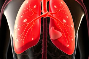
- The lungs are separated by the mediastinum and contain three lobes in the right lung and two in the left.
- The pleura, a serous membrane, surrounds each lung and reduces friction during ventilation.
- The blood supply to the lungs includes the pulmonary route for alveolar oxygenation and the bronchial route for lung tissues.
- Ventilation is controlled by skeletal muscles, and the rate and depth of breathing are regulated by the medulla in response to CO2, O2 levels, and emotions.
Lymphoid Organs and Tissues and the Lymphatic System
- Primary lymphoid organs and tissues are sites where lymphoid stem cells divide and mature, such as the red bone marrow and thymus gland.
- The secondary lymphoid organs and tissues, where most lymphocyte immune responses occur, include lymph nodes, spleen, and mucosal-associated lymphoid tissue (MALT).
- MALT includes tonsils, appendix, and lymphoid tissues on mucosal membranes of digestive, respiratory, reproductive, and urinary systems.
- Tonsils form a ring around the entrance to the pharynx and possess crypts that trap bacteria and debris.
- The respiratory system has main functions of ventilation and gas exchange, with upper and lower respiratory system subdivisions.
- The nasal cavity functions as an airway passage, for olfaction, and as a resonance chamber for speech.
- The respiratory area of the nasal cavity is lined by respiratory mucosa and contains goblet cells that secrete mucus.
- Thin, scroll-like projections called conchae protrude medially from each lateral wall of the nasal cavity.
- The olfactory area essential for smell is located on the roof of the nasal cavity and lacks columnar cell goblet cells.
- Paranasal sinuses are pairs of air-filled spaces in the frontal, ethmoid, sphenoid, and maxillae bones that open directly into the nasal cavity.
- The pharynx is divided into nasopharynx, oropharynx, and laryngopharynx, and is lined by mucous membrane.
- The larynx is composed of eight hyaline cartilages and one non-hyaline cartilage, including the epiglottis, thyroid cartilage, and cricoid cartilage.
Anatomy and Histology of the Digestive System
- Mucosa is the innermost layer of the digestive system, with epithelium, lamina propria, and muscularis mucosae, and it varies in structure in different parts of the digestive tract.
- The submucosa is an areolar connective tissue layer containing blood and lymphatic vessels and a network of nerve cells.
- The muscularis externa consists of two layers of smooth muscle separated by a network of nerve cells, forming the enteric nervous system.
- The serosa (or adventitia) is the superficial layer of the digestive system, and the largest serous membrane of the body is the peritoneum, which lines the abdominopelvic cavity and most of the organs.
- The mesentery is a fused, double-layer sheet of parietal peritoneum that binds organs to the abdominal cavity wall and helps hold them in place.
- The omenta are mesenteries formed of visceral peritoneum, including the greater omentum and the lesser omentum.
- Some abdominal organs are retroperitoneal, lying behind the peritoneum, and the inflammation of the peritoneum is called peritonitis.
- The oral cavity is lined by mucus membrane and contains structures such as the lips, cheeks, palate, tongue, and teeth.
- The teeth are classified into primary dentition (deciduous) and secondary dentition (permanent), each with different numbers of teeth and types of enamel.
- There are three paired salivary glands: parotid, submandibular, and sublingual, each secreting different types of saliva.
- The pharynx has a similar histology to the oral cavity and is connected to skeletal muscle.
- The stomach has distinct regions, unique histology in its muscularis externa, and forms folds (rugae) when empty to allow for expansion without tearing.
Studying That Suits You
Use AI to generate personalized quizzes and flashcards to suit your learning preferences.
Description
Test your knowledge of the respiratory system anatomy and function, as well as lymphoid organs and tissues and the lymphatic system. This quiz covers key structures, functions, and clinical applications in the respiratory and lymphatic systems. Sharpen your understanding of these essential physiological systems with this informative quiz.