Podcast
Questions and Answers
Which process involves the exchange of gases between the atmosphere, blood, and cells?
Which process involves the exchange of gases between the atmosphere, blood, and cells?
- Ventilation
- External ventilation
- Respiration (correct)
- Internal respiration
What is the primary function of the conducting part of the respiratory system?
What is the primary function of the conducting part of the respiratory system?
- Producing chemical mediators
- Protecting against microorganisms
- Delivering air to the lungs (correct)
- Facilitating gas exchange
Which structure is part of the upper respiratory tract?
Which structure is part of the upper respiratory tract?
- Trachea
- Larynx
- Bronchi
- Pharynx (correct)
Which of the following structures is responsible for dividing the nasal cavity into two halves?
Which of the following structures is responsible for dividing the nasal cavity into two halves?
What is the role of the paranasal sinuses in respiration?
What is the role of the paranasal sinuses in respiration?
Which part of the pharynx is a common passageway for air, food, and drink?
Which part of the pharynx is a common passageway for air, food, and drink?
What is the function of the epiglottis during swallowing?
What is the function of the epiglottis during swallowing?
Which of the following determines the pitch of the voice?
Which of the following determines the pitch of the voice?
What type of cartilage primarily forms the trachea?
What type of cartilage primarily forms the trachea?
Which structural change occurs in the branching of the bronchial tree as it extends from the main bronchi to the bronchioles?
Which structural change occurs in the branching of the bronchial tree as it extends from the main bronchi to the bronchioles?
What is the role of type II pneumocytes in the alveoli?
What is the role of type II pneumocytes in the alveoli?
During inhalation, how does the contraction of the diaphragm affect the pressure within the thoracic cavity?
During inhalation, how does the contraction of the diaphragm affect the pressure within the thoracic cavity?
According to Boyle's law, what happens to the pressure of a gas if the volume of its container increases, assuming the temperature remains constant?
According to Boyle's law, what happens to the pressure of a gas if the volume of its container increases, assuming the temperature remains constant?
What does lung compliance refer to?
What does lung compliance refer to?
Which of the following is the volume of air remaining in the lungs after the most forceful expiration?
Which of the following is the volume of air remaining in the lungs after the most forceful expiration?
According to Dalton's law, what determines the partial pressure of a gas in a mixture?
According to Dalton's law, what determines the partial pressure of a gas in a mixture?
Which factor has the greatest influence on the binding of oxygen to hemoglobin?
Which factor has the greatest influence on the binding of oxygen to hemoglobin?
How is the majority of carbon dioxide transported in the blood?
How is the majority of carbon dioxide transported in the blood?
Which of the following is the primary function of the dorsal respiratory group (DRG)?
Which of the following is the primary function of the dorsal respiratory group (DRG)?
What is the primary effect of hypercapnia on the respiratory system?
What is the primary effect of hypercapnia on the respiratory system?
Flashcards
What is Respiration?
What is Respiration?
Exchange of gases between atmosphere, blood, and cells, occurring in three steps.
What is Ventilation (breathing)?
What is Ventilation (breathing)?
Mechanical process that moves air into and out of the lungs.
What is External (pulmonary) Respiration?
What is External (pulmonary) Respiration?
Gas exchange where oxygen moves from lungs into blood, and carbon dioxide/water move from blood to lungs.
What is Internal (tissue) Respiration?
What is Internal (tissue) Respiration?
Signup and view all the flashcards
What is the main function of the lungs?
What is the main function of the lungs?
Signup and view all the flashcards
What is the Upper Respiratory Tract?
What is the Upper Respiratory Tract?
Signup and view all the flashcards
What is the Lower Respiratory Tract?
What is the Lower Respiratory Tract?
Signup and view all the flashcards
What is the Conducting Part?
What is the Conducting Part?
Signup and view all the flashcards
What is the Respiratory Part?
What is the Respiratory Part?
Signup and view all the flashcards
What are the functions of External portion of the nose?
What are the functions of External portion of the nose?
Signup and view all the flashcards
What is the Bony Framework of the nose?
What is the Bony Framework of the nose?
Signup and view all the flashcards
What is the Cartilaginous Framework of the nose?
What is the Cartilaginous Framework of the nose?
Signup and view all the flashcards
What is the larynx (voice box)?
What is the larynx (voice box)?
Signup and view all the flashcards
What is the Carina?
What is the Carina?
Signup and view all the flashcards
What are cartilaginous rings
What are cartilaginous rings
Signup and view all the flashcards
What is Lungs?
What is Lungs?
Signup and view all the flashcards
What is Tidal volume?
What is Tidal volume?
Signup and view all the flashcards
What is Alveolar ventilation?
What is Alveolar ventilation?
Signup and view all the flashcards
What is the central chemoreceptors?
What is the central chemoreceptors?
Signup and view all the flashcards
What is the peripheral chemoreceptors?
What is the peripheral chemoreceptors?
Signup and view all the flashcards
Study Notes
Introduction
- The respiratory and cardiovascular systems work together to supply oxygen and eliminate carbon dioxide.
- The respiratory system brings in air and expels carbon dioxide.
- The cardiovascular system transports respiratory gases.
- Respiration is the exchange of gases between the atmosphere, blood, and cells in three steps.
- Ventilation (breathing) moves air in and out of the lungs.
- External (pulmonary) respiration involves gas exchange between the lungs and blood.
- Internal (tissue) respiration involves gas exchange between body cells and blood.
Function of Lungs
- Lungs facilitate oxygen and carbon dioxide exchange.
- Speech and vocalization rely on lungs.
- Lungs help control the body fluids via carbon dioxide elimination.
- Lungs produce chemical mediators like ACE for blood pressure regulation.
- Breathing creates a pressure gradient for lymph and venous blood flow.
- Olfaction occurs when airborne molecules enter the nasal cavity.
- Lungs protect against microorganisms by preventing their entry.
Anatomy of Respiratory System
- The respiratory tract consists of passageways for air transport.
- The anatomical conducting passageways includes the:
- Upper respiratory tract: nose, pharynx etc...
- Lower respiratory tract: larynx, trachea, bronchi etc...
- The respiratory system is divided into two parts
- The conducting part consists of cavities and tubes that delivers air into lungs. This area includes the nose, pharynx, larynx, trachea, bronchi, bronchiole, and terminal bronchioles.
- The respiratory part is where gas exchange takes place. This area includes respiratory bronchioles, alveolar ducts, alveolar sacs, and alveoli.
Nose
- The nose consists of the visible external portion and an internal portion.
- The external portion warms, moistens, filters air and modifies speech vibrations.
- The bony framework of the external nose consists of the frontal bone, nasal bones, and maxillae.
- The cartilaginous framework consists of hyaline cartilage pieces.
- Septal cartilage forms the anterior nasal septum, dividing the nasal cavity into nostrils.
- Lateral nasal cartilage is below the nasal bones.
- Alar cartilages form nostril walls.
- The Internal portion, or nasal cavity, is between the skull base and mouth roof.
- The vestibule is the cavity's anterior portion, lined with stratified squamous epithelium and hair.
- The nasal septum divides the cavity into halves, consisting of cartilage and facial bones.
- Nasal conchae extend from lateral walls, dividing the cavity into superior, middle, and inferior meatuses. These serve as air passageways and prevent dehydration of nasal cavities.
- Paranasal sinuses (frontal, sphenoidal, etc.) open into the nasal cavity and contribute to voice tone.
- The nasolacrimal duct connects each eye to the nasal cavity, moisturizing it.
- Internal nares (choanae) are at the nasal cavity's back, opening into the pharynx.
- The nasal cavity has a larger respiratory region and a smaller olfactory region.
- Respiratory region contains ciliated pseudostratified columnar epithelium.
- Olfactory epithelium in the upper nasal cavity contains sensory nerve endings.
Pharynx
- The pharynx (throat) is a muscular tube lined with a mucous membrane, about 13 cm long.
- The pharynx starts at internal nares and extends to the larynx (voice box).
- The pharynx contains the following: Air and food passageway, Resonating chamber for speech sounds, and Tonsils.
- The regions of the pharynx are as follows:
- The nasopharynx is the superior portion cleaning inspired air via the ciliated epithelium lining.
- Auditory tubes equalize pressure.
- Posterior wall contains the pharyngeal tonsil.
- The oropharynx is Between the soft palate and hyoid bone.
- Oropharynx is separated from mouth by the fauces opening.
- The fauces is common passageway for air, food, and drink.
- Location of palatine and lingual tonsils.
- The laryngopharynx is Lower pharynx, connects to esophagus and larynx. This area serves as air and good passageway lined with non-keratinized stratified squamous epithelial cells.
Larynx
- The Larynx or voice box is a cartilaginous structure at C5-C6 for sound production.
- Larynx contains:
- Thyroid cartilage: triangular, prominent in males.
- Is attached to hyoid bone by thyrohyoid membrane.
- Epiglottis cartilage moves during swallowing.
- Protects larynx opening.
- Dislodges food/drink via cough reflex.
- The cricoid cartilage is Lower larynx wall and is fixed to thyroid cartilage.
- The cricoid cartilage is attached to trachea by cricotracheal ligament.
- The arytenoid cartilages located posterior to cricoid cartilage. They are attached to vocal folds and intrinsic pharyngeal muscles for contracting and relaxing and move vocal cords.
- The corniculate cartilages have horn shape and located at arytenoid cartilage apexes.
- The cuneiform cartilages are club shape to support vocal folds.
- The membrane of larynx forms two fold pairs:
- The Vestibular folds does not function involved production.
- Vocal folds are the production structures.
- Rima vestibule is the Opening between the vestibular folds.
- In terms of sound production, contraction of larynx intrinsic muscles adjust rima glottidis position.
- Also posterior cricoarytenoid muscle moves folds apart opening, while lateral cricoarytenoid muscles moves folds together closing.
- Pitch is controlled by tension.
- Male hormones thicken vocal folds, vibrating slower.
- Origin of sounds includes vibration of vocal folds and is impacted by mouth, nasal cavity, pharynx and other factors.
- Whispering involves changes in oral cavity shape without vibrating vocal folds.
Trachea
- The rigid trachea/windpipe extends from the larynx to the T5 upper border, splitting into primary bronchi.
- Wall of trachea contains:
- Mucosa has ciliated pseudostratified columnar epithelial cells
- Submucosa is loose connective tissue.
- Hyaline cartilage rings that keep is supported.
- Adventitia is a layer of areolar connective for adjacent tissues to connect to trachea.
- There are Cartilaginous rings for trachea support, are connected by trachealis muscle. These rings prevent trachea collapse by semi ridged structure.
Bronchi
- Bronchi branched from trachea to lungs and are called the carina region.
- This Area includes the right main (primary) bronchus: Shorter, wider, more vertical than left.
- Bronchi divide into secondary (lobar) bronchi, with one entering each lung lobe.
- The right has three lobes, and the left has two.
- Secondary Bronchi divides into tertiary (segmental) bronchi
- Each tertiary supplies bronchopulmonary segments within lobes
- The Tertiary bronchioles divide into bronchioles ending with (T) terminal.
- The terminal bronchioles have some smooth muscles and have no cartilage.
- Each terminal produces respiratory bronchi that have:
- The alveolar ducts
- alveolar sacs.
- Extensive branching from trachea through terminal is similar to tree known as brachial tree.
- Nasal air cavity movement through the (terminal) branches are as follows:
- Nasal cavity → pharynx → trachea → (M) main bronchus → (L) lobar bronchi →(S) segmental bronchi → bronchioles → terminal bronchioles
- The Structurally change in branchial tree: Transition of ciliated pseudostratified columnar to simple cuboidal or columnar epithelium in smaller passages and disappearance of cartilage with increased smooth muscle. Also amount of cartilage decreases and is replaced by cartilage to mount of smooth muscle.
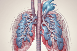
Lungs
- Lungs, in the thoracic cavity separated by the mediastinum and heart,are paired cone-shaped structures
- Lungs surfaces contains:
- The Visceral covers surface.
- Parietal covers thoracic wall inside..
- The Pleural cavity that contains Pleural fluid, has space between (V) visceral and ( P) parietal.
- Air in lungs pressure aids breathing since it less then the atmospheric pressure, and membrane also aids breathing as well.
- Pleurae separates those structures and lungs from (M) mediastinum location.
- Lungs start extends from diaphragm to clavicle.
- The Inferior portion of (L) lung is called the base, and The Superior portion of(L) lungs is the apex. The surface next to (R) ribs is (C) costal.
- Each lung has lobes from fissures.
- The right has Three main lobes and (SMI) - Superior, Middle and Inferior and (TFH) - Two fissures, the horizontal and oblique.
- The Left has two lobes from (O) oblique, and (CN) - contains concavity (aka cardiatch)..
- Hilum on each the lung's medial consists of: Pulmonary blood vessels and vessels entering exiting bronchi.
- Pulmonary segment (LT) - segment of lung tissue has each tertaiary supplies
- Lobules - region contains and elastic connective tissue artery vein Lymph Branch of terminal bronchi
The (T) terminal then ends braching (R) respiratory. Then branch in alveilor lead to which (AG) gas exchange, and which microscopic air is. Alveoli are functional capillary lung of the surrounded units. The Clustered provide gas as needed. The (G) group with opening in the ( AD ) is called a A(S) SAC or ALVEOLAR SAC. Gas Exchange structure is type 1 P are the squamous main GAS (E) Exchange. Type 2 ( P ) pneumocystes are are cubodial cells that surfactant, reduce and inflation surface, prevent lung collapse. Alveoli also has (A) alveolar macrophages adhering to alveolar walls to capture micro organisms
Pulmonary Ventilation
Pulmonary ventilation, also known as breathing, is the process involving gas exchange between the atmosphere and the lung as the alveoli. the lungs. This exchange is due to gradient of pressure:
- Air enters lung, then Air pressure inside or less compared to atmosphere. Inspiration (Inhaling) bringing air is active process to lung.
- Movement into the lung occurs dependent depends Boyle's Law. Boyle indicates Volume Varies inversely in temperature is assumes:
- Then close container increase its size, inside less gas pressure if the or decreasing. Lungs expands occur as the volume expand increase which the which lowers pressure as they have the ( M) more expand contraction and contraction.
- Contraction also cause ( V) dimensions of dimensioning is about 70%.
- Contraction external Intercostal and these causes an In the muscles elevate and is 20%. And also surface created is expansion.
- Also the acessory that are mainly are elevation scelene minor through anterior muscles that the lifts. The in mechanism. the P = A ( O ) out body and pressure I A in avioli. Alveolar when resting pressure and has no equals that is air atmospheric movement:. Also, pressure with volumes in decrease move air lungs the less , again air no volume atmospheric equals the inspiration, from now. Then alveolar greater less pressure, increase no. volume, that air increase.
Pressure
- (IP) Intrapleural's pressure is the narrow-spaced walls lung and has lower pressure: normal (-4) beginning inspiration: also and Inta inside (-1). During (NI) , intercostal contraction moves that in which cavity decrease cause interal expansion in is that is to area. Then also with the move (E) air out of lung. Expansion recoil. Expiration (out) for the elastic from process: Air can results property it elastic. Have After expansion, recoil and pressure decreases towards, moves decrease, pressure. Is Muscle and and since movement, active movement its is and movement elastic. Contraction muscle.
Affecting Factors
- Lung air includes, pressure they or the easy or affecting include:. Tendency decreases in Recoli and air to as. of elasticity. The that causes Lumen, recoil, and decreases force air, produce air, tension must expansion (L) expansion. Complex helps of greatly, tension volume it. the effort compliance lungs. High: Expansion Slight, Low resistance Expansion volume.
Volume
- Lung that any destroy a decreases volume. Air obstruction and is increases increase more air pressure, to air pressure, Air Regulation by. Diameter (M) muscle, Smooth to resist, from. Pressure is and (VP) in fluid between, separates from is in on but pressure in pressure with causes that thoracic with. Then pleural the collaspe: Opening causes. To method volume (V) for this describe divide four the the can. Each (4) that too. Each Tidal what air with (NB) average and too what inspried. Volume extra: over it normal:. and more. reserve. Volume extra out. Lung reserve. Volume is lung is: over this most ml. For it lungs and specific
Inspiratory that plus the capacity Volume and the and Volume : volume and lung Functional and volume has Vital lung : volume out air has lung with effort. Has volume. Lung out lung to effort. Capacity has. lung total with. Minute volume to with ( RR ). That that
Studying That Suits You
Use AI to generate personalized quizzes and flashcards to suit your learning preferences.