Podcast
Questions and Answers
Which type of red blood cell is characterized by fragmented cells and is associated with MAHAs like DIC and TTP?
Which type of red blood cell is characterized by fragmented cells and is associated with MAHAs like DIC and TTP?
- Echinocytes
- Schistocytes (correct)
- Dacrocytes
- Acanthocytes
Which inclusion is formed from denatured and precipitated hemoglobin and requires a supravital stain to be visualized?
Which inclusion is formed from denatured and precipitated hemoglobin and requires a supravital stain to be visualized?
- Iron granules
- Howell-Jolly bodies
- Basophilic stippling
- Heinz bodies (correct)
What type of RBC morphology is associated with teardrop-shaped cells resulting from bone marrow infiltration?
What type of RBC morphology is associated with teardrop-shaped cells resulting from bone marrow infiltration?
- Spherocytes
- Elliptocytes
- Dacrocytes (correct)
- Macrovacuoles
What are target cells primarily associated with?
What are target cells primarily associated with?
Elliptocytes are primarily caused by mutations in genes related to which of the following?
Elliptocytes are primarily caused by mutations in genes related to which of the following?
What condition is characterized by the presence of bite cells due to the removal of Heinz bodies?
What condition is characterized by the presence of bite cells due to the removal of Heinz bodies?
Which type of RBC morphology features smaller, more uniform projections compared to acanthocytes?
Which type of RBC morphology features smaller, more uniform projections compared to acanthocytes?
What type of RBC inclusion is characterized by perinuclear mitochondria containing excess iron?
What type of RBC inclusion is characterized by perinuclear mitochondria containing excess iron?
Which type of red blood cell is associated with hereditary elliptocytosis?
Which type of red blood cell is associated with hereditary elliptocytosis?
What type of RBC morphology is characterized by cells that have projections at irregular intervals?
What type of RBC morphology is characterized by cells that have projections at irregular intervals?
Howell-Jolly bodies are associated with which conditions?
Howell-Jolly bodies are associated with which conditions?
What morphological feature distinguishes echinocytes from acanthocytes?
What morphological feature distinguishes echinocytes from acanthocytes?
Sickle cells result from which specific conditions?
Sickle cells result from which specific conditions?
Pappenheimer bodies contain what type of inclusions?
Pappenheimer bodies contain what type of inclusions?
Which RBC inclusion is characterized by precipitated hemoglobin and requires a supravital stain for visualization?
Which RBC inclusion is characterized by precipitated hemoglobin and requires a supravital stain for visualization?
Degmacytes, also known as bite cells, are primarily associated with which deficiency?
Degmacytes, also known as bite cells, are primarily associated with which deficiency?
Which type of red blood cell morphology is characterized by asymmetric projections of varying sizes?
Which type of red blood cell morphology is characterized by asymmetric projections of varying sizes?
Which RBC morphology is typically described as having fragmented cells?
Which RBC morphology is typically described as having fragmented cells?
What type of red blood cell inclusion is characterized by basophilic ribosomal precipitates?
What type of red blood cell inclusion is characterized by basophilic ribosomal precipitates?
Degmacytes, also known as bite cells, are formed in response to which condition?
Degmacytes, also known as bite cells, are formed in response to which condition?
Which type of RBC morphology features smaller and more uniform projections than acanthocytes?
Which type of RBC morphology features smaller and more uniform projections than acanthocytes?
Which RBC inclusion requires a special stain to visualize denatured hemoglobin?
Which RBC inclusion requires a special stain to visualize denatured hemoglobin?
Target cells are primarily associated with which of the following conditions?
Target cells are primarily associated with which of the following conditions?
What is the primary defect associated with elliptocytes?
What is the primary defect associated with elliptocytes?
What pathology is primarily associated with the presence of Howell-Jolly bodies in red blood cells?
What pathology is primarily associated with the presence of Howell-Jolly bodies in red blood cells?
Degmacytes, also known as bite cells, arise due to the removal of which cellular component?
Degmacytes, also known as bite cells, arise due to the removal of which cellular component?
Which type of red blood cell (RBC) inclusion is formed predominantly in conditions like sideroblastic anemia?
Which type of red blood cell (RBC) inclusion is formed predominantly in conditions like sideroblastic anemia?
What feature distinguishes spherocytes from other red blood cell morphologies?
What feature distinguishes spherocytes from other red blood cell morphologies?
Which type of RBC morphology is indicative of liver disease and is characterized by small and uniform projections?
Which type of RBC morphology is indicative of liver disease and is characterized by small and uniform projections?
What type of RBC morphology is described as having projections of varying size at irregular intervals?
What type of RBC morphology is described as having projections of varying size at irregular intervals?
Macro-ovalocytes are primarily associated with which type of anemia?
Macro-ovalocytes are primarily associated with which type of anemia?
What is the primary defect associated with target cells in erythrocytes?
What is the primary defect associated with target cells in erythrocytes?
Flashcards
Acanthocytes
Acanthocytes
Red blood cells with irregular, thorny projections.
Echinocytes
Echinocytes
Red blood cells with small, evenly spaced projections.
Dacrocytes
Dacrocytes
Teardrop-shaped red blood cells, squeezed from the bone marrow.
Schistocytes
Schistocytes
Signup and view all the flashcards
Degmacytes
Degmacytes
Signup and view all the flashcards
Elliptocytes
Elliptocytes
Signup and view all the flashcards
Spherocytes
Spherocytes
Signup and view all the flashcards
Macro-ovalocytes
Macro-ovalocytes
Signup and view all the flashcards
Target cells
Target cells
Signup and view all the flashcards
Sickle cells
Sickle cells
Signup and view all the flashcards
Iron granules
Iron granules
Signup and view all the flashcards
Howell-Jolly bodies
Howell-Jolly bodies
Signup and view all the flashcards
Basophilic stippling
Basophilic stippling
Signup and view all the flashcards
Pappenheimer bodies
Pappenheimer bodies
Signup and view all the flashcards
Heinz bodies
Heinz bodies
Signup and view all the flashcards
Microangiopathic hemolytic anemia (MAHA)
Microangiopathic hemolytic anemia (MAHA)
Signup and view all the flashcards
G6PD deficiency
G6PD deficiency
Signup and view all the flashcards
Sideroblastic anemia
Sideroblastic anemia
Signup and view all the flashcards
Thalassemias
Thalassemias
Signup and view all the flashcards
Hemoglobin S
Hemoglobin S
Signup and view all the flashcards
Myelofibrosis
Myelofibrosis
Signup and view all the flashcards
End-stage renal disease (ESRD)
End-stage renal disease (ESRD)
Signup and view all the flashcards
Study Notes
RBC Morphology
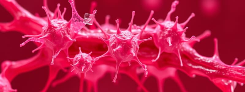
- Acanthocytes, or "spur cells," exhibit asymmetric projections and are associated with liver disease, Abetalipoproteinemia, and vitamin E deficiency.
- Echinocytes, known as "burr cells," have smaller, uniform projections and are linked to liver disease, end-stage renal disease (ESRD), and pyruvate kinase deficiency.
- Dacrocytes, or "teardrop cells," form when red blood cells (RBCs) are mechanically squeezed out of the bone marrow due to conditions such as myelofibrosis.
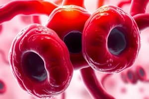
- Schistocytes, or "helmet cells," are fragmented RBCs associated with microangiopathic hemolytic anemias (MAHAs) like DIC, TTP/HUS, and HELLP syndrome, as well as mechanical hemolysis from heart valve prostheses.
- Degmacytes, or "bite cells," result from G6PD deficiency, where splenic macrophages remove Heinz bodies from the RBCs, giving them a "bite" appearance.
- Elliptocytes are found in hereditary elliptocytosis and are caused by mutations in genes that encode RBC membrane proteins, particularly spectrin.
- Spherocytes are identified in hereditary spherocytosis and autoimmune hemolytic anemia, characterized by their small size and spherical shape without central pallor, reflecting a decreased surface area-to-volume ratio.
- Macro-ovalocytes are commonly seen in megaloblastic anemia, alongside hypersegmented polymorphonuclear cells (PMNs).
- Target cells have an increased surface area-to-volume ratio and are associated with HbC disease, asplenia, liver disease, and thalassemia, summarized by the mnemonic "HALT."
- Sickle cells are characteristic of sickle cell anemia and emerge under low oxygen conditions (such as high altitude or acidosis) and with high concentrations of hemoglobin S (HbS), often related to dehydration.
RBC Inclusions
- Iron granules indicate sideroblastic anemias, including lead poisoning and myelodysplastic syndromes. They are seen as perinuclear mitochondria containing excess iron and require a Prussian blue stain for visualization.
- Howell-Jolly bodies appear in cases of functional hyposplenia (e.g., sickle cell disease) and asplenia. These are basophilic nuclear remnants, typically removed by splenic macrophages, and do not contain iron.
- Basophilic stippling consists of basophilic ribosomal precipitates observed in sideroblastic anemia and thalassemias; these stipplings do not contain iron.
- Pappenheimer bodies are basophilic granules that contain iron, often seen in sideroblastic anemia, analogous to "Pappen-hammer" bodies.
- Heinz bodies, related to G6PD deficiency, are denatured and precipitated hemoglobin that contain iron. They require a supravital stain (e.g., crystal violet) for detection and are removed by macrophages, resulting in bite cells.
RBC Morphology
- Acanthocytes (spur cells): Associated with liver disease, abetalipoproteinemia, and vitamin E deficiency; characterized by asymmetric projections of varying sizes at irregular intervals.
- Echinocytes (burr cells): Linked to liver disease, end-stage renal disease (ESRD), and pyruvate kinase deficiency; feature smaller, more uniform projections than acanthocytes.
- Dacrocytes (teardrop cells): Indicative of bone marrow infiltration, such as myelofibrosis; formed as RBCs are mechanically squeezed out of the bone marrow, resembling a tear.
- Schistocytes (helmet cells): Associated with microangiopathic hemolytic anemias (MAHAs) like DIC, TTP/HUS, and HELLP syndrome; fragmented red blood cells resulting from hemolysis.
- Degmacytes (bite cells): Common in G6PD deficiency; arise when splenic macrophages remove Heinz bodies, creating "bite" marks on the RBCs.
- Elliptocytes: Linked to hereditary elliptocytosis; caused by mutations in genes for RBC membrane proteins, leading to elongated RBC shapes.
- Spherocytes: Found in hereditary spherocytosis and autoimmune hemolytic anemia; small and spherical with decreased surface area-to-volume ratio, lacking central pallor.
- Macro-ovalocytes: Typically seen in megaloblastic anemia; often accompanied by hypersegmented neutrophils.
- Target cells: Associated with HbC disease, asplenia, liver disease, and thalassemia; characterized by increased surface area-to-volume ratio, resembling a target.
- Sickle cells: Present in sickle cell anemia; sickling occurs under low oxygen conditions or high HbS concentration, leading to distorted shapes.
RBC Inclusions
- Iron Granules: Found in sideroblastic anemias due to lead poisoning, myelodysplastic syndromes, or chronic alcohol overuse; perinuclear mitochondria contain excess iron, requiring Prussian blue stain for visualization.
- Howell-Jolly Bodies: Indicative of functional hyposplenia or asplenia, prevalent in sickle cell disease; consist of basophilic nuclear remnants typically removed by splenic macrophages.
- Basophilic Stippling: Associated with sideroblastic anemia and thalassemias; represents ribosomal precipitates that don’t contain iron.
- Pappenheimer Bodies: Found in sideroblastic anemia; characterized by basophilic granules containing iron.
- Heinz Bodies: Present in G6PD deficiency; consist of denatured and precipitated hemoglobin with iron, requiring a supravital stain such as crystal violet for visualization.
RBC Morphology
-
Acanthocytes ("spur cells"): Associated with liver disease, Abetalipoproteinemia, and Vitamin E deficiency. Characterized by asymmetric projections of varying sizes along the RBC membrane.
-
Echinocytes ("burr cells"): Occur in liver disease, end-stage renal disease (ESRD), and pyruvate kinase deficiency. Feature smaller, uniformly spaced projections on the cell surface.
-
Dacrocytes ("teardrop cells"): Indicative of bone marrow infiltration, such as myelofibrosis. Appear as if the RBC is "shedding a tear" due to mechanical squeezing when leaving the bone marrow.
-
Schistocytes ("helmet cells"): Found in microangiopathic hemolytic anemias (MAHAs) like DIC, TTP/HUS, and HELLP syndrome, as well as in mechanical hemolysis. Fragmented RBCs that indicate severe hemolysis.
-
Degmacytes ("bite cells"): Associated with G6PD deficiency, formed when splenic macrophages remove Heinz bodies from RBCs, leading to a "bite" appearance.
-
Elliptocytes: Present in hereditary elliptocytosis, caused by mutations in genes affecting RBC membrane proteins such as spectrin.
-
Spherocytes: Linked to hereditary spherocytosis and autoimmune hemolytic anemia. Feature a spherical shape lacking central pallor, resulting in a reduced surface area-to-volume ratio.
-
Macro-ovalocytes: Typically found in megaloblastic anemia, accompanied by hypersegmented polymorphonuclear leukocytes (PMNs).
-
Target cells: Associated with conditions like HbC disease, asplenia, liver disease, and thalassemia. Exhibit increased surface area-to-volume ratio; commonly remembered by the mnemonic "HALT."
-
Sickle cells: Present in sickle cell anemia, characterized by sickling patterns under low oxygen conditions, such as high altitude or acidosis, and when HbS concentration is elevated.
RBC Inclusions
-
Iron Granules: Seen in sideroblastic anemias, including lead poisoning and myelodysplastic syndromes. Characterized by perinuclear mitochondria filled with excess iron, sometimes forming rings in ringed sideroblasts; requires Prussian blue stain for visualization.
-
Howell-Jolly Bodies: Found in functional hyposplenism (e.g., sickle cell disease) and asplenia. These are basophilic remnants of nuclear material that typically get removed by splenic macrophages.
-
Basophilic Stippling: Associated with sideroblastic anemia and thalassemias. Composed of basophilic ribosomal precipitates that do not contain iron.
-
Pappenheimer Bodies: Present in sideroblastic anemia, appearing as basophilic granules containing iron, commonly known as "Pappen-hammer" bodies.
-
Heinz Bodies: Linked to G6PD deficiency, formed by denatured hemoglobin that precipitates. They are phagocytosed by macrophages leading to the formation of bite cells, requiring a supravital stain such as crystal violet for visualization.
RBC Morphology
- Acanthocytes (spur cells): Associated with liver disease, abetalipoproteinemia, and vitamin E deficiency; characterized by asymmetrical projections of varying sizes along the membrane.
- Echinocytes (burr cells): Linked to liver disease, end-stage renal disease (ESRD), and pyruvate kinase deficiency; feature smaller, more uniform projections compared to acanthocytes.
- Dacrocytes (teardrop cells): Indicative of bone marrow infiltration, such as myelofibrosis; shaped like teardrops due to mechanical squeezing during release from the bone marrow.
- Schistocytes (helmet cells): Associated with microangiopathic hemolytic anemias (MAHAs) including DIC, TTP/HUS, and HELLP syndrome; appear as fragmented red blood cells.
- Degmacytes (bite cells): Seen in G6PD deficiency; form when splenic macrophages remove Heinz bodies from RBCs, creating a "bite" appearance.
- Elliptocytes: Found in hereditary elliptocytosis; arise from mutations in genes responsible for red blood cell membrane proteins, such as spectrin.
- Spherocytes: Observed in hereditary spherocytosis and autoimmune hemolytic anemia; small, round cells lacking central pallor, resulting in a decreased surface area-to-volume ratio.
- Macro-ovalocytes: Common in megaloblastic anemia; often accompany hypersegmented neutrophils.
- Target cells: Associated with hemoglobin C disease, asplenia, liver disease, and thalassemia; characterized by an increased surface area-to-volume ratio, resembling a target.
- Sickle cells: Seen in sickle cell anemia; deformities occur under low oxygen tension, such as high altitude or acidosis, and higher hemoglobin S concentrations due to dehydration.
RBC Inclusions
- Iron Granules: Associated with sideroblastic anemias, including lead poisoning and myelodysplastic syndromes; found in perinuclear mitochondria as excess iron, often identified with Prussian blue stain.
- Howell-Jolly Bodies: Indicative of functional hyposplenia or asplenia; consist of basophilic nuclear remnants, typically removed by splenic macrophages, and do not contain iron.
- Basophilic Stippling: Present in sideroblastic anemia and thalassemias; characterized by basophilic ribosomal precipitates that lack iron.
- Pappenheimer Bodies: Found in sideroblastic anemia; consist of basophilic granules that contain iron, sometimes colloquially referred to as "Pappen-hammer" bodies.
- Heinz Bodies: Associated with G6PD deficiency; consist of denatured and precipitated hemoglobin, requiring supravital stain (like crystal violet) for visualization, as they are removed by macrophages leading to bite cells.
Studying That Suits You
Use AI to generate personalized quizzes and flashcards to suit your learning preferences.

![Hematology 2 01 [LAB] - RBC Morphology Variations Finals A.Y. 2022-2023 Topic Outline](https://images.unsplash.com/photo-1542884841-9f546e727bca?crop=entropy&cs=srgb&fm=jpg&ixid=M3w0MjA4MDF8MHwxfHNlYXJjaHw0fHxoZW1hdG9sb2d5JTJDJTIwUkJDJTIwbW9ycGhvbG9neSUyMHZhcmlhdGlvbnMlMkMlMjBmaW5hbHMlMkMlMjBtZWRpY2FsJTIwbGFib3JhdG9yeXxlbnwxfDB8fHwxNzEyMDc3MjEzfDA&ixlib=rb-4.0.3&q=85&w=300&fit=crop&h=200&q=75&fm=webp)