Podcast
Questions and Answers
Which of the following best describes the primary function of radiology?
Which of the following best describes the primary function of radiology?
- To administer medications for chronic illnesses.
- To use imaging techniques for diagnosis and treatment guidance. (correct)
- To perform physical therapy and rehabilitation.
- To surgically repair internal injuries.
Which of the following imaging modalities is NOT typically categorized under diagnostic imaging?
Which of the following imaging modalities is NOT typically categorized under diagnostic imaging?
- Radiographs
- Chemotherapy (correct)
- Ultrasound
- Magnetic Resonance Imaging (MRI)
What is the fundamental principle behind X-rays, concerning their interaction with atoms?
What is the fundamental principle behind X-rays, concerning their interaction with atoms?
- X-rays cause electrons to be ejected from atoms, creating ions. (correct)
- X-rays have no interaction with atoms.
- X-rays stabilize atoms by neutralizing their charge.
- X-rays cause atoms to gain electrons, creating negative ions.
Which statement accurately describes the behaviour of X-rays in relation to magnetic or electrical fields?
Which statement accurately describes the behaviour of X-rays in relation to magnetic or electrical fields?
What is a key distinction between radio waves and X-rays in terms of the electromagnetic spectrum and their potential effects?
What is a key distinction between radio waves and X-rays in terms of the electromagnetic spectrum and their potential effects?
Why is the term 'radiograph' more accurate than 'X-ray' when referring to the final image?
Why is the term 'radiograph' more accurate than 'X-ray' when referring to the final image?
What is the primary mechanism through which radiation exposure can lead to genetic abnormalities?
What is the primary mechanism through which radiation exposure can lead to genetic abnormalities?
Which of the following cell types is generally considered most susceptible to the harmful effects of radiation?
Which of the following cell types is generally considered most susceptible to the harmful effects of radiation?
What is the specific purpose of lead or lead-equivalent materials in personal protective equipment (PPE) used during radiology procedures?
What is the specific purpose of lead or lead-equivalent materials in personal protective equipment (PPE) used during radiology procedures?
What is the function of structural shielding, such as lead-impregnated doors and walls, in a radiology suite?
What is the function of structural shielding, such as lead-impregnated doors and walls, in a radiology suite?
In radiographic imaging, what does a 'black' area typically indicate on the image?
In radiographic imaging, what does a 'black' area typically indicate on the image?
In radiographic imaging, what causes different shades of gray to appear on the image?
In radiographic imaging, what causes different shades of gray to appear on the image?
How does the atomic number of a substance affect its radiopacity?
How does the atomic number of a substance affect its radiopacity?
What is the best definition of radiolucency?
What is the best definition of radiolucency?
Why might the appearance of tissues in a radiograph not always directly correlate with specific anatomical structures?
Why might the appearance of tissues in a radiograph not always directly correlate with specific anatomical structures?
What is the role of the collimator in an X-ray machine?
What is the role of the collimator in an X-ray machine?
In veterinary radiography, how are radiographic views typically named?
In veterinary radiography, how are radiographic views typically named?
What does the term 'ventrodorsal' (VD) signify when describing a radiographic view of the thorax?
What does the term 'ventrodorsal' (VD) signify when describing a radiographic view of the thorax?
When viewing lateral radiographs, how should the image be typically oriented for interpretation?
When viewing lateral radiographs, how should the image be typically oriented for interpretation?
Which way should you orient a ventrodorsal or dorsoventral radiograph for viewing?
Which way should you orient a ventrodorsal or dorsoventral radiograph for viewing?
Which way should you orient a radiograph for viewing limbs?
Which way should you orient a radiograph for viewing limbs?
In a right lateral thoracic radiograph, which anatomical structure is situated in the dependent position (closer to the table)?
In a right lateral thoracic radiograph, which anatomical structure is situated in the dependent position (closer to the table)?
Which anatomical structure can be well-evaluated in a ventrodorsal (VD) view of the thorax?
Which anatomical structure can be well-evaluated in a ventrodorsal (VD) view of the thorax?
In a right lateral view of the abdomen, what is the position of the ventral liver?
In a right lateral view of the abdomen, what is the position of the ventral liver?
What anatomical structures are generally best assessed in a ventrodorsal (VD) view of the abdomen?
What anatomical structures are generally best assessed in a ventrodorsal (VD) view of the abdomen?
How are radiographs named when imaging the equine carpus or other distal limb structures?
How are radiographs named when imaging the equine carpus or other distal limb structures?
In a lateromedial view of the equine carpus, which carpal bones are typically superimposed?
In a lateromedial view of the equine carpus, which carpal bones are typically superimposed?
What anatomical feature is best evaluated when using a dorsopalmar view of the equine carpus?
What anatomical feature is best evaluated when using a dorsopalmar view of the equine carpus?
Considering the case of 'Roxy,' a 6-month-old intact female dog presenting with vomiting, what radiographic finding would be most suggestive of a foreign body obstruction?
Considering the case of 'Roxy,' a 6-month-old intact female dog presenting with vomiting, what radiographic finding would be most suggestive of a foreign body obstruction?
Given the radiograph provided for 'Ally,' a 2-year old DSH cat, what is the most likely diagnosis based on the radiographic findings?
Given the radiograph provided for 'Ally,' a 2-year old DSH cat, what is the most likely diagnosis based on the radiographic findings?
Which of the following statements best relates to the use of personal shielding, specifically regarding tag placement and lead equivalence?
Which of the following statements best relates to the use of personal shielding, specifically regarding tag placement and lead equivalence?
What is a crucial consideration when assessing a radiograph for diagnostic purposes?
What is a crucial consideration when assessing a radiograph for diagnostic purposes?
Which of the following statements captures an essential concept underlying effective shielding?
Which of the following statements captures an essential concept underlying effective shielding?
What is a guiding principle one must consider when naming radiographic views?
What is a guiding principle one must consider when naming radiographic views?
Why, must cell types be meticulously understood to appreciate radiation hazards?
Why, must cell types be meticulously understood to appreciate radiation hazards?
How do atomic numbers primarily influence radiopacity vs radiolucency?
How do atomic numbers primarily influence radiopacity vs radiolucency?
Which of the following statements accurately clarifies essential imaging aspects related to diagnosing internal ailments by the practice of radiology?
Which of the following statements accurately clarifies essential imaging aspects related to diagnosing internal ailments by the practice of radiology?
Flashcards
What is Radiology?
What is Radiology?
The use of imaging to make diagnoses and guide treatment.
Diagnostic Imaging
Diagnostic Imaging
Radiographs, Ultrasound, CT, MRI, nuclear medicine.
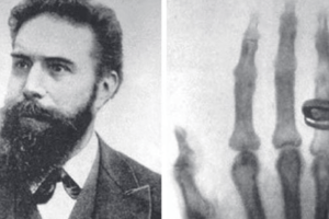
What are X-rays?
What are X-rays?
Form of radiation which involves the transfer of energy from one place to another.
Ionizing radiation
Ionizing radiation
Signup and view all the flashcards
Properties of X-rays
Properties of X-rays
Signup and view all the flashcards
X-ray Characteristics
X-ray Characteristics
Signup and view all the flashcards
Radiation Hazard
Radiation Hazard
Signup and view all the flashcards
Cell Sensitivity to Radiation
Cell Sensitivity to Radiation
Signup and view all the flashcards
Shielding
Shielding
Signup and view all the flashcards
Personal Shielding
Personal Shielding
Signup and view all the flashcards
Structural Shielding
Structural Shielding
Signup and view all the flashcards
Black Image
Black Image
Signup and view all the flashcards
White Radiograph Image
White Radiograph Image
Signup and view all the flashcards
Radiopacity
Radiopacity
Signup and view all the flashcards
Radiolucency
Radiolucency
Signup and view all the flashcards
How Radiographic Views are named
How Radiographic Views are named
Signup and view all the flashcards
Ventrodorsal View
Ventrodorsal View
Signup and view all the flashcards
Right Lateral View
Right Lateral View
Signup and view all the flashcards
Viewing Lateral Radiographs
Viewing Lateral Radiographs
Signup and view all the flashcards
ALARA principle
ALARA principle
Signup and view all the flashcards
Study Notes
- Radiology involves using imaging to diagnose and guide treatment.
- Diagnostic imaging methods: Radiographs, Ultrasound, CT, MRI, and nuclear medicine.
X-Rays
- X-rays involve energy or energetic particles being transferred from one place to another.
- Ionizing radiation causes electrons to leave an atom, creating ions.
Properties of X-Rays/Photons
- X-rays travel at the speed of light.
- X-rays cannot be refracted or reflected like light.
- X-rays have no electrical charge and are unaffected by magnetic or electrical fields.
- X-rays travel in a straight line.
- X-rays can penetrate matter to some degree.
- X-rays can cause fluorescence.
- X-rays can interact with photogenic emulsion.
- X-rays can cause ionization.
- X-rays cannot be felt and are invisible.
Electromagnetic Spectrum
- The electromagnetic spectrum ranges from harmless radio waves (low frequency, long wavelength) to harmful gamma rays (high frequency, short wavelength).
Hazards of Radiation
- Sufficient energy from radiation can transfer to living tissue, creating ions.
- Radiation hazards include changes in molecular structure and cells, loss or abnormal cell function, genetic/somatic abnormalities like mutations/cataracts/leukemia/cancer/death.
- Most sensitive cells have a high division rate, such as gonads and embryonic tissue.
- Damaged sperm/eggs carry defects to the next generation.
- The first trimester is very vulnerable leading to embryonic death, congenital abnormalities, or growth defects.
Shielding
- Personal shielding: Lead or lead equivalent impregnated clothing protects against radiation.
- Lead aprons and gloves contain 0.5 mm Pb equivalent.
- Thyroid shields contain 0.25 mm or 0.5 mm Pb equivalent.
- Eye glasses contain 0.75 mm Pb equivalent.
- Structural shielding uses screens and lead-impregnated doors/walls for protection.
Image Formation
- Black images are formed when all X-rays hit the receiver (digital plate/cassette and film).
- White images are formed when all X-rays are absorbed by the patient.
- Various shades of gray occur when some X-rays hit the receiver, while others are absorbed by the patient.
Radiopacities
- Radiopacity is the relative inability of structures to be penetrated by X-rays.
- High atomic number metals prevent X-ray penetration.
- Radiolucency describes substances that allow X-rays to penetrate them, leaving darkened images.
- Air is the most radiolucent substance in radiographs.
- Radiographic opacities aren't tied to specific structures; tissue thickness plays a role in tissue appearance.
X-Ray Machine
- X-ray machines have an X-ray housing and tube.
- Collimators turn lead shutters internally to adjust the image size.
- A grid and imaging plate/cassette contain a metal plate to prevent scatter.
Naming the Views
- Radiographic views are named for the direction of the primary beam.
- The name starts with the point of entry, followed by the point of exit of the primary beam.
- Ventrodorsal(VD) view of the thorax: The patient is in dorsal recumbency, as the beam enters the ventral portion and exits via the dorsum.
- Right Lateral(Lat) view of the thorax: The patient is in right lateral recumbency, when lying down on the right side.
- True name = Left to right lateral view.
- When viewing Lateral radiographs, the cranial portion is to the viewer's left.
- Ventrodorsal/dorsoventral views: The head should be pointing upwards, and the left side of the patient should be on the viewer's right.
- Limbs: The proximal portion faces upwards, the distal portion faces the floor, and the cranial/dorsal portion faces the viewer's left.
- Radiographs are named by the photon direction through the patient.
- Lateromedial refers to lateral/medial positioning.
- Radiographs are most sensitive on the edges.
Anatomical Examples in Radiographs
- Right lateral thorax radiograph depicts the trachea, descending aorta, right/left crus of diaphragm, pulmonary arteries/veins, and caudal vena cava.
- Ventrodorsal thorax radiograph illustrates the cranial mediastinum, left cranial pulmonary artery/vein, and tracheal bifurcation.
- Right lateral abdomen radiograph depicts the descending colon and urinary bladder.
- Ventrodorsal abdomen radiograph displays the liver, fundus of the stomach, head of the spleen, left kidney, and descending colon.
- A Lateromedial view of the Equine carpus shows superimposed radial and intermediate carpal bones with accessory and ulnar carpal bones.
- A Dorsopalmar view of the Equine carpus includes the distal radius, intermediate and radial carpal bones, as well as MC2/3/4.
Studying That Suits You
Use AI to generate personalized quizzes and flashcards to suit your learning preferences.