Podcast
Questions and Answers
What is a characteristic feature of the diaphragm?
What is a characteristic feature of the diaphragm?
- Left side is higher than the right
- The diaphragm is not affected by any conditions
- Both sides are at the same level
- Right side is higher than the left (correct)
What is the percentage of adult cases where the CXR is normal in primary tuberculosis?
What is the percentage of adult cases where the CXR is normal in primary tuberculosis?
- 10%
- 15% (correct)
- 20%
- 5%
What is a characteristic feature of primary tuberculosis in the parenchyma?
What is a characteristic feature of primary tuberculosis in the parenchyma?
- Consolidation only in the upper lobe
- Dense consolidation in any lobe (correct)
- Consolidation only in the lower lobe
- Sparse consolidation in any lobe
What is the percentage of adult cases where scarring occurs in primary tuberculosis?
What is the percentage of adult cases where scarring occurs in primary tuberculosis?
What is the percentage of cases where primary tuberculosis resolves without sequelae?
What is the percentage of cases where primary tuberculosis resolves without sequelae?
What is a characteristic feature of miliary tuberculosis?
What is a characteristic feature of miliary tuberculosis?
What is a characteristic feature of postprimary tuberculosis?
What is a characteristic feature of postprimary tuberculosis?
What is a characteristic feature of COPD?
What is a characteristic feature of COPD?
What is the purpose of the lateral decubitus position in chest X-ray?
What is the purpose of the lateral decubitus position in chest X-ray?
What makes a chest X-ray image good?
What makes a chest X-ray image good?
What is the main pulmonary artery located in?
What is the main pulmonary artery located in?
What is the purpose of the PA projection in chest X-ray?
What is the purpose of the PA projection in chest X-ray?
What is the characteristic of the trachea on a chest X-ray?
What is the characteristic of the trachea on a chest X-ray?
What is the purpose of the dorsal vertebra being barely seen through the heart on a chest X-ray?
What is the purpose of the dorsal vertebra being barely seen through the heart on a chest X-ray?
What is the characteristic of the costophrenic angle on a chest X-ray?
What is the characteristic of the costophrenic angle on a chest X-ray?
Flashcards are hidden until you start studying
Study Notes
Radiological Examination of the Chest
- Learning objectives:
- Evaluate the quality of a chest X-ray (CXR)
- Learn basic radiographic anatomy on a CXR
- Be aware of the "hidden areas"
- Learn CXR imaging findings in common and emergency conditions
Methods Used
- Chest X-ray (CXR)
- Computerised Tomography (CT)
- Magnetic Resonance (MR) or Ultrasound (US) (rarely)
- Angiography (very rarely)
Chest X-ray (CXR)
- Two projections:
- Posterior-Anterior (PA)
- Lateral
- Lateral projection:
- Taken with the left side on the film side
- Magnification effect is reduced
- Heart appears smaller
- Lateral decubitus position:
- Detects pleural effusion
- Not frequently used anymore
Quality of a Good Image
- Full inspiration
- Penetration:
- Dorsal vertebra barely seen through the heart
- Vessels are visible
- Rotation:
- Clavicles are not rotated
- Superposition:
- Scapulae are not superimposed on the lungs
Anatomy
Trachea and Bronchi
- Trachea:
- Located to the right of the aortic arch
- Radiolucent (black)
- Bronchi:
- Large airways are radiolucent (black)
- Trachea at the midline
The Hilum
- Main pulmonary bronchi
- Main pulmonary arteries
Lung Zones
- CXR is interpreted in zones, not lobes
Pleura
- Only visible in pathologic conditions
Lung Lobes and Fissures
- Both lungs have oblique fissures
- Right lung has a horizontal fissure
- Accessory fissures:
- Azygos fissure and lobe (most common, 1-2%)
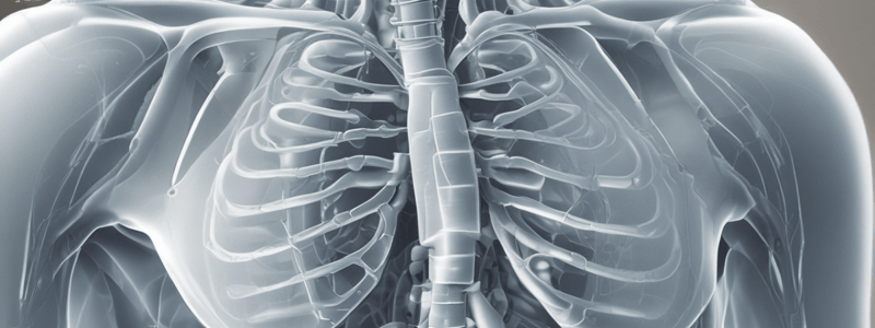
Costophrenic Angle and Recess
- Between the lateral chest wall and diaphragm
- Should be sharp and clearly visible
- Diaphragm:
- Right side is higher than left
- Stomach air is below the left diaphragm
Heart
- Cardiothoracic Ratio:
- Lower than CT
Pneumonia
- Air Bronchogram
Tuberculosis
Primary Tuberculosis
- Initial infection
- Mostly in children, but rate increases in adults (25-35% of all adult cases)
- CXR is normal in 15%
- Features may overlap with reactivation
- Differentiation not always possible
- Parenchymal focus:
- Dense consolidation in any lobe
- Similar appearance to bacterial pneumonia
- Lack of response to antibiotics
- Lymphadenopathy:
- Seen in 96% of children and 43% of adult cases
- Can be the only finding, especially in children
- Calcified LAP + Ghon focus = Ranke complex
- Miliary Disease:
- 1-7% of all forms of TBC
- Elderly, infants, immunocompromised patients
- Evenly distributed 2-3 mm sized nodules
Postprimary Tuberculosis
- Primarily a disease of adolescence and adulthood
- Re-infection or reactivation
- Progressive
- Cavitation
- Causes hematogenous spread
- Endobronchial:
- Tree-in-bud pattern
Chronic Obstructive Pulmonary Disease (COPD)
- Resistance to expiratory airflow
- CXR is highly specific
- Hyper-expanded lungs with flattening of both hemi-diaphragms
- Barrel-shaped chest
Foreign Body Aspiration
- Symptoms: Asymptomatic > Choking
- CXR:
- Foreign body may or may not be seen
Studying That Suits You
Use AI to generate personalized quizzes and flashcards to suit your learning preferences.


