Podcast
Questions and Answers
What does the acronym VQ stand for in nuclear medicine?
What does the acronym VQ stand for in nuclear medicine?
- Ventilation and Quantification
- Ventilation Quantitative
- Ventilation and Perfusion (correct)
- Vascular Quality
What is the primary purpose of conducting lung scans?
What is the primary purpose of conducting lung scans?
- To rule out pulmonary embolism (correct)
- To assess lung capacity
- To monitor post-operative lung function
- To evaluate for chronic obstructive pulmonary disease
Which statement is true about the performance of lung scans?
Which statement is true about the performance of lung scans?
- They should only be performed in outpatient settings.
- They must be performed in a single step only.
- They can be scheduled weeks in advance for any condition.
- They are usually ordered as immediate emergency procedures. (correct)
How does a low probability VQ scan differ from a high probability VQ scan for pulmonary embolism?
How does a low probability VQ scan differ from a high probability VQ scan for pulmonary embolism?
What technique is used for a quantitative lung scan as part of nuclear medicine?
What technique is used for a quantitative lung scan as part of nuclear medicine?
During a VQ scan, what specific symptoms might a patient typically exhibit?
During a VQ scan, what specific symptoms might a patient typically exhibit?
What is one of the steps to be followed when performing a VQ scan?
What is one of the steps to be followed when performing a VQ scan?
What distinguishes a quantitative lung scan from traditional lung scans?
What distinguishes a quantitative lung scan from traditional lung scans?
What is the approximate number of small capillaries in the lungs?
What is the approximate number of small capillaries in the lungs?
What is the typical ratio of right lung uptake to left lung uptake?
What is the typical ratio of right lung uptake to left lung uptake?
What is a potential complication of injecting micro emboli into the lungs?
What is a potential complication of injecting micro emboli into the lungs?
What does the VQ consist of?
What does the VQ consist of?
What might cause the left lung to show less uptake than the right lung?
What might cause the left lung to show less uptake than the right lung?
Which of the following is NOT typically included in a VQ scan?
Which of the following is NOT typically included in a VQ scan?
How many regions are commonly compared in lung segment analysis?
How many regions are commonly compared in lung segment analysis?
When comparing total counts in lung segments, what is measured?
When comparing total counts in lung segments, what is measured?
What might affect uptake percentages in lung studies?
What might affect uptake percentages in lung studies?
What study is usually a standalone examination in lung diagnostics?
What study is usually a standalone examination in lung diagnostics?
What is usually indicated by the presence of a triangular wedge during a lung perfusion study?
What is usually indicated by the presence of a triangular wedge during a lung perfusion study?
Which lung condition may show clumpy areas due to particle deposition?
Which lung condition may show clumpy areas due to particle deposition?
In a ventilation study, what does normal ventilation with abnormal perfusion indicate?
In a ventilation study, what does normal ventilation with abnormal perfusion indicate?
What might be a consequence of using a larger amount of the isotope in patients with pulmonary hypertension?
What might be a consequence of using a larger amount of the isotope in patients with pulmonary hypertension?
How do obstruction presentations in lung imaging typically manifest?
How do obstruction presentations in lung imaging typically manifest?
What does accelerated clearance of aerosols in lung imaging suggest?
What does accelerated clearance of aerosols in lung imaging suggest?
Why is the perfusion portion crucial in lung studies?
Why is the perfusion portion crucial in lung studies?
Which of the following is a contraindication for lung perfusion studies?
Which of the following is a contraindication for lung perfusion studies?
What is the significance of the ventilation study in diagnosing conditions like COPD?
What is the significance of the ventilation study in diagnosing conditions like COPD?
What is the risk of using too many particles in the perfusion study?
What is the risk of using too many particles in the perfusion study?
What happens to ventilation findings when a patient has a significant perfusion issue?
What happens to ventilation findings when a patient has a significant perfusion issue?
What can significantly alter the imaging time in a perfusion study?
What can significantly alter the imaging time in a perfusion study?
For evaluating particulate deposition, which lung disorders are typically observed?
For evaluating particulate deposition, which lung disorders are typically observed?
What is the biological half-life of xenon as described?
What is the biological half-life of xenon as described?
Why is patient positioning crucial before administering the ventilation process?
Why is patient positioning crucial before administering the ventilation process?
What should be the expected appearance of the left lung during a normal ventilation scan?
What should be the expected appearance of the left lung during a normal ventilation scan?
What is the characteristic gamma energy level for xenon used in lung scans?
What is the characteristic gamma energy level for xenon used in lung scans?
Which position is ideally preferred for the patient during the scanning process?
Which position is ideally preferred for the patient during the scanning process?
What is the primary purpose of using a lead shielded container in this procedure?
What is the primary purpose of using a lead shielded container in this procedure?
What is a common result indicative of an abnormal ventilation scan?
What is a common result indicative of an abnormal ventilation scan?
How long should radioactive materials typically be stored before disposal?
How long should radioactive materials typically be stored before disposal?
How long should the mask remain on during the xenon lung scan?
How long should the mask remain on during the xenon lung scan?
What imaging orientation is not typically performed during ventilation imaging?
What imaging orientation is not typically performed during ventilation imaging?
In the context of nuclear medicine, what does a physical half-life of 5.3 days imply?
In the context of nuclear medicine, what does a physical half-life of 5.3 days imply?
What occurs during the 'wash out' phase of the lung scan?
What occurs during the 'wash out' phase of the lung scan?
What could cause decreased activity in the lungs during imaging?
What could cause decreased activity in the lungs during imaging?
Why is it preferable to conduct ventilation imaging before perfusion imaging?
Why is it preferable to conduct ventilation imaging before perfusion imaging?
What ensures effective visualization during a lung scan?
What ensures effective visualization during a lung scan?
What issue might arise if a patient swallows a lot of contaminated saliva during the procedure?
What issue might arise if a patient swallows a lot of contaminated saliva during the procedure?
What does a ventilation scan typically show in cases of pulmonary embolism?
What does a ventilation scan typically show in cases of pulmonary embolism?
What is a common visual artifact found in the imaging process?
What is a common visual artifact found in the imaging process?
What is one common challenge faced during the xenon administration process?
What is one common challenge faced during the xenon administration process?
What technological device is utilized to survey radioactive materials before disposal?
What technological device is utilized to survey radioactive materials before disposal?
What range of activity in the lungs should be expected during a normal xenon scan?
What range of activity in the lungs should be expected during a normal xenon scan?
What happens if the oxygen flow is inadequate during the ventilation process?
What happens if the oxygen flow is inadequate during the ventilation process?
What is the role of the isotope marked on the patient during preparation?
What is the role of the isotope marked on the patient during preparation?
What is considered low gamma energy according to the information provided?
What is considered low gamma energy according to the information provided?
Which of the following might be visualized in addition to the lungs during imaging?
Which of the following might be visualized in addition to the lungs during imaging?
What effect do ventilation and perfusion studies typically aim to highlight?
What effect do ventilation and perfusion studies typically aim to highlight?
What is an acceptable practice in nuclear medicine regarding image presentation?
What is an acceptable practice in nuclear medicine regarding image presentation?
During the imaging process, what is an important step to minimize unwanted results?
During the imaging process, what is an important step to minimize unwanted results?
What condition may cause a significant decrease in ventilation and slight decrease in perfusion?
What condition may cause a significant decrease in ventilation and slight decrease in perfusion?
What indicates fatty liver prevalent in certain individuals?
What indicates fatty liver prevalent in certain individuals?
What may be indicated if one lung shows poor presentation in imaging?
What may be indicated if one lung shows poor presentation in imaging?
Which isotopes are commonly designed to target the lungs?
Which isotopes are commonly designed to target the lungs?
How long is the biological half-life of xenon in ventilation imaging?
How long is the biological half-life of xenon in ventilation imaging?
What is the typical dose range for aerosol used in lung studies?
What is the typical dose range for aerosol used in lung studies?
What is a recommended patient position during aerosol ventilation?
What is a recommended patient position during aerosol ventilation?
What is the significant advantage of using DTA aerosol over xenon for imaging?
What is the significant advantage of using DTA aerosol over xenon for imaging?
What body part is most commonly targeted by isotopes during lung studies?
What body part is most commonly targeted by isotopes during lung studies?
What should be avoided during the inhalation procedure of aerosol?
What should be avoided during the inhalation procedure of aerosol?
How does oxygen contribute to the aerosolization process during imaging?
How does oxygen contribute to the aerosolization process during imaging?
In patients with certain lung conditions, what is an important logistical consideration?
In patients with certain lung conditions, what is an important logistical consideration?
What can high liver uptake during lung studies signify?
What can high liver uptake during lung studies signify?
What is a common weight limit for tables used in lung scans?
What is a common weight limit for tables used in lung scans?
What might a reverse mismatch in lung scans indicate?
What might a reverse mismatch in lung scans indicate?
During a lung scan, how does scanning in an upright position affect the process?
During a lung scan, how does scanning in an upright position affect the process?
What is the primary benefit of administering a lower dose of particles to a patient?
What is the primary benefit of administering a lower dose of particles to a patient?
What type of lung defect is characterized by wedge-shaped areas in the lungs?
What type of lung defect is characterized by wedge-shaped areas in the lungs?
In which scenario should a chest X-ray be performed within one hour?
In which scenario should a chest X-ray be performed within one hour?
What is the most common reason patients visit a nuclear medicine technologist?
What is the most common reason patients visit a nuclear medicine technologist?
What is the likelihood of pulmonary embolism at the low probability level?
What is the likelihood of pulmonary embolism at the low probability level?
What is the preferred method of injection for the radiopharmaceutical?
What is the preferred method of injection for the radiopharmaceutical?
How many large mismatch segmental perfusion defects indicate a high probability of pulmonary embolism?
How many large mismatch segmental perfusion defects indicate a high probability of pulmonary embolism?
What procedure is commonly performed in conjunction with a lung scan?
What procedure is commonly performed in conjunction with a lung scan?
What is the consequence of injecting the radiopharmaceutical through tubing?
What is the consequence of injecting the radiopharmaceutical through tubing?
In what situations might a lung scan still be performed despite advancements in CT technology?
In what situations might a lung scan still be performed despite advancements in CT technology?
What typically correlates with high probability of pulmonary embolism in lung scans?
What typically correlates with high probability of pulmonary embolism in lung scans?
What is indicated by a striped sign in lung imaging?
What is indicated by a striped sign in lung imaging?
What is the typical dose range for TEC Museum 99 M macro aggregated albumin?
What is the typical dose range for TEC Museum 99 M macro aggregated albumin?
Why is it critical to use a negatively pressurized room when administering xenon gas?
Why is it critical to use a negatively pressurized room when administering xenon gas?
What is a significant risk associated with the longer half-life of xenon gas?
What is a significant risk associated with the longer half-life of xenon gas?
What is the role of the physician when interpreting lung scan data?
What is the role of the physician when interpreting lung scan data?
Why should patients be maintained in a supine position during the perfusion study?
Why should patients be maintained in a supine position during the perfusion study?
What percentage of the population is affected by subsegment defects?
What percentage of the population is affected by subsegment defects?
What could happen if blood is drawn into the syringe before injecting the radiopharmaceutical?
What could happen if blood is drawn into the syringe before injecting the radiopharmaceutical?
Which component helps contain excess xenon during the procedure?
Which component helps contain excess xenon during the procedure?
What result is considered normal after administering the radiopharmaceutical?
What result is considered normal after administering the radiopharmaceutical?
What characterizes intermediate probability for pulmonary embolism?
What characterizes intermediate probability for pulmonary embolism?
What should a patient do during the 'wash in' phase when ventilation with xenon gas is initiated?
What should a patient do during the 'wash in' phase when ventilation with xenon gas is initiated?
Which lung condition correlates with a negative chest x-ray and matched ventilation defects?
Which lung condition correlates with a negative chest x-ray and matched ventilation defects?
During which phase is dynamic imaging taken while using xenon gas for lung scans?
During which phase is dynamic imaging taken while using xenon gas for lung scans?
What should be done if intravenous administration is the only option for injection?
What should be done if intravenous administration is the only option for injection?
How long does the entire process of the xenon lung scan typically take?
How long does the entire process of the xenon lung scan typically take?
What might be the impact of a patient's claustrophobia during lung scanning?
What might be the impact of a patient's claustrophobia during lung scanning?
What is the primary imaging concern if a patient has a pulmonary embolism?
What is the primary imaging concern if a patient has a pulmonary embolism?
What is a disadvantage of using xenon gas for lung imaging compared to CT scans?
What is a disadvantage of using xenon gas for lung imaging compared to CT scans?
What defines a normal result in the context of pulmonary embolism assessment?
What defines a normal result in the context of pulmonary embolism assessment?
How should the patient be positioned following the injection to promote proper blood flow?
How should the patient be positioned following the injection to promote proper blood flow?
What is the main purpose of ventilation in nuclear medicine lung scans?
What is the main purpose of ventilation in nuclear medicine lung scans?
What is the primary purpose of asking patients to take deep breaths before injection?
What is the primary purpose of asking patients to take deep breaths before injection?
Which type of imaging is typically performed in conjunction with the ventilation portion?
Which type of imaging is typically performed in conjunction with the ventilation portion?
Which patient population may not be suitable for a CT scan for lung imaging?
Which patient population may not be suitable for a CT scan for lung imaging?
What flow rate is commonly set for delivering xenon gas during lung imaging?
What flow rate is commonly set for delivering xenon gas during lung imaging?
What imaging method has largely replaced lung scans for diagnosing pulmonary embolism?
What imaging method has largely replaced lung scans for diagnosing pulmonary embolism?
What is the primary treatment for a patient diagnosed with a pulmonary embolism?
What is the primary treatment for a patient diagnosed with a pulmonary embolism?
Why might a physician be cautious in diagnosing a pulmonary embolism?
Why might a physician be cautious in diagnosing a pulmonary embolism?
What does a quantitative lung scan primarily evaluate?
What does a quantitative lung scan primarily evaluate?
In the case of a quantitative lung scan, why is the posterior perfusion view considered most important?
In the case of a quantitative lung scan, why is the posterior perfusion view considered most important?
What is necessary to make a proper diagnosis using imaging for pulmonary embolism?
What is necessary to make a proper diagnosis using imaging for pulmonary embolism?
What is the minimum number of particles needed for effective imaging in a quantitative lung scan?
What is the minimum number of particles needed for effective imaging in a quantitative lung scan?
Which condition makes results from a quantitative lung study indeterminate?
Which condition makes results from a quantitative lung study indeterminate?
What imaging technique is most commonly used for assessing airflow in the lungs?
What imaging technique is most commonly used for assessing airflow in the lungs?
Why do patients need to remove items like jewelry during a quantitative lung scan?
Why do patients need to remove items like jewelry during a quantitative lung scan?
What would indicate a normal ventilation scan?
What would indicate a normal ventilation scan?
What is the typical post-treatment requirement for patients treated with blood thinners for pulmonary embolism?
What is the typical post-treatment requirement for patients treated with blood thinners for pulmonary embolism?
What should the positioning of patients be during the injection for a quantitative lung scan?
What should the positioning of patients be during the injection for a quantitative lung scan?
Which of the following lung scan views are usually included in a complete assessment?
Which of the following lung scan views are usually included in a complete assessment?
Which imaging result indicates areas of concern for lung perfusion?
Which imaging result indicates areas of concern for lung perfusion?
Flashcards
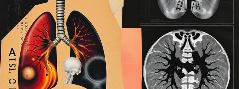
VQ Scan
VQ Scan
A nuclear medicine procedure used to evaluate lung function by looking at both ventilation and perfusion. It helps diagnose pulmonary embolism (PE), a serious blood clot in the lungs.
Ventilation Study
Ventilation Study
A component of a VQ scan that assesses air movement in the lungs. It involves inhaling a radioactive gas (Xenon 133) and imaging its distribution.
Perfusion Study
Perfusion Study
A component of a VQ scan evaluating blood flow to the lungs. It involves injecting a radioactive substance (technetium-99m macroaggregated albumin) into the veins, allowing for visualization of its distribution.
ISM (Inhaled Technetium-99m DTPA Aerosol)
ISM (Inhaled Technetium-99m DTPA Aerosol)
Signup and view all the flashcards
VQ Scan for Pulmonary Embolism
VQ Scan for Pulmonary Embolism
Signup and view all the flashcards
High Probability VQ Scan
High Probability VQ Scan
Signup and view all the flashcards
Low Probability VQ Scan
Low Probability VQ Scan
Signup and view all the flashcards
Quantitative Lung Scan
Quantitative Lung Scan
Signup and view all the flashcards
Physical Half-Life
Physical Half-Life
Signup and view all the flashcards
Biological Half-Life
Biological Half-Life
Signup and view all the flashcards
Ventilation Phase
Ventilation Phase
Signup and view all the flashcards
Wash-in
Wash-in
Signup and view all the flashcards
Equilibrium
Equilibrium
Signup and view all the flashcards
Wash-out
Wash-out
Signup and view all the flashcards
Normal Ventilation Results
Normal Ventilation Results
Signup and view all the flashcards
Abnormal Ventilation Results
Abnormal Ventilation Results
Signup and view all the flashcards
Cardiac Notch
Cardiac Notch
Signup and view all the flashcards
Perfusion Phase
Perfusion Phase
Signup and view all the flashcards
Pulmonary Embolism (PE)
Pulmonary Embolism (PE)
Signup and view all the flashcards
Ventilation-Perfusion Mismatch
Ventilation-Perfusion Mismatch
Signup and view all the flashcards
Patient Positioning
Patient Positioning
Signup and view all the flashcards
Radioactive Marking
Radioactive Marking
Signup and view all the flashcards
Xenon Administration
Xenon Administration
Signup and view all the flashcards
Ventilation Scan
Ventilation Scan
Signup and view all the flashcards
Supine Position
Supine Position
Signup and view all the flashcards
Lead Shielded Container
Lead Shielded Container
Signup and view all the flashcards
Half-Life
Half-Life
Signup and view all the flashcards
GM Survey Meter
GM Survey Meter
Signup and view all the flashcards
Anterior-Posterior (AP) Images
Anterior-Posterior (AP) Images
Signup and view all the flashcards
Lateral Images
Lateral Images
Signup and view all the flashcards
Oblique Images
Oblique Images
Signup and view all the flashcards
Perfusion Scan
Perfusion Scan
Signup and view all the flashcards
Air Trapping
Air Trapping
Signup and view all the flashcards
Contaminated Saliva Swallowing
Contaminated Saliva Swallowing
Signup and view all the flashcards
Masking
Masking
Signup and view all the flashcards
Trachea and Bronchi Visualization
Trachea and Bronchi Visualization
Signup and view all the flashcards
Areas of Decreased Activity
Areas of Decreased Activity
Signup and view all the flashcards
Ventilation/Perfusion Lung Scan
Ventilation/Perfusion Lung Scan
Signup and view all the flashcards
Xenon Ventilation Lung Scan
Xenon Ventilation Lung Scan
Signup and view all the flashcards
Negatively Pressurized Room
Negatively Pressurized Room
Signup and view all the flashcards
Xenon Gas
Xenon Gas
Signup and view all the flashcards
Xenon Charcoal Trap
Xenon Charcoal Trap
Signup and view all the flashcards
Ventilation System
Ventilation System
Signup and view all the flashcards
Mask
Mask
Signup and view all the flashcards
Wash In Phase
Wash In Phase
Signup and view all the flashcards
Equilibrium Phase
Equilibrium Phase
Signup and view all the flashcards
Wash Out Phase
Wash Out Phase
Signup and view all the flashcards
Disadvantage of Xenon Gas
Disadvantage of Xenon Gas
Signup and view all the flashcards
Correlation
Correlation
Signup and view all the flashcards
Claustrophobia
Claustrophobia
Signup and view all the flashcards
Ventilation Study in Pulmonary Embolism
Ventilation Study in Pulmonary Embolism
Signup and view all the flashcards
Xenon Ventilation Study
Xenon Ventilation Study
Signup and view all the flashcards
Lung Perfusion Study
Lung Perfusion Study
Signup and view all the flashcards
Ventilation Study: Obstruction
Ventilation Study: Obstruction
Signup and view all the flashcards
Ventilation Study: Obstructive Lung Disease
Ventilation Study: Obstructive Lung Disease
Signup and view all the flashcards
Lung Perfusion Study: Pulmonary Embolism
Lung Perfusion Study: Pulmonary Embolism
Signup and view all the flashcards
Importance of Lung Perfusion Study
Importance of Lung Perfusion Study
Signup and view all the flashcards
Ventilation Study: Accelerated Clearance
Ventilation Study: Accelerated Clearance
Signup and view all the flashcards
Technetium-99m Macroaggregated Albumin (MAA)
Technetium-99m Macroaggregated Albumin (MAA)
Signup and view all the flashcards
Lung Scan Indications: Pulmonary Embolism
Lung Scan Indications: Pulmonary Embolism
Signup and view all the flashcards
Lung Scan Resolution
Lung Scan Resolution
Signup and view all the flashcards
Lung Scan History
Lung Scan History
Signup and view all the flashcards
Lung Perfusion Study Applications
Lung Perfusion Study Applications
Signup and view all the flashcards
Lung Perfusion Study Contraindication: Pulmonary Hypertension
Lung Perfusion Study Contraindication: Pulmonary Hypertension
Signup and view all the flashcards
Lung Perfusion Study Modification: Reduced MAA Dose
Lung Perfusion Study Modification: Reduced MAA Dose
Signup and view all the flashcards
COPD and Emphysema in Ventilation Scan
COPD and Emphysema in Ventilation Scan
Signup and view all the flashcards
Bacterial Pneumonia in Ventilation Scan
Bacterial Pneumonia in Ventilation Scan
Signup and view all the flashcards
Fatty Liver in Ventilation Scan
Fatty Liver in Ventilation Scan
Signup and view all the flashcards
Tracer Uptake in Other Organs (Ventilation Imaging)
Tracer Uptake in Other Organs (Ventilation Imaging)
Signup and view all the flashcards
Alcoholism in Ventilation Scan
Alcoholism in Ventilation Scan
Signup and view all the flashcards
Atelelectasis or Bronchial Obstruction in Ventilation Scan
Atelelectasis or Bronchial Obstruction in Ventilation Scan
Signup and view all the flashcards
Xenon Ventilation Scan
Xenon Ventilation Scan
Signup and view all the flashcards
Aerosol Ventilation (DTA) Scan
Aerosol Ventilation (DTA) Scan
Signup and view all the flashcards
DTA Aerosol Delivery
DTA Aerosol Delivery
Signup and view all the flashcards
Importance of Tight Seal (DTA Aerosol)
Importance of Tight Seal (DTA Aerosol)
Signup and view all the flashcards
Breathing Technique (DTA Aerosol)
Breathing Technique (DTA Aerosol)
Signup and view all the flashcards
Ventilation Scan Duration (DTA Aerosol)
Ventilation Scan Duration (DTA Aerosol)
Signup and view all the flashcards
Oxygen Flow Rate (DTA Aerosol)
Oxygen Flow Rate (DTA Aerosol)
Signup and view all the flashcards
Patient Positioning (DTA Aerosol)
Patient Positioning (DTA Aerosol)
Signup and view all the flashcards
Ventilation Scan in Upright Position
Ventilation Scan in Upright Position
Signup and view all the flashcards
Table Weight Limit
Table Weight Limit
Signup and view all the flashcards
Upright Lung Scan
Upright Lung Scan
Signup and view all the flashcards
Perfusion Defect
Perfusion Defect
Signup and view all the flashcards
Matched Ventilation Perfusion Defect
Matched Ventilation Perfusion Defect
Signup and view all the flashcards
Mismatched Ventilation Perfusion Defect
Mismatched Ventilation Perfusion Defect
Signup and view all the flashcards
Pipe Head Protocol
Pipe Head Protocol
Signup and view all the flashcards
No Probability of Pulmonary Embolism
No Probability of Pulmonary Embolism
Signup and view all the flashcards
Low Probability Pulmonary Embolism
Low Probability Pulmonary Embolism
Signup and view all the flashcards
Intermediate Probability Pulmonary Embolism
Intermediate Probability Pulmonary Embolism
Signup and view all the flashcards
High Probability Pulmonary Embolism
High Probability Pulmonary Embolism
Signup and view all the flashcards
Probability of Pulmonary Embolism
Probability of Pulmonary Embolism
Signup and view all the flashcards
Physician's Diagnosis of Pulmonary Embolism
Physician's Diagnosis of Pulmonary Embolism
Signup and view all the flashcards
Technologist's Role in Pulmonary Embolism Diagnosis
Technologist's Role in Pulmonary Embolism Diagnosis
Signup and view all the flashcards
What is a VQ scan?
What is a VQ scan?
Signup and view all the flashcards
What is a ventilation study?
What is a ventilation study?
Signup and view all the flashcards
What is a perfusion study?
What is a perfusion study?
Signup and view all the flashcards
What is a high probability VQ scan?
What is a high probability VQ scan?
Signup and view all the flashcards
What is a low probability VQ scan?
What is a low probability VQ scan?
Signup and view all the flashcards
What is a quantitative lung scan?
What is a quantitative lung scan?
Signup and view all the flashcards
What is half-life?
What is half-life?
Signup and view all the flashcards
What is the typical right lung to left lung ventilation/perfusion ratio?
What is the typical right lung to left lung ventilation/perfusion ratio?
Signup and view all the flashcards
What is the supine position?
What is the supine position?
Signup and view all the flashcards
What are the four quadrants of the lungs?
What are the four quadrants of the lungs?
Signup and view all the flashcards
Activity and Acquisition Time
Activity and Acquisition Time
Signup and view all the flashcards
VQ Scan for Lung Disorders
VQ Scan for Lung Disorders
Signup and view all the flashcards
Chest X-ray in VQ Scan
Chest X-ray in VQ Scan
Signup and view all the flashcards
Pre-Injection Preparation for VQ Scan
Pre-Injection Preparation for VQ Scan
Signup and view all the flashcards
Ventilation Study in VQ Scan
Ventilation Study in VQ Scan
Signup and view all the flashcards
Perfusion Study in VQ Scan
Perfusion Study in VQ Scan
Signup and view all the flashcards
Direct Injection in VQ Scan
Direct Injection in VQ Scan
Signup and view all the flashcards
Tubing Injection for VQ Scan
Tubing Injection for VQ Scan
Signup and view all the flashcards
Supine Position for VQ Scan
Supine Position for VQ Scan
Signup and view all the flashcards
Avoiding Blood Mixing in VQ Scan
Avoiding Blood Mixing in VQ Scan
Signup and view all the flashcards
Normal VQ Scan Results
Normal VQ Scan Results
Signup and view all the flashcards
Upright Position for VQ Scan
Upright Position for VQ Scan
Signup and view all the flashcards
MRA Properties in VQ Scan
MRA Properties in VQ Scan
Signup and view all the flashcards
MRA Dose in VQ Scan
MRA Dose in VQ Scan
Signup and view all the flashcards
Ventilation Images
Ventilation Images
Signup and view all the flashcards
Perfusion Images
Perfusion Images
Signup and view all the flashcards
Wedge-shaped Areas
Wedge-shaped Areas
Signup and view all the flashcards
Nuclear Medicine
Nuclear Medicine
Signup and view all the flashcards
Inhaled Technetium-99m DTPA Aerosol
Inhaled Technetium-99m DTPA Aerosol
Signup and view all the flashcards
Study Notes
Learning Objectives
- Differentiate Xenon 133 gas ventilation from ISM 99m DTA aerosol ventilation techniques.
- Describe the perfusion lung scan process for pulmonary embolism (PE).
- Explain the combined ventilation and perfusion process for VQ scans.
- Differentiate between low and high probability VQ scans for PE.
- Describe the process for a quantitative lung scan.
Ventilation Scan
- VQ scan assesses ventilation and perfusion (blood flow).
- "VQ" originally had quantitative data for perfusion; now separate.
- Primary purpose: rule out PE, often a medical emergency.
- Performed as a stat procedure; sometimes scheduled.
Ventilation Methods
- Xenon Gas:
- Used in a negatively pressurized room with exhaust to prevent contamination.
- Gas can escape easily.
- Administered via mask; 10-20 millicuries (mCi).
- Room walls and surfaces can be contaminated due to escaping gas.
- Long physical half-life (5.3 days); short biological half-life (30 seconds) impacts contamination risks.
- Charcoal filter traps escaping gas.
- Tight patient seal required to prevent contamination.
- Gas flow is generally 12 liters per minute.
- Patient takes deep breath for initial Xenon uptake.
- Dynamic images during wash-in, equilibrium, and wash-out phases.
- Scan takes approximately 60 seconds.
- Proper patient positioning vital for lung visibility. Markers used on top of shoulders, left and right, and slightly below lungs.
- DTA Aerosol:
- Higher energy (140 keV) than Xenon.
- Liquid radiopharmaceutical vaporized into an aerosol.
- Administered via tight-seal mask and oxygen flow (10-12 liters/minute).
- Typically lasts 5 minutes, allowing for re-administration if needed.
- Patient can be positioned upright or supine; upright for better view, sometimes useful for larger patients.
Perfusion Scan
- Injects TEC-99m-macroaggregated albumin (MRA; radiopharmaceutical) into vein.
- Straight stick injection preferred to avoid tubing contamination.
- Administered 2-6 millicuries (mCi).
- Patient placed in supine position.
- Requires 75,000-700,000 particles for optimal viewing, avoiding clots when possible.
- Images acquired for each acquisition view (anterior, posterior, lateral, oblique).
- Acquisition views are matched with corresponding ventilation view.
- Important to have patient take deep breaths and cough to distribute the isotope through the lungs.
Quantitative Lung Scan
- Measures lung function.
- Usually a perfusion-only scan.
- Injects MRA radiopharmaceutical.
- Patient in supine position (to prevent gravity effects).
- Regions of interest (ROIs) drawn on images.
- Software quantifies relative blood flow to different lung segments.
- Normal ratio: Approximately 55% right lung, 45% left lung (left lung often slightly lower due to heart overlay).
Results Interpretation (for all scans)
- Normal Results: Uniform blood flow and vent activity throughout lungs.
- Abnormal Results (Ventilation): In cases of pulmonary embolism, ventilation should be normal; perfusion will show abnormalities. (low flow areas)
- Abnormal Results (Perfusion): Triangular-shaped defects (wedges), decreased blood flow. These indicate likely PE.
- Other Results:
- COPD/Emphysema: patchy uptake, clumping.
- Bacterial pneumonia: ventilation and perfusion decreased.
- Fatty liver (alcoholism, obesity): isotope uptake in liver.
- Collapsed lung: missing or very weak vent activity on one side.
- Bronchial obstruction, less ventilation distal to the obstruction.
Diagnosis Probability
- Low (0-10%), moderate (20-79%) or high (>80% probability): Based on extent/location of perfusion defects in combination with other radiographic evidence (X-ray, etc.).
Importance of Correlation
- Ventilation and perfusion scans must be interpreted together with a chest X-ray. Chest X-ray is extremely important for correlation
Additional Precautions for All Scans
- Maintain tight seals.
- Ensure patient's head and lungs remain in the proper position during scans for ventilation and perfusion.
- Shield radiopharmaceuticals appropriately.
- Follow departmental protocols for isotope disposal.
Studying That Suits You
Use AI to generate personalized quizzes and flashcards to suit your learning preferences.


