Podcast
Questions and Answers
What is the primary muscle of inspiration in the respiratory system?
What is the primary muscle of inspiration in the respiratory system?
- Intercostal muscles
- Serratus anterior
- Pectoralis major
- Diaphragm (correct)
What is the primary method used to diagnose pulmonary embolism?
What is the primary method used to diagnose pulmonary embolism?
- Multidetector CT pulmonary angiography (CTPA) (correct)
- MRI of the chest
- Radionuclide scanning
- Plain film chest radiograph
Which of the following structures is NOT found in the mediastinum?
Which of the following structures is NOT found in the mediastinum?
- Trachea
- Left lung (correct)
- Thymus gland
- Esophagus
Which of the following is NOT a symptom of pulmonary embolism?
Which of the following is NOT a symptom of pulmonary embolism?
What function do the C-shaped rings of cartilage in the trachea serve?
What function do the C-shaped rings of cartilage in the trachea serve?
What is the role of intravenous contrast medium in a CT pulmonary angiogram?
What is the role of intravenous contrast medium in a CT pulmonary angiogram?
When performing a CT pulmonary angiogram, how is the contrast administered?
When performing a CT pulmonary angiogram, how is the contrast administered?
How many lobes does the right lung have?
How many lobes does the right lung have?
Which imaging method is primarily used for assessing pleural and chest wall diseases?
Which imaging method is primarily used for assessing pleural and chest wall diseases?
What condition may be discovered alongside the presence of thrombus during a CT pulmonary angiogram?
What condition may be discovered alongside the presence of thrombus during a CT pulmonary angiogram?
What separates the superior and middle lobes of the right lung?
What separates the superior and middle lobes of the right lung?
What traditional imaging method has been superseded by multidetector CTPA for detecting pulmonary emboli?
What traditional imaging method has been superseded by multidetector CTPA for detecting pulmonary emboli?
During the sampling of pleural fluid, when should the needle be advanced?
During the sampling of pleural fluid, when should the needle be advanced?
Which of the following methods allows for imaging blood flow in the respiratory system?
Which of the following methods allows for imaging blood flow in the respiratory system?
What denotes the region between the two lungs?
What denotes the region between the two lungs?
What is a common origin of a pulmonary embolism?
What is a common origin of a pulmonary embolism?
What is one of the indications for a CT scan of the thorax?
What is one of the indications for a CT scan of the thorax?
Which of the following is a contraindication for a CT scan requiring intravenous contrast?
Which of the following is a contraindication for a CT scan requiring intravenous contrast?
What type of CT scan is typically performed with a delay of 25-40 seconds after intravenous contrast administration?
What type of CT scan is typically performed with a delay of 25-40 seconds after intravenous contrast administration?
Which technique is used to maximize spatial resolution in high-resolution CT (HRCT)?
Which technique is used to maximize spatial resolution in high-resolution CT (HRCT)?
Low Dose CT is most advantageous in which of the following scenarios?
Low Dose CT is most advantageous in which of the following scenarios?
What type of scan is beneficial for differentiating early interstitial fibrosis from reversible-dependent changes?
What type of scan is beneficial for differentiating early interstitial fibrosis from reversible-dependent changes?
Which of the following describes a feature of Low Dose CT compared to regular CT?
Which of the following describes a feature of Low Dose CT compared to regular CT?
Which application does CT angiography primarily serve in thoracic evaluation?
Which application does CT angiography primarily serve in thoracic evaluation?
What is the primary purpose of windowing in CT imaging?
What is the primary purpose of windowing in CT imaging?
How does the window width affect the CT image?
How does the window width affect the CT image?
Which window width range is defined as narrow?
Which window width range is defined as narrow?
Why is a soft tissue window not suitable for lung parenchyma imaging?
Why is a soft tissue window not suitable for lung parenchyma imaging?
Which of the following is a contraindication for performing a CT-guided lung biopsy?
Which of the following is a contraindication for performing a CT-guided lung biopsy?
What is a recommended action regarding clopidogrel before a CT-guided lung biopsy?
What is a recommended action regarding clopidogrel before a CT-guided lung biopsy?
What is the purpose of performing a CT-guided lung biopsy?
What is the purpose of performing a CT-guided lung biopsy?
What does the window level or center represent in CT imaging?
What does the window level or center represent in CT imaging?
What is the primary indication for performing pulmonary arteriography?
What is the primary indication for performing pulmonary arteriography?
Which imaging technique is primarily recommended for assessing the mediastinum and chest wall?
Which imaging technique is primarily recommended for assessing the mediastinum and chest wall?
Which technique is used to introduce the catheter during pulmonary arteriography?
Which technique is used to introduce the catheter during pulmonary arteriography?
What is the maximum volume of contrast medium recommended for pulmonary arteriography?
What is the maximum volume of contrast medium recommended for pulmonary arteriography?
Why is MRI challenging for imaging pulmonary embolism?
Why is MRI challenging for imaging pulmonary embolism?
What is the typical position for the catheter tip during pulmonary arteriography?
What is the typical position for the catheter tip during pulmonary arteriography?
In PET-CT imaging, what does the Standardized Uptake Value (SUV) below 2.4 typically indicate?
In PET-CT imaging, what does the Standardized Uptake Value (SUV) below 2.4 typically indicate?
Which of the following is a recommended consideration for using MRPA?
Which of the following is a recommended consideration for using MRPA?
Flashcards
What is the thorax?
What is the thorax?
The upper part of the trunk between the neck and abdomen, containing organs essential for breathing and blood circulation.
What is the bony thorax?
What is the bony thorax?
The bony framework that protects the organs within the chest, like a ribcage.
What is the respiratory system?
What is the respiratory system?
A system of organs responsible for breathing and gas exchange with the blood.
What is the diaphragm?
What is the diaphragm?
Signup and view all the flashcards
What is the trachea?
What is the trachea?
Signup and view all the flashcards
How many lobes do the lungs have?
How many lobes do the lungs have?
Signup and view all the flashcards
What is the mediastinum?
What is the mediastinum?
Signup and view all the flashcards
What are plain films?
What are plain films?
Signup and view all the flashcards
Windowing
Windowing
Signup and view all the flashcards
Window Width (WW)
Window Width (WW)
Signup and view all the flashcards
Window Level (WL)
Window Level (WL)
Signup and view all the flashcards
Soft Tissue (Mediastinal) Window
Soft Tissue (Mediastinal) Window
Signup and view all the flashcards
CT-Guided Lung Biopsy
CT-Guided Lung Biopsy
Signup and view all the flashcards
Investigation of new or progressing lung opacity
Investigation of new or progressing lung opacity
Signup and view all the flashcards
Investigation of a new lung/pleural/chest wall lesion in a patient with known malignancy
Investigation of a new lung/pleural/chest wall lesion in a patient with known malignancy
Signup and view all the flashcards
To obtain material for culture when other techniques have failed
To obtain material for culture when other techniques have failed
Signup and view all the flashcards
CT Scan
CT Scan
Signup and view all the flashcards
MRI Scan
MRI Scan
Signup and view all the flashcards
PET Scan
PET Scan
Signup and view all the flashcards
High-Resolution CT (HRCT)
High-Resolution CT (HRCT)
Signup and view all the flashcards
Low-Dose CT
Low-Dose CT
Signup and view all the flashcards
CT Angiography
CT Angiography
Signup and view all the flashcards
CT with Contrast
CT with Contrast
Signup and view all the flashcards
Indications for CT Scan
Indications for CT Scan
Signup and view all the flashcards
What is a pulmonary embolism?
What is a pulmonary embolism?
Signup and view all the flashcards
What is a CT pulmonary angiogram?
What is a CT pulmonary angiogram?
Signup and view all the flashcards
How is a CT pulmonary angiogram performed?
How is a CT pulmonary angiogram performed?
Signup and view all the flashcards
What are the technical aspects of a CT pulmonary angiogram?
What are the technical aspects of a CT pulmonary angiogram?
Signup and view all the flashcards
How does a normal CTPA scan look?
How does a normal CTPA scan look?
Signup and view all the flashcards
What can a CTPA scan reveal besides a pulmonary embolism?
What can a CTPA scan reveal besides a pulmonary embolism?
Signup and view all the flashcards
Why is CTPA preferred for pulmonary emboli diagnosis?
Why is CTPA preferred for pulmonary emboli diagnosis?
Signup and view all the flashcards
What is the contrast injection process during a CTPA?
What is the contrast injection process during a CTPA?
Signup and view all the flashcards
Pulmonary Arteriography
Pulmonary Arteriography
Signup and view all the flashcards
Pulmonary Embolism
Pulmonary Embolism
Signup and view all the flashcards
MRI of the Respiratory System
MRI of the Respiratory System
Signup and view all the flashcards
Magnetic Resonance Pulmonary Angiography (MRPA)
Magnetic Resonance Pulmonary Angiography (MRPA)
Signup and view all the flashcards
Positron Emission Tomography (PET)
Positron Emission Tomography (PET)
Signup and view all the flashcards
PET-CT
PET-CT
Signup and view all the flashcards
Standardized Uptake Value (SUV)
Standardized Uptake Value (SUV)
Signup and view all the flashcards
Catheter-Directed Chemotherapy
Catheter-Directed Chemotherapy
Signup and view all the flashcards
Study Notes
Introduction to Respiratory System Imaging
- The chest, or thorax, is the upper portion of the torso between the neck and abdomen.
- Radiographic anatomy of the chest is divided into three sections: bony thorax, respiratory system, and mediastinum.
Bony Thorax
- The bony thorax is part of the skeletal system providing a protective framework for the structures involved in breathing and blood circulation within the chest.
- The bony structure includes ribs, sternum, and thoracic spine.
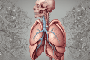
Respiratory System
- The respiratory system includes the pharynx, trachea, bronchi, and lungs.
- The dome-shaped diaphragm is a critical muscle for inspiration, the process of drawing air into the lungs.
- The diaphragm moves down, creating a "sucking" action. Air is drawn through the nose, mouth, pharynx, larynx, trachea, and bronchi. This fills the lungs.
Trachea
- The trachea is a fibrous muscular tube, approximately 2 cm in diameter and 11 cm long.
- It contains C-shaped rings of cartilage that prevent it from collapsing during exhalation.
- The trachea sits anterior to the esophagus. It branches into the right and left primary bronchi.
Lungs
- Lungs are located on each side of the thoracic cavity.
- The right lung is divided into three lobes (superior, middle, and inferior), separated by fissures (horizontal and oblique).
- The left lung is divided into two lobes (superior and inferior), separated by a single oblique fissure.
Mediastinum
- The mediastinum is the medial portion of the thoracic cavity between the two lungs.
- Four radiographically important structures within the mediastinum are the thymus gland, heart and great vessels, trachea, and esophagus.
Methods of Imaging the Respiratory System
- Plain films
- Computed tomography (CT)
- Radionuclide imaging (V/Q scans)
- Ultrasound (US) - for pleural, chest wall, and diaphragm disease
- Magnetic resonance imaging (MRI)
- Positron emission tomography (PET)
CT Scan of the Thorax - Indications
- Assessing pulmonary masses, chest wall, or pleural disease.
- Staging and follow-up of malignancy in the chest.
- Evaluation of known/suspected thoracic vascular issues, including pulmonary embolus, aortic dissection, or arteriovenous malformations.
- Assessing airway disease like bronchiectasis and large airway stenosis.
- Evaluating trauma to the chest.
- Performing CT-guided interventions, such as lung, pleura, or chest wall mass biopsy.
CT Scan of the Thorax - Contraindications
- Intravenous (IV) contrast medium administration requires evaluation for impaired renal function, use of nephrotoxic drugs, and history of previous adverse reactions to iodinated contrast media.
Technique CT Chest - Types
- Standard/Conventional CT (Volume scan): Typically performed with intravenous contrast enhancement at a rate of 3-5 mL/sec with a delay of 25-40 seconds. This quickly covers the entire lung. Delayed imaging at 60-65 seconds can assist in assessing otherwise hidden pleural thickening, particularly in cases of large pleural effusions.
- High-resolution CT (HRCT): Noncontrast scan acquired during full inspiration. 1mm (or thinner) slices at 10-20mm intervals (or volumetric acquisitions) are ideal. High resolution image reconstruction techniques are important to maximize visualization of detailed lung anatomy. Additional expiratory images can assist in evaluating suspected air-trapping. Prone images might be needed to distinguish reversible changes from fibrosis.
- Low-dose CT: Utilizes reduced radiation doses while maintaining diagnostic quality. This technique is useful for follow-up exams (infections, post-lung transplant, metastases).
CT Angiography (CT with contrast)
- Evaluation of mediastinum (lymph nodes, infection)
- Assessment of suspected cancer
- Evaluation of pleural masses
Windowing (CT)
- Windowing (grey-level mapping, contrast stretching, histogram modification) is used to manipulate the CT image grayscale to highlight certain structures. It adjusts brightness (Window level) and contrast (Window width) of the image.
Window Width (WW)
- Wide window: displays a broader range of CT numbers, including more tissues. (400-2000 HU)
- Narrow window: displays a limited range of CT numbers, showing fewer tissues. (50-350 HU)
Window Level/Center (WL)
- WL or window center is the midpoint of the range of CT numbers displayed.
Windowing Examples
- Soft Tissue (mediastinal) window
- Lung window
- Bone window
CT-Guided Lung Biopsy
- Investigation of new or progressive pulmonary opacities
- Assessing new lung, pleural, or chest wall lesions in patients with existing malignancies.
- Obtaining tissue samples when other procedures fail to yield sufficient diagnostic material.
- Genetic testing of tissue samples in patients with known lung malignancy, to identify epidermal growth factor receptor (EGFR) mutations, for example
Patient Preparation
- Procedures can often occur on an outpatient basis with several hours of observation afterwards.
- Patients should have clotting screen (INR and platelets assessments).
- Certain medications, like aspirin and clopidogrel, may need to be stopped before the procedure.
- Pulmonary function tests (like spirometry) may be ordered.
Pulmonary Embolism Methods of Imaging
- Plain film chest radiograph
- Radionuclide scanning
- Multidetector CT pulmonary angiography (CTPA); consider the gold standard nowadays
- Pulmonary arteriography (less common now)
- MR angiography (alternative for scenarios where radiation or IV contrast is contraindicated.)
Pulmonary Embolism Technique CTPA
- Patients receive IV contrast using an injector pump at a high rate.
- Images are taken when the contrast reaches the pulmonary arteries.
- Normal scan shows contrast filling pulmonary blood vessels. An embolus appears as a filling defect (dark shadow).
Pulmonary Arteriography
- Procedures are aimed at identifying emboli and other peripheral abnormalities.
- Treatment planning and performing interventions like catheter-directed thrombolysis or embolectomy for pulmonary embolism.
Contrast Medium (for Pulmonary Embolism Imaging)
- LOCM 370 mg I/mL; 0.75 mL kg-1 at 20-25 mL/sec (max. 40 mL).
Equipment (for Pulmonary Embolism Imaging)
- Digital subtraction angiography facilities
- Pump injector
- Catheter: pigtail (coiled, end-hole and side holes)
Seldinger Technique
- The Seldinger technique is used to introduce catheters via the right femoral vein; the catheters are advanced through an introducer sheath, then placed within 1-3 cm of the pulmonary valve under fluoroscopic guidance.
- Pulmonary pressures are routinely monitored during the procedure. Lower injection rates should be used if pressures are elevated.
MRI of the Respiratory System
- MRI has a limited role in the respiratory system due to artefacts from air in the lungs.
- Mediastinum and chest wall can be assessed well because of MRI's ability to image in coronal and sagittal planes.
MRI of Pulmonary Embolism
- Technically adequate images are often difficult to achieve with MRI for pulmonary embolism.
- MR pulmonary angiography (MRPA) is generally reserved for specific situations; most often, when standard imaging tests are contraindicated (e.g. pregnant patients, patients allergic to contrast media).
PET and PET-CT of the Respiratory System
- Positron emission tomography (PET), a noninvasive technique, measures radioactivity in vivo. This is useful in showing radioactivity distribution, post injection of a radiopharmaceutical, to help identify issues.
- PET/CT scan together give both morphological (CT) and metabolic (PET) information about a lesion.
- Typical radiopharmaceutical used is F-18 fluorodeoxyglucose (FDG).
- A standardized uptake value (SUV) < 2.4 suggests less chance of malignancy compared to SUV > 3.5.
PET-CT Procedure
- Patients must fast 4-6 hours before the scan. Diabetic patients should fast for 12 hours.
- Blood glucose levels are usually under 150 mg/dL.
- Strenuous exercise should be avoided 24 hours prior to the scan.
- Scan is carried out 60 minutes after FDG injection.
PET-CT Applications
- Oncological applications include baseline staging, treatment planning, assessing treatment response, and monitoring changes post-therapy.
- Non-oncological applications include cardiology, neurology.
Studying That Suits You
Use AI to generate personalized quizzes and flashcards to suit your learning preferences.