Podcast
Questions and Answers
What is the primary mechanism by which unfractionated heparin (UFH) prevents clot formation?
What is the primary mechanism by which unfractionated heparin (UFH) prevents clot formation?
- Binding to antithrombin and enhancing its inactivation of factor Xa. (correct)
- Inhibiting the synthesis of vitamin K-dependent clotting factors in the liver.
- Blocking the conversion of fibrinogen to fibrin.
- Directly dissolving existing clots in pulmonary arteries.
A patient receiving intravenous unfractionated heparin (UFH) for a pulmonary embolism has a supratherapeutic aPTT. What is the most important initial nursing intervention?
A patient receiving intravenous unfractionated heparin (UFH) for a pulmonary embolism has a supratherapeutic aPTT. What is the most important initial nursing intervention?
- Administer Vitamin K intravenously.
- Increase the heparin infusion rate to maintain patency of the IV line.
- Prepare to administer protamine sulfate. (correct)
- Monitor blood pressure every 15 minutes.
Which of the following is a key advantage of using low-molecular-weight heparin (LMWH) over unfractionated heparin (UFH) in the treatment of pulmonary embolism?
Which of the following is a key advantage of using low-molecular-weight heparin (LMWH) over unfractionated heparin (UFH) in the treatment of pulmonary embolism?
- LMWH does not require routine laboratory monitoring. (correct)
- LMWH has a shorter half-life, allowing for quicker reversal in case of bleeding.
- LMWH is administered intravenously, ensuring faster onset of action.
- LMWH directly activates antithrombin to dissolve clots more rapidly than UFH.
A patient with a confirmed pulmonary embolism is started on both intravenous unfractionated heparin and oral warfarin. What primary rationale supports this 'bridging' therapy?
A patient with a confirmed pulmonary embolism is started on both intravenous unfractionated heparin and oral warfarin. What primary rationale supports this 'bridging' therapy?
During a rheolytic catheter embolectomy, which action poses the greatest risk to the patient?
During a rheolytic catheter embolectomy, which action poses the greatest risk to the patient?
What is a key difference between rheolytic and rotational catheter embolectomy?
What is a key difference between rheolytic and rotational catheter embolectomy?
A patient with a pulmonary embolism (PE) exhibits tachypnea and reports chest pain. Which of the following nursing interventions would be MOST appropriate to address the immediate respiratory distress?
A patient with a pulmonary embolism (PE) exhibits tachypnea and reports chest pain. Which of the following nursing interventions would be MOST appropriate to address the immediate respiratory distress?
What laboratory value is most important to monitor in a patient receiving intravenous unfractionated heparin?
What laboratory value is most important to monitor in a patient receiving intravenous unfractionated heparin?
A patient with a confirmed pulmonary embolism (PE) is receiving a heparin infusion. Which laboratory value is MOST critical for the nurse to monitor regularly?
A patient with a confirmed pulmonary embolism (PE) is receiving a heparin infusion. Which laboratory value is MOST critical for the nurse to monitor regularly?
Which potential adverse effect requires monitoring of platelet levels in patients receiving unfractionated heparin?
Which potential adverse effect requires monitoring of platelet levels in patients receiving unfractionated heparin?
A patient with a massive pulmonary embolism (PE) is hypotensive despite initial fluid resuscitation. Which intervention should the nurse anticipate the MOST immediate need for?
A patient with a massive pulmonary embolism (PE) is hypotensive despite initial fluid resuscitation. Which intervention should the nurse anticipate the MOST immediate need for?
A patient with a pulmonary embolism (PE) is started on warfarin. Which of the following instructions is MOST important for the nurse to include in the patient's discharge teaching?
A patient with a pulmonary embolism (PE) is started on warfarin. Which of the following instructions is MOST important for the nurse to include in the patient's discharge teaching?
A patient with a pulmonary embolism (PE) is experiencing metabolic acidosis. Which of the following factors MOST likely contributes to this acid-base imbalance?
A patient with a pulmonary embolism (PE) is experiencing metabolic acidosis. Which of the following factors MOST likely contributes to this acid-base imbalance?
Which assessment finding would be MOST indicative of right ventricular overload in a patient with a pulmonary embolism (PE)?
Which assessment finding would be MOST indicative of right ventricular overload in a patient with a pulmonary embolism (PE)?
After administering a thrombolytic medication to a patient with a massive pulmonary embolism (PE), which nursing intervention is the HIGHEST priority?
After administering a thrombolytic medication to a patient with a massive pulmonary embolism (PE), which nursing intervention is the HIGHEST priority?
A patient presents with sudden-onset chest pain, dyspnea, and tachypnea. Which of the following nursing actions is MOST crucial for the nurse to perform FIRST?
A patient presents with sudden-onset chest pain, dyspnea, and tachypnea. Which of the following nursing actions is MOST crucial for the nurse to perform FIRST?
Which of the following best explains why fractured ribs compromise effective ventilation?
Which of the following best explains why fractured ribs compromise effective ventilation?
A patient with a flail chest exhibits paradoxical chest-wall movement. What does this entail?
A patient with a flail chest exhibits paradoxical chest-wall movement. What does this entail?
What is the primary difference in pressure application between Continuous Positive Airway Pressure (CPAP) and invasive positive-pressure ventilation?
What is the primary difference in pressure application between Continuous Positive Airway Pressure (CPAP) and invasive positive-pressure ventilation?
In the context of chest trauma, what is the primary effect of a pneumothorax on lung function?
In the context of chest trauma, what is the primary effect of a pneumothorax on lung function?
A patient is prescribed warfarin following a pulmonary embolism (PE). Which dietary recommendation is MOST important to include in their discharge teaching?
A patient is prescribed warfarin following a pulmonary embolism (PE). Which dietary recommendation is MOST important to include in their discharge teaching?
A patient being discharged after treatment for a pulmonary embolism (PE) is prescribed a factor Xa inhibitor. What key point should the nurse include in the patient's discharge education regarding medication management?
A patient being discharged after treatment for a pulmonary embolism (PE) is prescribed a factor Xa inhibitor. What key point should the nurse include in the patient's discharge education regarding medication management?
Which of the following is the most common injury associated with chest trauma?
Which of the following is the most common injury associated with chest trauma?
Positive End-Expiratory Pressure (PEEP) is primarily applied during which phase of mechanical ventilation, and what is its effect?
Positive End-Expiratory Pressure (PEEP) is primarily applied during which phase of mechanical ventilation, and what is its effect?
What is the underlying physiological principle that is disrupted by a pneumothorax?
What is the underlying physiological principle that is disrupted by a pneumothorax?
Which lifestyle modification would MOST directly address the underlying pathophysiology of venous thromboembolism (VTE) and provide the MOST benefit?
Which lifestyle modification would MOST directly address the underlying pathophysiology of venous thromboembolism (VTE) and provide the MOST benefit?
A nurse is monitoring a patient with COPD receiving oxygen therapy. Which respiratory drive characteristic necessitates careful observation in this patient population?
A nurse is monitoring a patient with COPD receiving oxygen therapy. Which respiratory drive characteristic necessitates careful observation in this patient population?
Inhaled bronchodilators improve airflow in acute respiratory failure by primarily targeting which physiological mechanism?
Inhaled bronchodilators improve airflow in acute respiratory failure by primarily targeting which physiological mechanism?
A patient with a history of PE is prescribed an anticoagulant. Which instruction regarding bleeding precautions is MOST appropriate?
A patient with a history of PE is prescribed an anticoagulant. Which instruction regarding bleeding precautions is MOST appropriate?
A patient who has suffered blunt chest trauma has a flail chest. Which of the following is the greatest concern regarding this condition?
A patient who has suffered blunt chest trauma has a flail chest. Which of the following is the greatest concern regarding this condition?
In the context of acute respiratory failure, inhaled steroids are administered to achieve which primary therapeutic effect?
In the context of acute respiratory failure, inhaled steroids are administered to achieve which primary therapeutic effect?
A patient who has had a pulmonary embolism (PE) is being discharged. What should comprise the MOST important element of the discharge instructions?
A patient who has had a pulmonary embolism (PE) is being discharged. What should comprise the MOST important element of the discharge instructions?
A patient presents with a pneumothorax following a motor vehicle accident. Which of the following assessment findings would be most indicative of a tension pneumothorax, requiring immediate intervention?
A patient presents with a pneumothorax following a motor vehicle accident. Which of the following assessment findings would be most indicative of a tension pneumothorax, requiring immediate intervention?
The synergistic effect of combining bronchodilators and inhaled steroids in treating respiratory failure implies which of the following?
The synergistic effect of combining bronchodilators and inhaled steroids in treating respiratory failure implies which of the following?
In a patient with fractured ribs, pain management is crucial to prevent complications. Which of the following is the most important reason for effective pain control in this scenario?
In a patient with fractured ribs, pain management is crucial to prevent complications. Which of the following is the most important reason for effective pain control in this scenario?
Which exercise regimen is MOST beneficial for a patient recovering from a pulmonary embolism (PE)?
Which exercise regimen is MOST beneficial for a patient recovering from a pulmonary embolism (PE)?
Diuretics are used in the management of acute respiratory failure primarily when which underlying condition is present?
Diuretics are used in the management of acute respiratory failure primarily when which underlying condition is present?
Following a massive pulmonary embolism (PE), a patient is at highest risk for developing which of the following complications?
Following a massive pulmonary embolism (PE), a patient is at highest risk for developing which of the following complications?
If acute respiratory failure progresses despite oxygen therapy, mechanical ventilation, and medications, which of the following is a significant and life-threatening potential complication?
If acute respiratory failure progresses despite oxygen therapy, mechanical ventilation, and medications, which of the following is a significant and life-threatening potential complication?
What is the primary reason for advising a patient recovering from a pulmonary embolism (PE) to maintain adequate fluid intake?
What is the primary reason for advising a patient recovering from a pulmonary embolism (PE) to maintain adequate fluid intake?
A patient with ARDS is receiving supplemental oxygen. Despite increasing the fraction of inspired oxygen (FiO2), their PaO2 remains persistently low. This condition is best described as:
A patient with ARDS is receiving supplemental oxygen. Despite increasing the fraction of inspired oxygen (FiO2), their PaO2 remains persistently low. This condition is best described as:
In the context of ARDS, decreased lung compliance directly leads to which of the following changes during mechanical ventilation?
In the context of ARDS, decreased lung compliance directly leads to which of the following changes during mechanical ventilation?
During the fibrotic phase of ARDS, pulmonary hypertension and right-sided heart failure can develop. Which of the following hemodynamic changes is a direct consequence of right-sided heart failure in this phase?
During the fibrotic phase of ARDS, pulmonary hypertension and right-sided heart failure can develop. Which of the following hemodynamic changes is a direct consequence of right-sided heart failure in this phase?
Severe tissue hypoxia and lactic acidosis are significant complications in the later stages of ARDS. These are primarily caused by:
Severe tissue hypoxia and lactic acidosis are significant complications in the later stages of ARDS. These are primarily caused by:
What is the primary pathological mechanism responsible for the pulmonary edema observed in patients with ARDS?
What is the primary pathological mechanism responsible for the pulmonary edema observed in patients with ARDS?
In the early diagnostic phase of ARDS, a chest X-ray is crucial. Which of the following findings is most characteristic of ARDS on a chest X-ray?
In the early diagnostic phase of ARDS, a chest X-ray is crucial. Which of the following findings is most characteristic of ARDS on a chest X-ray?
An initial arterial blood gas (ABG) analysis is performed on a patient suspected of developing ARDS. Which of the following sets of ABG values is most likely to be observed in the early stages?
An initial arterial blood gas (ABG) analysis is performed on a patient suspected of developing ARDS. Which of the following sets of ABG values is most likely to be observed in the early stages?
A complete blood count (CBC) with differential is ordered as part of the diagnostic workup for a patient with suspected ARDS. What is the primary reason for including a CBC in this evaluation?
A complete blood count (CBC) with differential is ordered as part of the diagnostic workup for a patient with suspected ARDS. What is the primary reason for including a CBC in this evaluation?
Flashcards
Embolectomy
Embolectomy
Surgical removal of an embolus.
Rheolytic Catheter Embolectomy
Rheolytic Catheter Embolectomy
Uses pressurized saline to erode a clot.
Rotational Catheter Embolectomy
Rotational Catheter Embolectomy
Uses a rotating tool to break down a clot.
Unfractionated Heparin: Mechanism of Action
Unfractionated Heparin: Mechanism of Action
Signup and view all the flashcards
SQ Heparin Use
SQ Heparin Use
Signup and view all the flashcards
IV Heparin Use
IV Heparin Use
Signup and view all the flashcards
Heparin Reversal Agent
Heparin Reversal Agent
Signup and view all the flashcards
LMWH: Mechanism of Action
LMWH: Mechanism of Action
Signup and view all the flashcards
Anticoagulants/Thrombolytics
Anticoagulants/Thrombolytics
Signup and view all the flashcards
PE Prevention
PE Prevention
Signup and view all the flashcards
Exercise for PE
Exercise for PE
Signup and view all the flashcards
Cardiac-Prudent Diet
Cardiac-Prudent Diet
Signup and view all the flashcards
Adequate Fluid Intake
Adequate Fluid Intake
Signup and view all the flashcards
Smoking Cessation
Smoking Cessation
Signup and view all the flashcards
Bleeding Precautions
Bleeding Precautions
Signup and view all the flashcards
Recurrent PE/DVT Symptoms
Recurrent PE/DVT Symptoms
Signup and view all the flashcards
Impaired Gas Exchange (PE)
Impaired Gas Exchange (PE)
Signup and view all the flashcards
Tachypnea in PE
Tachypnea in PE
Signup and view all the flashcards
Decreased Cardiac Output (PE)
Decreased Cardiac Output (PE)
Signup and view all the flashcards
Bleeding Risk (PE Treatment)
Bleeding Risk (PE Treatment)
Signup and view all the flashcards
Pulse Oximetry Changes (PE)
Pulse Oximetry Changes (PE)
Signup and view all the flashcards
HOB Elevation for PE
HOB Elevation for PE
Signup and view all the flashcards
Anticoagulants (PE)
Anticoagulants (PE)
Signup and view all the flashcards
Thrombolytics in PE
Thrombolytics in PE
Signup and view all the flashcards
CPAP
CPAP
Signup and view all the flashcards
Invasive Positive-Pressure Ventilation
Invasive Positive-Pressure Ventilation
Signup and view all the flashcards
PEEP (Positive End-Expiratory Pressure)
PEEP (Positive End-Expiratory Pressure)
Signup and view all the flashcards
Bronchodilators
Bronchodilators
Signup and view all the flashcards
Inhaled Steroids
Inhaled Steroids
Signup and view all the flashcards
Synergistic Response (Respiratory)
Synergistic Response (Respiratory)
Signup and view all the flashcards
Sedation (in Respiratory Failure)
Sedation (in Respiratory Failure)
Signup and view all the flashcards
Changes in Mental Status (Respiratory)
Changes in Mental Status (Respiratory)
Signup and view all the flashcards
Refractory Hypoxemia
Refractory Hypoxemia
Signup and view all the flashcards
Increased Peak Inspiratory Pressures
Increased Peak Inspiratory Pressures
Signup and view all the flashcards
Causes of Refractory Hypoxemia in ARDS
Causes of Refractory Hypoxemia in ARDS
Signup and view all the flashcards
Results of Refractory Hypoxemia
Results of Refractory Hypoxemia
Signup and view all the flashcards
Cause of Pulmonary Edema in ARDS
Cause of Pulmonary Edema in ARDS
Signup and view all the flashcards
Hallmark Sign of ARDS (Imaging)
Hallmark Sign of ARDS (Imaging)
Signup and view all the flashcards
"Ground-Glass Appearance"
"Ground-Glass Appearance"
Signup and view all the flashcards
ABG Findings in Early ARDS
ABG Findings in Early ARDS
Signup and view all the flashcards
Chest Trauma Injuries
Chest Trauma Injuries
Signup and view all the flashcards
Fractured Ribs
Fractured Ribs
Signup and view all the flashcards
Impact of Fractured Ribs
Impact of Fractured Ribs
Signup and view all the flashcards
Flail Chest
Flail Chest
Signup and view all the flashcards
Paradoxical Chest-Wall Movement
Paradoxical Chest-Wall Movement
Signup and view all the flashcards
Flail Chest Severity
Flail Chest Severity
Signup and view all the flashcards
Pneumothorax
Pneumothorax
Signup and view all the flashcards
Pneumothorax Impact
Pneumothorax Impact
Signup and view all the flashcards
Study Notes
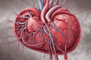
Pulmonary Embolism (PE) Definition
- PE is the obstruction of one or more pulmonary artery branches by particulate matter originating elsewhere in the body.
- Thrombi are the most common cause of pulmonary emboli.
- Tumor fragments, amniotic fluid, air, or fat can also cause a pulmonary embolus - referred to as nonthrombotic pulmonary emboli (NTPE).
- NTPEs are very dangerous; amniotic fluid emboli (AFE) has a 17% failure to rescue rate, with the mother dying in two-thirds of cases.
- The failure to rescue rate rises above 30% when AFE occurs alongside another complication.
Epidemiology of PE
- Deep vein thrombosis (DVT) is the greatest risk factor for PE.
- Virchow's triad, consisting of venous stasis, vessel wall damage, and hypercoagulability, is the major predisposing factor for the development of DVT.
- Prolonged immobility is the most common cause of DVT.
Sources of Pulmonary Emboli
- Deep vein thrombus: A clot that breaks loose from its origin (often the deep veins of the leg or pelvis) and travels to the pulmonary vasculature.
- Fat: Typically from a long-bone fracture.
- Osteomyelitis.
- Liposuction.
- Air: From central venous catheter (CVC) insertion due to negative intrathoracic pressure allowing air entry or disconnection from a fluid source.
- Cardiopulmonary bypass.
- Hemodialysis.
- Amniotic fluid: Can enter the vascular space during delivery through placental vessels.
- Tumor: Sloughs off, and tumor particles travel to the pulmonary vasculature.
Virchow's Triad Causes
- Hypercoagulability can be caused by cancer, oral contraceptives, dehydration/hemoconcentration, sickle cell anemia, polycythemia vera, or abrupt discontinuation of anticoagulants.
- Prolonged bedrest/immobility.
- Obesity.
- Burns.
- Pregnancy.
- Vasculitis/thrombophlebitis.
- Bacterial endocarditis.
- Any postoperative patient.
- Intimal damage of vessels is caused by trauma or IV drug use.
- Atherosclerosis.
Other Risk Factors for DVT
- Obesity.
- Smoking.
- Chronic heart or vascular disease.
- Fracture (hip or leg).
- Hip or knee replacement.
- Major surgery or trauma.
- Spinal cord injury.
- History of previous venous thromboembolism.
- Malignancy.
- Age >50 years.
- Estrogen use (i.e., oral contraceptives).
- Pregnancy.
Incidence of PE
- Prior to 2020, the incidence of PE was approximately 1 to 2 per 1,000 persons in the United States; 50,000 to 100,000 patients died from PE.
- PE was a common complication of SARS-CoV-2 virus, with >15% of patients developing a PE; >40% of patients diagnosed with PE also had DVT.
- Venous thromboembolism (VTE) occurred 30 days after COVID-19 infection in 50.99 per 1,000 persons, compared to 2.37 per 1,000 persons in noninfected patients.
- PE is the third most common cause of death in hospitalized patients.
- Older adult patients are the most likely recipients of knee or hip replacement surgery with higher DVT risk due to limited mobility and aging.
Pathophysiology of PE
- A blood clot or particulate matter lodges in the pulmonary artery (PA), blocking blood flow.
- PE leads to an impaired ventilation-to-perfusion ratio (V/Q ratio), causing a V/Q mismatch with decreased blood flow to functioning alveoli.
- Prevents gas exchange at the alveolar level, leading to hypoxemia and vasoconstriction in the affected pulmonary vascular bed.
- Pulmonary vascular resistance (PVR) increases due to blood flow obstruction.
- If the right ventricle cannot overcome the increased PVR, left ventricular preload is reduced, leading to decreased oxygenation, cardiac output, and hypotension.
- Inadequate tissue perfusion and hypoxia result from decreased oxygenation and cardiac output.
- Increased PVR leads to pulmonary hypertension causing blood backflow into the right ventricle and right heart failure.
- Increased vascular obstruction exacerbates the condition.
- Classification of acute pulmonary emboli: massive (high risk), submassive (intermediate risk), or low risk.
- Massive PE: Sudden symptom onset following obstruction and can rapidly cause right ventricular heart failure and death.
- Present if >50% of blood flow through the PA is obstructed.
- Submassive PE: Right heart dysfunction seen on echocardiogram but without hemodynamic instability.
- Low-risk PE: No indications of heart dysfunction, elevated biomarkers, or hypotension.
- Saddle PE: Straddles the bifurcation of the PA, obstructing both branches and often results in sudden death.
- Central PE: Embolus found in the main branch of the PA or its right or left branch.
- Peripheral PE: Embolism found in the peripheral or smaller branches of the pulmonary arteries.
Clinical Manifestations of Pulmonary Embolism
- Sudden onset of intense dyspnea, pleuritic chest pain, and tachypnea is indicative of an acute PE.
- Suspect PE in any postoperative patient, especially after long-bone surgeries with new shortness of breath onset; pain is attributed to inflammatory mediators released.
- Massive PE: May present with jugular venous distention (JVD) and acute right ventricular failure.
- Decreased cardiac output may lead to hypotension and tachycardia.
- Hypoxemia-induced tachycardia increases dead-space ventilation.
- Compromised cerebral perfusion may cause anxiety, restlessness, and confusion.
- Pulmonary infarction: Hemoptysis (bloody sputum) may occur due to tissue hypoxia.
Pulmonary Embolism Classification and Manifestations
- Massive (High Risk)
- Acute pulmonary embolism with prolonged hypotension requiring pharmacological support.
- Right and left ventricular dysfunction.
- Shock and/or cardiac arrest.
- Submassive (Intermediate Risk)
- Acute pulmonary embolism with normal blood pressure
- Right ventricular dysfunction evidenced by echocardiogram
- Myocardial necrosis indicated by elevated troponin I and elevated brain natriuretic peptide (BNP)
- Low Risk
- Acute pulmonary embolism with normal blood pressure
- No right ventricular dysfunction
- No elevated Biomarkers (troponin or BNP)
Common Signs and Symptoms of Pulmonary Embolism
- Dyspnea.
- Accessory muscle use.
- Pleuritic chest pain.
- Tachycardia.
- Tachypnea.
- Crackles upon auscultation.
- Cough.
- Hemoptysis.
- Unilateral lower extremity edema due to DVT, pain in extremity with redness and warmth
Diagnosis of Pulmonary Embolism
- Imaging and laboratory studies should be performed.
- If chest pain is present, an electrocardiogram (ECG) should be performed immediately to rule out myocardial infarction (MI).
- ECG changes may be nonspecific and insensitive in massive PE, including ischemic changes (inverted T waves, ST changes) and myocardial damage (new Q waves, right bundle branch blocks).
- ECG's predominant value is to rule out MI.
- Chest x-ray: May be done to rule out the cause of respiratory distress.
- X-rays may show atelectatic changes.
- Alone, a chest x-ray is not sufficient to diagnose a PE.
- Spiral computed tomography (CT) scan with IV contrast is the most ordered test to diagnose PE and can effectively identify central and peripheral emboli.
- Nuclear medicine ventilation-perfusion scan (V/Q scan) can identify ventilated but not effectively perfused areas of the lungs.
- Phrase "high probability" indicates a V/Q mismatch.
- V/Q scan can take up to 60 minutes
- Pulmonary angiography is the most definitive study for PE diagnosis and allows for the visualization of the pulmonary vasculature, detecting obstructions.
- Used only if other studies are inconclusive and only in stable patients.
Additional Diagnostic tests
- Lower extremity venous ultrasound determines the presence and extent of any DVT in stabilized patients.
- A plasma D-dimer level is tested; elevated levels indicate the possibility of the presence of a thrombus in the body.
- A negative D-dimer rules out a clot.
- A positive D-dimer requires further testing.
- Arterial blood gas (ABG) evaluation reveals hypoxemia (PaO2 less than 80 mm Hg) and respiratory alkalosis (PaCO2 less than 35) due to increased respiratory rate.
- As the condition progresses, ABGs may indicate metabolic acidosis.
- B-type natriuretic peptide (BNP) may be elevated due to ventricular strain, with levels above 100 pg/mL indicating heart failure.
- Troponin I and troponin T may also rise transiently, unlike in MI.
Treatment of Pulmonary Embolism
- Supportive care includes ensuring oxygenation, removing or reducing the clot, and preventing further clot growth and formation.
- Providers choose treatment based on patient presentation, risk factors, and comorbidities.
- Anticoagulation is the primary medication therapy for acute PE; it does not reduce the clot size but prevents it from enlarging and other clots from forming.
- For asymptomatic patients with a low-risk clot, anticoagulation usually consists of an oral factor Xa inhibitor and does not require hospitalization.
- Symptomatic patients are hospitalized, and IV heparin therapy is initiated for any symptomatic pulmonary embolus. This is because platelets will aggregate and adhere to the obstruction, making it larger.
- IV heparin is typically administered with a bolus, followed by a continuous drip, with weight-based dosages.
- Heparin therapy is monitored through the activated partial thromboplastin time (aPTT); therapeutic goal is 1.5 to 2.5 times the normal value, or 40 to 90 seconds.
- An additional heparin bolus may be given with an increased infusion rate if the initial aPTT is below the therapeutic level. If the aPTT is above therapeutic, the infusion rate is reduced or held.
- Once the aPTT is in the therapeutic range for two consecutive iterations, it is evaluated every 12 or 24 hours. Reaching a therapeutic level within 24 hours has been shown to have better outcomes.
- Protamine sulfate is the reversal agent to heparin.
- Subcutaneous low-molecular-weight heparin, fondaparinux, or unfractionated heparin may be prescribed instead of a weight-based heparin protocol.
- Patients need to be on anticoagulation for at least 3 months post-discharge like subcutaneous low-molecular-weight heparin, factor Xa inhibitors, or warfarin.
- Warfarin (Coumadin) therapy is monitored using the international normalized ratio (INR), with a therapeutic anticoagulation goal of 2.0 to 3.0
- Takes approximately 3 to 5 days to reach a therapeutic level.
- If the patient is hemodynamically compromised, thrombolytic therapy (alteplase) should be considered to lyse or remove the clot.
- Risk of bleeding.
- Heparin administration is discontinued during alteplase infusion and restarted after infusion completion.
- Patients who are hemodynamically unstable may be candidates for catheter-directed thrombolysis (CDT), using a low-dose hourly infusion of tissue plasminogen activator (tPa) or urokinase which administers directly at clot site.
- IV Isotonic fluid.
- Caution with right ventricular compromise as IV fluid may be held and dobutamine administered.
- Hypotension can be managed with vasopressors like norepinephrine and vasopressin.
Surgical Management of PE
- Surgical is considered for acute massive PE resulting in hemodynamic instability .
- Procedure: embolectomy to remove the clot.
- Types of embolectomy: catheter or surgical.
- Rheolytic catheter embolectomy: Pressurized saline is used to erode the clot requiring venous catheter or sheath.
- Rotational embolectomy: Rotating tool used to break down the clot via a standard cardiac catheter.
Medications Used in the Treatment of Pulmonary Embolism
- Unfractionated Heparin - Heparin
- Binds to antithrombin and stimulates it to inactivate factor Xa, stopping the clotting cascade
- SQ- Used for VTE prevention, treatment for uncomplicated or asymptomatic VTE. No laboratory monitoring required.
- IV- Used for treatment of symptomatic or unsymptomatic VTE and PE. Adjustments required to maintain a therapeutic range for aPTT. Blood draws every 4-6 hours to measure aPTT. May be used as a “bridge” to maintain anticoagulation while patient is initiating warfarin. Supratherapeutic aPTT increases the risk for bleeding complications.
- Nursing: maintain adequate IV access, bleeding precautions, educate patient/family about bleeding risk, precautions, and s/s of bleeding, protamine sulfate is the reversal agent, monitor aPTT and platelets, and BUN/Creatinine.
- Low-Molecular Weight Heparin (LMWH)
- Enoxaparin, Dalteparin
- Binds to antithrombin and stimulates it to inactivate factor Xa, stopping the clotting cascade; more specific than unfractionated heparin because it does not bond to thrombin and has less activity against factor IIa; more bioavailable than unfractionated heparin
- Nursing: Used for PE treatment, no laboratory monitoring required, initiate bleeding precautions, Educate patient/family about bleeding risk, precautions, and s/s of bleeding, protamine sulfate is the reversal agent, Monitor platelets and BUN/Creatinine, Patient/family education with teach-back if patient will be self-administering LMWH at home. -Vitamin K Antagonist - Warfarin
- Interfere with the vitamin K synthesis needed in the clotting cascade, inhibiting factors II, VII, IX, and X
- Given PO
- Nursing: Takes a few days to reach therapeutic level,Monitored using INR (therapeutic level is 2-3), Routine blood testing is required, Vitamin K is reversal agent, and Educate patient/family about bleeding risk, precautions, and s/s of bleeding Xa Inhibitors -Rivaroxaban (PO), Apixaban (PO), Fondaparinux (SQ)
- Selectively binds to factor Xa, inhibiting its role in the clotting cascade, inhibiting the generation of thrombin
- Can be seen on a lab draw, not routinely necessary, Andexanet alfa is Reversal agent and educate patient/family about bleeding risk, precautions, and s/s of bleeding Thrombolytics - Streptokinase, Urokinase, Alteplase (rt-PA)
- Enzymes which break down fibrin and dissolve clots
- used for emergent, diagnosis must be confirmed prior,Higher risk of bleeding - including intracerebral, breaks down targeting including clot, Strict inclusion and exclusion criteria, Astute monitoring to assess complication
Absolute Contraindications to Thrombolytic Therapy
- History of hemorrhagic stroke.
- Active intracranial neoplasm.
- Recent surgery or trauma.
- Active or recent internal bleeding.
- Severe hypertension.
- Nonhemorrhagic stroke.
- Surgery in past 10 days.
- Thrombocytopenia.
- History of bleeding tendencies.
Surgical Management of PE
- Surgical embolectomy: Complex thoracic surgery requiring cardiopulmonary bypass which is usually when thrombolytics is contraindicated.
- Inferior vena cava (IVC) filters prevent recurrent PEs and allow for blood passage but designed to trap emoboli.
- Indication for IVC: Active bleeding that disqualifies anticoagulation therapy, recurrent PE despite anticoagulation therapy and sever hemodynamic or dysfunction.
- Complications: Filter migration erosion, obstruction due to thrombosis
- Retrievable IVC filters are now able to remove when anticoagulation is advisable.
Nursing Management for PE
- Assess and Analysis
- The manifestation is nonspecific
- Clinical Manifestation:
- Sudden Pleurtic chest pain/Dyspnea/Tachypnea
- Cardiac:
- elevated PVR indicate right ventricular such as JVD/ reduced to left side lead decrease cardiac output and Hypotension
- Mental status (impending doom/sudden cardiac arrest)
- Nursing Diagnoses/Problem List
- Impaired gas exchange related to interruption of pulmonary blood flow
- Ineffective breathing pattern: tachypnea related to pain and hypoxemia
- Decreased cardiac output related to increased PVR
- Risk for bleeding related to anticoagulant/thrombolytic therapy -Nursing Interventions -Assessment: is breathing effective to maintain oxygenation, pulse oximitery decline decrease from baseline, frequent vital and laboratory level
Interventions for PE
- Elevate the head of the bed.
- Administer IV fluids.
- Administer anticoagulation medications as ordered.
- Administer thrombolytic medications if ordered.
- Administer inotropic agents if ordered.
- Administer norepinephrine or vasopressin as ordered.
- Institute bleeding precautions.
- Be prepared for intubation and resuscitation.
Teaching for PE
- Disease process/lifestyle modifications
- Exercise regimen -includes aerobic exercise
- Cardiac-prudent diet that minimizes saturated fats
- Adequate fluid intake
- Smoking cessation
- Medications, mechanism, and follow up
- Bleeding precautions
- Diet: Limit foods high in vitamin K
- Signs and symptoms of a recurrent PE/DVT
Acute Respiratory Failure
- Compromised gas exchange functions of the lungs related to oxygenation and ventilation
- Two types: hypoxemia, and hypercapnic
Hypoxemic Respiratory Failure (Type I)
- Characterized by a PaO2 of less than 60 mm Hg despite increased inspired oxygen with a normal or low PaCO2
Hypercapnic Respiratory Failure (Type II)
- Characterized by respiratory acidosis, a PaCO2 greater than 50 mm Hg and pH < 7.35.
Risk factors for hypoxemic failure
- Disease processes that produce a V/Q mismatch or impair oxygen diffusion at the alveolar level such as pulmonary edema, pneumonia, or pulmonary embolus.
Risk Factors for hypercapnic failure
- Disease that impair ventilation
- Airway obstruction
- Respiratory muscle weaknedd/paralysis
- Chest-wall injury
- Anesthesia
- Opioid administration
- COPD
- Restictive lung disease Hypoxemic failure Patho- Is characterizated imparied exchanged and oxygenation because of the v/q mistmatch Hypercapnic- impaired ventailtaion occurs when there is a reduced ability of lungs adequately expand
Clinical Manifestations of Acute respiratory Failure:
- Result in (Hypercapnia/Hypoxemia) headache, confusion, somnolence/ Dizzy (Pink Colring) increase Heart rate, respiratory rate and blood pressure Effort for oxygen, restlessness, anxiety
Diagnosis of Respiratory Failure
- ABG/venous saturation:
- Blood gas show adequacy for both oxygenation and ventilation in lungs
- Check Xray and sputum culture
Treatment of Respiratory Failure
- Give Oxygen; nasal cannula or venturi mask/ Nonrebreather mask oxygen(100%), High fow- nasal (100%).
- Meds: Bronchodilators (stimulate Beta 2 Receptors help increase airflow/decrease inflamatory response, Decrease broncho constriction, and increase, diurectica (decrease pulmonmary congestion esp pulm edema: Sedation (control agitate and anxiety that oxygen consume and mechanical ventilation)
Complications of Respiratory Failure Medications
- Cardiac Failure
- Multiple orghan dysfunction
- Death
ARDS (Acute Respiratory Distress Syndrome)
- Direct damage or disruption to respiratory: caused by aspiration, chest trauma or pneumonia
- Characteristics: * acute (acute onset less than 7/Refeactory hypoxemia *Bilareal infiltrate ruling pulmonarry edema/ ratio severity-paO2/FiO2
ARDS phase process
- 3 phases:
- Exudative: 24 to 48 after injury disruption of act
- Interstitual edema/damage
- Clinic Hyperventilation, respiratory output increases as attempted increase blood flow through lungs, test X-ray alvelopar increases
- proliferative: Inflmatory mediator
- Exudative: 24 to 48 after injury disruption of act
- hypercardis
- Increase blood levels: -Fibrotic- Scaning/Imparied gas/change compliance decrease lead hypertension
ARDS Treatment
- X-ray for identify and guide treatment for ARDS
- ABGs
- CBC determine infection
- Mechanical Ventilation is primary treatment and oxygen decrease - Ventilation/PEEP, reduce lung
ARDS positioning/Medications
- Sedal -prone : improve Oxygen,
- Med. - antibiotic, corticosteroid, NEUROMUSCULAR BLOCKING AGENTS
- Fluid mangement: adeq hydration avoid thickness of lungs
- Nutrition -Complications - Ventilator: assoicaited VAE, infecion
Best interventions for ventilator and related events:
- 2022 GUIDELINE (VAP) AVOID INTUBLATION, DAY BRUSHING,
Clinical manifestations for ards:
- Pulmonary edema: lung, anxiety, altermental status
Chest Trauma Overview
- Thoracic/chest trauma acounts 1600, accounts 20 to 25% related death with MVC due to increase airbags and older Drivers
- Patho - Result: object that chest striking (steering wheels seat belt ) that cause sharp forces in bodies/tissue:
Chest trauma type
- (1Blunt injuries: ( acceleration and decerlation
- blunt force diffuse -Penrating trauma: sharp objects /shampriel more penetrate depends on location of angle
common injuries from chest trauma
- Fractur rib
- Pneumothorax: air collect
- Hemothorax: space with blood
clinical Manifestated in Traumatic :
- decrease oxygen/Tachnpea, Comporomies, aglation, Hypoexmia, Air Under the skin, pain
Clinical test for chest trauma :
- Chect XRAY (fractur/mediistinal cheezy
- Ultra sound Chest CCT used in emergency decearl/ rib fracture
labs and analysis to include testing
test that need on emergency:
- test for abgs
- serum lactics
- CBC metabolic profile to transufions
Test for airwat?
- Evaluate airway
- Breating help
if Chect is becomes disconnected :
- submere water to prevent
if Dislodged from chest:
- applu trodium gauge
Chect test plate if:
- hemo and pneumonia occurs as results of truama: place high at midclavicular- due rise/ due gravity fluid to drain
Medications
-Medications help manage- pain -antibitoics given incress for infection
- Compomcation: Tension pneomothrax ( air collects, high level increases. MediastinalShift occur ,
Tension pneumothorax (air collect in Pleural space:
-Large bore -needle between
- 2nd/3rd innerspace( insert chest)
- Cardia tamponeade
-fluid collext in penricadail sac when heart csnnot adequately
- beck triad
- Hypertension, pulsesNarrow / Disten Neckes
surgical menagenment in Chest: Flast chest, (surgery, exicsions) Componsed:
- ** Tachypeha
- Short of breatg
- Decreae oxygenation and consciousness- abgs to include/pulse check as well
Studying That Suits You
Use AI to generate personalized quizzes and flashcards to suit your learning preferences.