Podcast
Questions and Answers
Which representation of a protein structure is MOST suitable for quickly identifying the overall shape and domain arrangement?
Which representation of a protein structure is MOST suitable for quickly identifying the overall shape and domain arrangement?
- Space-filling model, illustrating atomic radii.
- Stick model, showing all bonds and atoms.
- Ball-and-stick model, highlighting specific residues.
- Ribbon diagram, emphasizing secondary structures. (correct)
A researcher wants to investigate the specific interactions between a protein and a small molecule drug. Which type of protein structure representation would be MOST useful?
A researcher wants to investigate the specific interactions between a protein and a small molecule drug. Which type of protein structure representation would be MOST useful?
- A space-filling model to observe the atoms.
- A surface model to represent the protein's shape.
- A stick model showing all atoms and bonds, including those involved in binding. (correct)
- A ribbon diagram to illustrate the overall fold.
When comparing different protein structures, which representation would be BEST for identifying conserved structural motifs across multiple proteins?
When comparing different protein structures, which representation would be BEST for identifying conserved structural motifs across multiple proteins?
- Ribbon diagram. (correct)
- Stick model.
- Surface model.
- Space-filling model.
If you want to visualize the surface of a protein and how it interacts with other molecules, which representation is the MOST appropriate?
If you want to visualize the surface of a protein and how it interacts with other molecules, which representation is the MOST appropriate?
Why do scientists use different representations of protein structures instead of relying on just one?
Why do scientists use different representations of protein structures instead of relying on just one?
Which of the following statements is true regarding the use of computer-based tools in visualizing protein structures?
Which of the following statements is true regarding the use of computer-based tools in visualizing protein structures?
A researcher is studying a protein with a known mutation that affects its function. Which type of protein structure representation would BEST help visualize the impact of this mutation on protein folding?
A researcher is studying a protein with a known mutation that affects its function. Which type of protein structure representation would BEST help visualize the impact of this mutation on protein folding?
Given a large multi-protein complex, what is the primary advantage of using simplified representations like ribbon diagrams?
Given a large multi-protein complex, what is the primary advantage of using simplified representations like ribbon diagrams?
What role do chaperone proteins play in protein folding?
What role do chaperone proteins play in protein folding?
How do some chaperone proteins prevent aggregation of newly synthesized polypeptide chains?
How do some chaperone proteins prevent aggregation of newly synthesized polypeptide chains?
What is the primary source of energy utilized by chaperone proteins during protein folding?
What is the primary source of energy utilized by chaperone proteins during protein folding?
Which of the following statements accurately describes the function of chaperone proteins?
Which of the following statements accurately describes the function of chaperone proteins?
Why is the isolation chamber mechanism of some chaperone proteins beneficial?
Why is the isolation chamber mechanism of some chaperone proteins beneficial?
Which event is most closely associated with the function of chaperone proteins that utilize isolation chambers?
Which event is most closely associated with the function of chaperone proteins that utilize isolation chambers?
A cell is mutated such that it can no longer produce functional chaperone proteins. What is the most likely consequence?
A cell is mutated such that it can no longer produce functional chaperone proteins. What is the most likely consequence?
What would happen if a mutation impairs the ATP hydrolysis activity of chaperone proteins?
What would happen if a mutation impairs the ATP hydrolysis activity of chaperone proteins?
What structural feature allows transmembrane proteins to be shielded from the hydrophobic lipid environment within the alpha helix?
What structural feature allows transmembrane proteins to be shielded from the hydrophobic lipid environment within the alpha helix?
Why is the alpha helix structure particularly well-suited for the portion of transmembrane proteins that crosses the lipid bilayer?
Why is the alpha helix structure particularly well-suited for the portion of transmembrane proteins that crosses the lipid bilayer?
Approximately how many amino acids are required for an alpha helix to span a typical cell membrane?
Approximately how many amino acids are required for an alpha helix to span a typical cell membrane?
In transmembrane proteins with alpha-helical segments, where are the hydrophilic portions of the polypeptide backbone primarily located?
In transmembrane proteins with alpha-helical segments, where are the hydrophilic portions of the polypeptide backbone primarily located?
What is the primary role of the extensive alpha-helical regions found in membrane-bound proteins?
What is the primary role of the extensive alpha-helical regions found in membrane-bound proteins?
How does the arrangement of amino acids in the α helix that crosses the lipid bilayer contribute to the protein's function?
How does the arrangement of amino acids in the α helix that crosses the lipid bilayer contribute to the protein's function?
Which characteristic of amino acids is crucial for their presence in the alpha-helical region that spans the cell membrane?
Which characteristic of amino acids is crucial for their presence in the alpha-helical region that spans the cell membrane?
What happens to the hydrophilic polypeptide backbone within the alpha helix as it traverses the hydrophobic lipid environment of the cell membrane?
What happens to the hydrophilic polypeptide backbone within the alpha helix as it traverses the hydrophobic lipid environment of the cell membrane?
What characteristic is most indicative of proteins belonging to the same family?
What characteristic is most indicative of proteins belonging to the same family?
Serine proteases, such as chymotrypsin and trypsin, are examples of:
Serine proteases, such as chymotrypsin and trypsin, are examples of:
Which statement is true regarding the three-dimensional conformations of serine proteases?
Which statement is true regarding the three-dimensional conformations of serine proteases?
What is the primary function of serine proteases?
What is the primary function of serine proteases?
If two proteins have highly similar amino acid sequences, what can be inferred?
If two proteins have highly similar amino acid sequences, what can be inferred?
Elastase is a type of serine protease. Based on this information, it is most likely involved in:
Elastase is a type of serine protease. Based on this information, it is most likely involved in:
How do serine proteases demonstrate the concept of evolutionary divergence?
How do serine proteases demonstrate the concept of evolutionary divergence?
Which of the following is NOT a characteristic of protein families?
Which of the following is NOT a characteristic of protein families?
What primary factor determines the stability of a protein's folded shape?
What primary factor determines the stability of a protein's folded shape?
Which type of interaction is NOT primarily involved in stabilizing the three-dimensional structure of a protein?
Which type of interaction is NOT primarily involved in stabilizing the three-dimensional structure of a protein?
Why are numerous noncovalent bonds required to hold regions of a polypeptide chain together?
Why are numerous noncovalent bonds required to hold regions of a polypeptide chain together?
Where do noncovalent bonds form within a protein to influence its folding?
Where do noncovalent bonds form within a protein to influence its folding?
If a protein's stability relies on a multitude of weak interactions, what is the likely consequence of a significant increase in temperature?
If a protein's stability relies on a multitude of weak interactions, what is the likely consequence of a significant increase in temperature?
Which of the following correctly ranks the interactions in terms of strength, from strongest to weakest?
Which of the following correctly ranks the interactions in terms of strength, from strongest to weakest?
Consider a mutation that replaces a polar amino acid with a nonpolar amino acid in the protein's interior. How might this affect protein folding and stability?
Consider a mutation that replaces a polar amino acid with a nonpolar amino acid in the protein's interior. How might this affect protein folding and stability?
A drug molecule binds to a protein and disrupts the electrostatic attractions within it. What is the most likely consequence?
A drug molecule binds to a protein and disrupts the electrostatic attractions within it. What is the most likely consequence?
What characteristic of the covalent bonds in a polypeptide backbone allows proteins to fold in numerous ways?
What characteristic of the covalent bonds in a polypeptide backbone allows proteins to fold in numerous ways?
If a scientist wants to study the properties of a specific amino acid side chain at neutral pH, what characteristic should they consider?
If a scientist wants to study the properties of a specific amino acid side chain at neutral pH, what characteristic should they consider?
Which statement accurately describes the balance of polar and nonpolar amino acids commonly found in proteins?
Which statement accurately describes the balance of polar and nonpolar amino acids commonly found in proteins?
An amino acid with a hydrophobic side chain would most likely be found where in a properly folded protein in an aqueous solution?
An amino acid with a hydrophobic side chain would most likely be found where in a properly folded protein in an aqueous solution?
What is the significance of knowing both the three-letter and one-letter abbreviations for amino acids?
What is the significance of knowing both the three-letter and one-letter abbreviations for amino acids?
Which of the following is true regarding the flexibility of polypeptide chains?
Which of the following is true regarding the flexibility of polypeptide chains?
How does the distribution of charged amino acids on a protein's surface affect its interactions in an aqueous solution at neutral pH?
How does the distribution of charged amino acids on a protein's surface affect its interactions in an aqueous solution at neutral pH?
A mutation results in a nonpolar amino acid being replaced by a polar amino acid on the surface of a protein. What is the most likely consequence?
A mutation results in a nonpolar amino acid being replaced by a polar amino acid on the surface of a protein. What is the most likely consequence?
Flashcards
Amino Acids
Amino Acids
The basic building blocks of proteins, each with a unique side chain.
Amino Acid Abbreviations
Amino Acid Abbreviations
A list that shows three-letter and one-letter codes for amino acids, along with the side chain properties.
Side Chain Character
Side Chain Character
Describes whether an amino acid side chain is attracted to (polar) or repelled by water (nonpolar).
Polar (Hydrophilic)
Polar (Hydrophilic)
Signup and view all the flashcards
Nonpolar (Hydrophobic)
Nonpolar (Hydrophobic)
Signup and view all the flashcards
Polypeptide Chains
Polypeptide Chains
Signup and view all the flashcards
Polypeptide Backbone Flexibility
Polypeptide Backbone Flexibility
Signup and view all the flashcards
Protein Folding
Protein Folding
Signup and view all the flashcards
Noncovalent Bonds
Noncovalent Bonds
Signup and view all the flashcards
Electrostatic Attractions
Electrostatic Attractions
Signup and view all the flashcards
Van der Waals Attractions
Van der Waals Attractions
Signup and view all the flashcards
Multiple Noncovalent Bonds
Multiple Noncovalent Bonds
Signup and view all the flashcards
Location of Noncovalent Bonds
Location of Noncovalent Bonds
Signup and view all the flashcards
Covalent Bond
Covalent Bond
Signup and view all the flashcards
Hydrogen Bonds
Hydrogen Bonds
Signup and view all the flashcards
Cumulative Strength
Cumulative Strength
Signup and view all the flashcards
Chaperone Proteins
Chaperone Proteins
Signup and view all the flashcards
Final Protein Shape
Final Protein Shape
Signup and view all the flashcards
Protein Folding Process
Protein Folding Process
Signup and view all the flashcards
Chaperone Binding
Chaperone Binding
Signup and view all the flashcards
ATP Role in Chaperones
ATP Role in Chaperones
Signup and view all the flashcards
Isolation Chamber
Isolation Chamber
Signup and view all the flashcards
Preventing Aggregation
Preventing Aggregation
Signup and view all the flashcards
ATP Hydrolysis Purpose
ATP Hydrolysis Purpose
Signup and view all the flashcards
Protein Conformation
Protein Conformation
Signup and view all the flashcards
Protein Representation
Protein Representation
Signup and view all the flashcards
N-terminus
N-terminus
Signup and view all the flashcards
C-terminus
C-terminus
Signup and view all the flashcards
Multiprotein Complexes
Multiprotein Complexes
Signup and view all the flashcards
Computer-Based Tools
Computer-Based Tools
Signup and view all the flashcards
Protein Conformation Representation
Protein Conformation Representation
Signup and view all the flashcards
Bacterial Transport Protein HPr
Bacterial Transport Protein HPr
Signup and view all the flashcards
α Helix
α Helix
Signup and view all the flashcards
Membrane Proteins
Membrane Proteins
Signup and view all the flashcards
Transmembrane α Helix
Transmembrane α Helix
Signup and view all the flashcards
Nonpolar Amino Acids
Nonpolar Amino Acids
Signup and view all the flashcards
Polypeptide Backbone
Polypeptide Backbone
Signup and view all the flashcards
Hydrophilic
Hydrophilic
Signup and view all the flashcards
Hydrophobic
Hydrophobic
Signup and view all the flashcards
Lipid Bilayer Interaction
Lipid Bilayer Interaction
Signup and view all the flashcards
Protein Families
Protein Families
Signup and view all the flashcards
Serine Proteases
Serine Proteases
Signup and view all the flashcards
Proteolytic Enzymes
Proteolytic Enzymes
Signup and view all the flashcards
Chymotrypsin, Trypsin, Elastase
Chymotrypsin, Trypsin, Elastase
Signup and view all the flashcards
Amino Acid Sequence
Amino Acid Sequence
Signup and view all the flashcards
Three-Dimensional Conformation
Three-Dimensional Conformation
Signup and view all the flashcards
Enzymatic Activity
Enzymatic Activity
Signup and view all the flashcards
Peptide Bonds
Peptide Bonds
Signup and view all the flashcards
Study Notes
- Proteins have a sophisticated molecular structure, with fine tuning of the structure and activity developed over billions of years.
- The position of amino acids in a protein determines its 3D conformation which is stabilized by noncovalent interactions, with structure at the atomic level defining function.
Amino Acid Sequences
- Proteins consist of a chain of 20 different amino acids held together by covalent peptide bonds, forming polypeptides or polypeptide chains.
- Each protein has a unique amino acid sequence that dictate order.
- Millions of different proteins from different species are identified, having unique amino acid sequences.
Polypeptide Chains
- Each includes a backbone with chemical side chains.
- The polypeptide backbone consists of repeating core atoms (-N-C-C-).
- Each has two chemically different ends: an amino group (NH3+) and a carboxyl group (COO-).
- Each chain has directionality, with an amino terminus (N-terminus) and a carboxyl terminus (C-terminus).
- The amino acid side chains project, giving the protein their distinct properties like being nonpolar, polar uncharged, positively charged, or negatively charged.
- The sequence is read starting with the N-terminus, going left to right.
- Long chains are very flexible due to free rotation of carbon atoms in the polypeptide backbone.
- The shape of folded chains is constrained by weak noncovalent bonds.
- Atoms include polypeptide backbone and amino acid side chains.
- Noncovalent bonds include hydrogen bonds, electrostatic attractions, and van der Waals attractions.
- Many noncovalent bonds are required to tightly hold polypeptide regions together, which determines stability of the folded shape.
- Hydrophobic forces also determine protein shape.
Aqueous environments
- Nonpolar side chains of amino acids are forced together to minimize disruptive effect on hydrogen-bonded network of surrounding water molecules.
- Distribution of polar and nonpolar amino acids governs protein folding.
- Nonpolar side chains cluster inside of the folded protein to avoid contact with the aqueous environment.
- Polar side chains arrange near the outside of the folded protein to form hydrogen bonds with water and other polar molecules.
- Polar amino acids buried within the protein are hydrogen-bonded to other polar amino acids or to the polypeptide backbone.
- Numerous bonds and forces yield protein’s final folded structure.
Protein Folding
- Each type of protein has a particular 3D structure determined by the order of amino acids in its polypeptide chain.
- The final folded structure/conformation, is adopted by any polypeptide chain and is determined by energetic considerations.
- Proteins generally fold into a shape in free energy (G) is minimized.
- The folding process is energetically favorable.
- Protein folding has been studied in the lab using purified proteins.
- Proteins can be denatured using solvents that disrupt noncovalent bonds, folding the chain together.
- Protein spontaneously refolds into its original conformation when the denaturing solvent is removed, which is called renaturation.
- Denatured protein can refold into the correct conformation indicates that all the information to specify the 3D shape of a protein exists in its amino acid sequence.
- Protein folding in a living cell is generally assisted by chaperone proteins.
- Some chaperones bind to partly folded chains to help them fold along the most energetically favorable pathway.
- Other chaperones form "isolation chambers" where single polypeptide chains can fold without the risk of forming aggregates in the crowded cytoplasm.
- Chaperones make the process more efficient and reliable.
Protein Conformation
- Most proteins normally fold into a single, stable conformation.
- This will change slightly when it interacts with other molecules in the cell.
- Proteins are the most structurally diverse macromolecules in the cell.
- They range in size from 30 amino acids to more than 10,000, however, most are between 50 and 2000 amino acids long.
- Proteins can be globular or fibrous, and they can form filaments, sheets, rings, or spheres.
- The structures of more than 100,000 different proteins are determined.
- Most proteins have a three-dimensional conformation so intricate and irregular.
- HPr is a small bacterial transport protein that is only 88 amino acids long, which transports sugar into bacterial cells.
- The backbone model shows the overall organization of the polypeptide chain.
- The ribbon model shows the polypeptide backbone, emphasizing its most conspicuous folding patterns.
- The wire model includes the positions of the side chains useful for predicting which amino acids might be involved in the protein’s activity.
- The space-filling model provides a contour map of the protein surface which revels which amino acids are exposed on the surface to water, or other macromolecules.
- Structures of larger proteins or multiprotein complexes are even more complicated.
Protein Folding Patterns
- Scientists use computer-based tools to emphasize different protein features.
- Comparisons reveal that overall conformation is unique with regular folding patterns.
- Scientists studying hair and silk discovered a helices and β sheets.
- The a helix discovered was a-keratin protein which is abundant in skin and its derivatives.
- The ẞ sheet discovered were found in the protein fibroin, which is the major constituent of silk
- They result from hydrogen bonds that form between the N-H and C=O groups in the polypeptide backbone.
- Amino acid side chains are not involved in forming these hydrogen bonds, hence, a helices and β sheets are generated by many different amino acid sequences.
- The protein chain adopts a regular, repeating form.
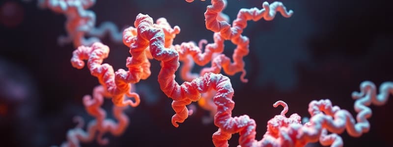
Helices
- The abundance in proteins is generated by placing similar subunits next to one another, each in the same repeated relationship.
- Result in a structure which resembles a spiral staircase.
- Can be either right-handed or left-handed. which depends on the way it twists.
- The a helix is generated when a single polypeptide chain turns around itself to form a structurally rigid cylinder.
- Hydrogen bonds are made between every fourth amino acid, linking the C=O of one peptide bond to the N-H of another.
- Transmembrane proteins usually form an α helix, made of amino acids with nonpolar side chains.
- The polypeptide backbone which is hydrophilic is hydrogen-bonded to itself inside the a helix, shielded from lipid environment.
- Two or three a helices will wrap around one another to form a coiled-coil.
Beta Sheets
- Hydrogen bonds form between segments of a polypeptide chain that lie side by side.
- Neighboring segments run in the same orientation structure and form a parallel sheet.
- When segments run in opposite directions forming an antiparallel sheet.
- Both types produce produce a very rigid, pleated structure, forming the core of many proteins.
- Give silk fibers their extraordinary tensile strength.
- Form the basis of amyloid structures where sheets are stacked like the teeth of a zipper.
Misfolded Proteins and Amyloid Structures
- Cells specialized for secretion store peptide, structure allows for efficient packaging, and they unfold once they reach the cell exterior.
- Can also form amyloid structures which are harmful to cells.
- Damage cells and tissues.
- Contributes to neurodegenerative disorders, including Alzheimer's, Parkinson's, and Huntington's diseases.
- Prions are considered "infectious" because the amyloid form can convert properly folded molecules of the protein into the abnormal, disease-causing confirmation.
- The abnormal form causes the conversion of normal proteins in the host's brain into the misfolded prion form.
- Protein aggregation results causing neurodegenerative disorder.
- Amyloid disrupts brain-cell function.
Levels of Protein Organization
- Does not begin and end with a helices and ẞ sheets.
- Confirmation includes interdependent levels and builds one another.
- The amino acid sequence is the considered its primary structure.
- The a helices and ẞ sheets that form within certain segments of the polypeptide chain are the secondary structure, which is produced by backbone hydrogen bonding.
- The full, three-dimensional conformation formed by an entire is the tertiary structure.
- Noncovalent bonds form along the polypeptide chain, between amino acid side chains, and between side chains and the polypeptide backbone.
- If the protein molecule exists as more than one polypeptide chain, then interacting polypeptides form its quaternary structure and are held together by noncovalent bonds.
- The protein domain unit is defined as any segment of a that can fold independently into a compact, stable structure.
- A domain contains 40 and 350 amino acids.
Protein Domains
- Are the modular unit in proteins which can be constructed.
- Elements of secondary structure pack together into stable, independently folding, globular elements.
- A typical protein molecule consists of at least one domain which are linked by polypeptide chain.
- Different domains are often associated with different functions
- For example, the bacterial catabolite activator protein (CAP) has a domain which binds to DNA and another that binds cyclic AMP, a small intracellular signaling molecule.
- Binding cAMP causes a conformational change in the protein which enables small domains to bind a with DNA and promote expression of an adjacent gene.
- Small protein molecules a single.
Protein Regions
- Larger proteins have connected domains between polypeptide chain.
- Short, unstructured ubiquity sequences in proteins can bend and be flexible via thermal buffering.
- Sequences may have important function within a cell.
Polypeptide Chains
- Vast of chains theoretically made from amino acids.
- Potential chains have functional proteins which should be "well-behaved" and not engage in unwanted associations.
Protein Confirmation
- Diverse organisms have protein and consistuent confirmations and domains remain effective and the structure is preserved throughout evolution.
Protein Classifications
- proteins can be grouped into protein families if the amino acids resemble the structure from the original protein in which each family member has an amino acid sequence a three-dimensional conformation that closely resemble those of the other family members.
- Serine, Protein-cleaving, digestive enzymes which include chymotrypsin, trypsin, and elastase,.
- When two enzymes are compared, amino acid sequences nearly the same but activities have distinct enzymatic activity.
Large Protein Molecules
- Types of weak noncovalent bonds enable a poplypeptide.
- binding sites recognizes surface pf a second protein the binding will be a larger protein with a a precisely defined and organized, Each poplypeptide chain is a sunbit they contain one or more.doman
- dimer a complex of two the bacterail protein with one protein sun bit 80th contain two domain protein complexes multi ple copys of sunbit
- enyme neuraninadase consist og for indetucal protein subunit
- protein molecules contain two protein subunits and multiple subunits
- Protein sassemble in filaments sheets of splinters, the binding as the protien which is complementary to create other region on
Protein filaments
- Actan long heculical structure
- Protune assoiciate for tubes such is protein coat virus or spherical shell
- subunits can assemble and complex structure and have inly of the structure binding, which are dentinal sites.
- Fibrils and filament form protein shape most
Protein Shapes
- Most proteins, we have been referring to are globular proteins in which the poplypeptide folds into a compact shape, like a ball with and irregular surface while enzymes tended to be globular proteins, but even though They are large and complicated with multiple.
- units most have have a cordially structure with an
Overall rounded shape but other proteins have roles in the cell and require them to expand large distance and generally have a relatively shapes.
Elongated Fibrous Proteins
- An akeratin molecule a dimer of two identical subunits with the long alicia's which form coiled
- These coiled coil region kept eat in by globular
Domains contents binding sites to allow them to assemble and ropelike international fillament and the cell mechanical - In multicellular the function of the extra are called
- Each has one glycine
- Matrix to help bind cells together to perform their job
- the function and structure, Each has been well
Studying That Suits You
Use AI to generate personalized quizzes and flashcards to suit your learning preferences.