Podcast
Questions and Answers
In staining techniques, what role does a mordant play in indirect staining?
In staining techniques, what role does a mordant play in indirect staining?
- It directly colors the tissue section without needing a dye.
- It prevents any non-specific binding of the dye to the tissue.
- It accelerates the staining reaction without participating in it.
- It acts as a bridge between the dye and the tissue, forming a dye lake. (correct)
Which of the following is NOT a method of artificial ripening of hematoxylin?
Which of the following is NOT a method of artificial ripening of hematoxylin?
- Adding potassium permanganate.
- Adding mercuric oxide.
- Exposure to air and sunlight. (correct)
- Adding hydrogen peroxide.
In the context of staining, what is the primary function of an auxochrome?
In the context of staining, what is the primary function of an auxochrome?
- To enable the dye to form a salt with another compound, retaining the color. (correct)
- To provide the initial color to a dye.
- To prevent the dye from fading over time.
- To act as a mordant, linking the dye to the tissue.
Which of the following describes the process of regressive staining?
Which of the following describes the process of regressive staining?
What is the role of differentiation (or decolorization) in staining techniques?
What is the role of differentiation (or decolorization) in staining techniques?
In metachromatic staining, what causes the change in color observed in certain tissue substances?
In metachromatic staining, what causes the change in color observed in certain tissue substances?
What is the purpose of metallic impregnation techniques in histology?
What is the purpose of metallic impregnation techniques in histology?
Which component determines whether a dye is classified as an acid dye or a basic dye?
Which component determines whether a dye is classified as an acid dye or a basic dye?
What is the main advantage of using Celestine Blue-Haemalum sequence staining over Iron Hematoxylin?
What is the main advantage of using Celestine Blue-Haemalum sequence staining over Iron Hematoxylin?
In immunohistochemistry, which of the following components is detected and visualized under a microscope?
In immunohistochemistry, which of the following components is detected and visualized under a microscope?
Why is it important to avoid saline water when rinsing tissues that have been stained for carbohydrates?
Why is it important to avoid saline water when rinsing tissues that have been stained for carbohydrates?
What is the significance of performing a PAS reaction with and without diastase digestion?
What is the significance of performing a PAS reaction with and without diastase digestion?
In lipid histochemistry, what is the purpose of using Formol-Calcium as a fixative?
In lipid histochemistry, what is the purpose of using Formol-Calcium as a fixative?
Why are controls important for the specificity of antibodies in immunohistochemistry?
Why are controls important for the specificity of antibodies in immunohistochemistry?
What is the general purpose of oil-soluble dyes (lysochromes) in staining?
What is the general purpose of oil-soluble dyes (lysochromes) in staining?
What is the role of antigen retrieval in immunohistochemistry?
What is the role of antigen retrieval in immunohistochemistry?
Which of the following is the most commonly used antibody in Immunohistochemistry?
Which of the following is the most commonly used antibody in Immunohistochemistry?
Why is polarized microscopy important in histopathology?
Why is polarized microscopy important in histopathology?
What is the purpose of vital staining?
What is the purpose of vital staining?
Which type of staining uses a combination of immunologic and histological staining?
Which type of staining uses a combination of immunologic and histological staining?
Flashcards
What is Staining?
What is Staining?
Applying dyes to sections to study tissue architecture and physical characteristics.
Chemical Affinity (Acidic)
Chemical Affinity (Acidic)
Acidic cell parts attract basic dyes (like the nucleus).
Chemical Affinity (Basic)
Chemical Affinity (Basic)
Basic cell parts attract acidic dyes (like cytoplasm).
Histological Staining
Histological Staining
Signup and view all the flashcards
Histochemical Staining
Histochemical Staining
Signup and view all the flashcards
Direct Staining
Direct Staining
Signup and view all the flashcards
What is a Mordant?
What is a Mordant?
Signup and view all the flashcards
What is an Accentuator?
What is an Accentuator?
Signup and view all the flashcards
Progressive Staining
Progressive Staining
Signup and view all the flashcards
Regressive Staining
Regressive Staining
Signup and view all the flashcards
Differentiation/Decolorization
Differentiation/Decolorization
Signup and view all the flashcards
Metachromatic Staining
Metachromatic Staining
Signup and view all the flashcards
Metallic Impregnation
Metallic Impregnation
Signup and view all the flashcards
Counterstaining
Counterstaining
Signup and view all the flashcards
Vital Staining
Vital Staining
Signup and view all the flashcards
Intravital Staining
Intravital Staining
Signup and view all the flashcards
Supravital Staining
Supravital Staining
Signup and view all the flashcards
H&E Staining Technique
H&E Staining Technique
Signup and view all the flashcards
Artificial/Synthetic Dyes
Artificial/Synthetic Dyes
Signup and view all the flashcards
Hematoxylin
Hematoxylin
Signup and view all the flashcards
Study Notes
Principles of Staining
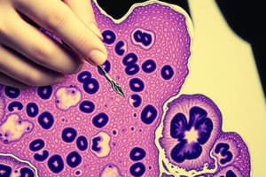
- Staining involves applying dyes to sections to visualize their architectural patterns and physical characteristics.
Chemical Affinity
- Acidic cell parts have a higher affinity for basic dyes, while basic cell parts prefer acidic dyes.
3 Major Groups of Staining
Histological Staining
- Tissue components are stained through direct interaction with a dye or solution.
- Examples include microanatomic, bacterial, and specific tissue stains.
Histochemical Staining
- Histochemical staining involves chemical reactions between specific tissue substances and dyes.
- Some examples include Perl's Prussian Blue for Hemoglobin and Periodic Acid Schiff for Carbohydrates.
Immunohistochemical Staining
- This staining combines immunologic and histological methods to detect phenotypic markers microscopically.
- It involves using polyclonal and monoclonal, fluorescent-labeled or enzyme-labeled antibodies.
Methods of Staining
Direct Staining
- Direct Staining involves coloring sections using aqueous or alcoholic solutions like Eosin and Methylene Blue.
Indirect Staining
- Indirect involves using a mordant or an accentuator.
- Mordants link tissue and dye.
- Mordants combine with the dye to form a "lake".
- The dye lake then combines with the tissue, forming the Tissue-Mordant-Dye Complex.
- Examples of mordants: potassium alum with Hematoxylin and iron in Weigert's Hematoxylin.
- Accentuators accelerate the staining reaction, but do not participate in it.
- Examples include potassium hydroxide (in Loeffler's Methylene Blue) and phenol (in Carbol Thionine and Carbol fuchsin).
Progressive Staining
- Stains are applied until the desired intensity is achieved.
- The distinction of tissue detail relies on the dye's selective affinity for different cellular elements.
Regressive Staining
- Regressive staining involves overstaining the tissue, and then removing or decolorizing excess stain until the desired intensity is attained.
Differentiation or Decolorization
- This process involves removing excess stain from the tissue during regressive staining, so specific substances are stained distinctly from their surroundings.
- Washing the section in solutions like water or alcohol accomplishes this.
- If the primary stain is basic, differentiation must come from an acid stain, and vice versa.
- Alcohol differentiate both acidic and basic dyes
- Mordants can also act as differentiators.
- Iron Alum oxidizes Hematoxylin into a colorless, soluble compound.
Metachromatic Staining
- Metachromatic is by using specific dyes that differentiate particular substances by staining them with a color different from the dye.
- Alcohol causes metachromasia to be lost once the tissue is dehydrated in alcohol after staining.
Counterstaining
Cytoplasmic Stains
- Red: Eosin Y, Eosin B, Phloxine B
- Yellow: Picric acid, Acid orange G, Rose Bengal
- Green: Light green SF, Lissamine green
Nuclear Stains
- Red: Neutral Red, Safranin O, Carmine, Hematoxylin
- Blue: Methylene Blue, Toluidine Blue, Celestine Blue
Metallic Impregnation
- Involves using metallic salts reduced by the tissue to produce an opaque or black deposit on the tissue surface.
Vital Staining
- Vital staining involves in the selective staining of living cell constituents
- Structures are demonstrated via phagocytosis of the dye particle.
- Reticulo-Endothelial System - stained with Trypan Blue
- Mitochondria - stained with Janus Green
- Nucleus of a living cell - resistant to vital stains
Intravital Staining
- Dye injected into any part of the animal body.
- Lithium, Carmine, and India ink are examples
Supravital Staining
- Supravital, staining of living cells immediately after removal from a living body
- Best vital stain = Neutral Red; Mitochondria = Janus Green; 1 gram of dye in 100mL = Trypan blue solution. Caution: if used more than 1 hour, it is toxic. Nile blue, Thionine, and Toluidine are examples.
Hematoxylin and Eosin Staining Technique
- Most common technique for microanatomical studies.
- Employs regressive staining technique in paraffin-embedded tissues.
- Involves overstaining of nuclei and removing excessive color from tissue constituents through acid differentiation.
- Nuclei - blue to blue black; Karyosome - Dark Blue; Cytoplasm - Pale pink
Other Staining Techniques
Heidenhain's Iron Hematoxylin Method
- Cell nuclei, cytoplasmic inclusions, and muscle striations appear black.
Celestine Blue-Haemalum Sequence Staining
- Uses an oxazine dye as an alternative to iron hematoxylin for nuclear staining.
- Celestine blue combines with iron alum, acting as a mordant to bind Hematoxylin.
- Stains Cell nuclei blue.
Mallory's Phloxine Methylene Blue Staining
- Originally known as Eosin-Methylene Blue method.
Types of Dye
Natural Dyes
- Obtained from plants and animals, and previously used for wool or nylon.
Hematoxylin
- Derived from the heartwood of Hematoxylin campechianum (Mexican Tree).
- The active coloring agent is Hematin, from the oxidation of hematoxylin or ripening.
- Natural Ripening - exposing the extract to air or sunlight for 3-4 months.
- Artificial Ripening - Adding oxidizing agents.
- Ripened Hematoxylin needs mordants like Alum, Iron, Chromium and Copper.
Cochineal Dyes
- An old histologic dye from the female cochineal bug (Coccus Cacti).
- Treats with Alum to produce the dye Carmine
- Add Picric acid (Picrocarmine) in neuropathological studies
- Add Aluminum Chloride (Best Carmine) - for glycogen
Orcein
- Comes from vegetable dye from certain lichens which are normally colorless.
- Treatment with Ammonia and exposure to air produces blue to violet colors
- Soluble with Alkali, for Elastic fibers
- Used for exposed to ammonia and air.
Artificial/Synthetic Dyes
- Derived from hydrocarbon Benzene, known as aniline dye.
Chromophores
- Substances with definite atomic groupings, capable for visible colors
Auxochrome
- Auxiliary radical that imparts electrolytic dissociation, alters the shade of the dye.
Three Groups of Artificial/Synthetic Dyes
Acid Dye
- Coloring agent is on the acidic component.
- Basic cell structures have affinity to Acidic Stains
- Examples - Acid Fuchsin; Picric Acid;
Basic Dye
- Coloring agent is on the basic component
- Acidic structures have affinity to basic stains
- Methylene blue can be used as an indicator and a dye for bacterial staining
Neutral Dye
- Formed by combining aqueous solutions of acidic and basic dyes
- Can stain cytoplasm and Nucleus simultaneously and differentially
Common Staining Solutions
Hematoxylin
- For routine histologic studies, needs the mordant Iron and Alum
Aluminum Hematoxylin
- For progressive staining and Regressive Staining
- Aluminum salts give a blue lake
Erlich's Hematoxylin
- Regressive staining.
Harris Hematoxylin
- Good regressive stain routine nuclear staining.
Cole's Hematoxylin
- Used in sequence with Celestine Blue
Carazzi's Hematoxylin
- Ripened by potassium iodide, frozen sections
Iron Hematoxylin
- For differential and regressive staining; uses acid alcohol as differentiating agent
Weigert's Hematoxylin
- Standard iron hematoxylin (Muscle fibers and Connective Tissues)
Heidenhain's Hematoxylin
- Ferric Ammonium Sulfate is the mordant
- Cytological staining, regressive staining
Loyez Hematoxylin
- Frozen Sections
Verhoeff Hematoxylin
- Mordant used is 1% Aqueous Phosphotungstic Acid
- Progressive Stain
Lead Hematoxylin
- For granules of endocrine cells of the alimentary tract
Eosin
- Eosin differentiates connective tissues and cytoplasm
- Used as a counterstain after hematoxylin and before methylene blue
Eosin Forms
- Eosin Y - Most commonly used
- Eosin B - Or Erythrosin B
- Eosin S - Or Ethyl Eosin, alcohol soluble
Carbohydrates
- Staining for carbohydrates - Carbohydrates stored in pure form (Glycogen) or bound to substances (Mucin)
- Glycogen - a polysaccharide of glucose stored in liver, heart and skeletal muscles
- Mucin - made up of hexosamines or neutral mucopolysaccharides
Periodic Acid Schiff
- Periodic acid oxidizes the 1,2 Glycol group of polysaccharides and mucin
- It liberates the aldehydes that will color the Schiff reagent - produces the red magenta or purplish pink color
Schiff Reagent
- Essential component is basic fuchsin (Made up of 3 stains, Rosanilin, Paratosanilin and Magenta II)
Staining of Mucin
- Forms the ground substance of connective tissues
Acid Mucopolysaccharides
- Bound to sulfuric acid esters and proteins
- hyaluronic acid
Proteins, Enzymes and Nucleic Acids
- Demonstration of Proteins through Histologic methods, acid histochemical methods, enzyme histochemical methods and immunocytochemical methods.
Protein Techniques
Connective Tissue
Immunohistochemistry
- Identifies specific or highly selective cellular epitopes or antigens in frozen or paraffin embedded tissues.
Epithelial Tumor Markers
Studying That Suits You
Use AI to generate personalized quizzes and flashcards to suit your learning preferences.


