Podcast
Questions and Answers
What is the primary management approach for diaphragmatic paralysis?
What is the primary management approach for diaphragmatic paralysis?
- Identification and treatment of the underlying cause (correct)
- Chest X-Ray to monitor progression
- Immediate intubation and invasive ventilation
- Symptomatic relief with oxygen therapy
In a bilateral diaphragmatic paralysis, which of the following may be required?
In a bilateral diaphragmatic paralysis, which of the following may be required?
- Non-invasive positive ventilation (correct)
- Non-invasive negative ventilation
- Chest physiotherapy
- Pneumonia vaccination
What is the characterization of lung function tests in diaphragmatic paralysis?
What is the characterization of lung function tests in diaphragmatic paralysis?
- Obstructive deficit
- Mixed deficit
- Normal lung function
- Restrictive deficit (correct)
What is the term for the condition where both sides of the diaphragm are paralyzed?
What is the term for the condition where both sides of the diaphragm are paralyzed?
What is the main symptom experienced by patients with bilateral diaphragmatic paralysis?
What is the main symptom experienced by patients with bilateral diaphragmatic paralysis?
What is the anatomical structure located between the two lungs?
What is the anatomical structure located between the two lungs?
What happens to the margins of the base of the lung during deep inspiration?
What happens to the margins of the base of the lung during deep inspiration?
What is the name of the dome of parietal pleura that extends into the root of the neck?
What is the name of the dome of parietal pleura that extends into the root of the neck?
Which structure does the scalenus anterior muscle envelop?
Which structure does the scalenus anterior muscle envelop?
What is the direction of the vertebral artery in relation to the subclavian artery?
What is the direction of the vertebral artery in relation to the subclavian artery?
What is the direction of the internal mammary artery from the subclavian artery?
What is the direction of the internal mammary artery from the subclavian artery?
What is the relationship between the costocervical trunk and the dome of the pleura?
What is the relationship between the costocervical trunk and the dome of the pleura?
What is the location of the superior intercostal branch in relation to the dome of the pleura?
What is the location of the superior intercostal branch in relation to the dome of the pleura?
What is the lateral boundary of the mediastinum?
What is the lateral boundary of the mediastinum?
What is the main function of the pleurae?
What is the main function of the pleurae?
What is the term for the layer of simple squamous cells that lines the pleura?
What is the term for the layer of simple squamous cells that lines the pleura?
What is the primary source of arterial supply to the lungs?
What is the primary source of arterial supply to the lungs?
What is the term for the condition where air or gas is present in the pleural space?
What is the term for the condition where air or gas is present in the pleural space?
What is the characteristic of the affected side in a pneumothorax upon percussion?
What is the characteristic of the affected side in a pneumothorax upon percussion?
What is the term for a pneumothorax that occurs without a specific cause?
What is the term for a pneumothorax that occurs without a specific cause?
What is the procedure used to remove excess air/gas in a pneumothorax?
What is the procedure used to remove excess air/gas in a pneumothorax?
What is the name of the nerve network that provides autonomic innervation to the lungs?
What is the name of the nerve network that provides autonomic innervation to the lungs?
What is the diameter of the ascending aorta?
What is the diameter of the ascending aorta?
Where does the ascending aorta begin?
Where does the ascending aorta begin?
At what level of the costal cartilage does the lung display the cardiac notch and lies 2.5 cm from the edge of the pleura and sternum?
At what level of the costal cartilage does the lung display the cardiac notch and lies 2.5 cm from the edge of the pleura and sternum?
What is the only branch of the ascending aorta?
What is the only branch of the ascending aorta?
What is the position of the arch of the aorta relative to the trachea and esophagus?
What is the position of the arch of the aorta relative to the trachea and esophagus?
What is the level of the vertebral column where the posterior borders of the lungs begin?
What is the level of the vertebral column where the posterior borders of the lungs begin?
What is the location of the arch of the aorta relative to the root of the left lung?
What is the location of the arch of the aorta relative to the root of the left lung?
What is the recess formed behind the dome of the diaphragm due to the separation of visceral and parietal pleura?
What is the recess formed behind the dome of the diaphragm due to the separation of visceral and parietal pleura?
Which lung lobe is produced by the additional transverse or minor fissure?
Which lung lobe is produced by the additional transverse or minor fissure?
What is the usual branch of the arch of the aorta associated with the right side of the body?
What is the usual branch of the arch of the aorta associated with the right side of the body?
What is the ligamentum arteriosum attached to?
What is the ligamentum arteriosum attached to?
What is the level where the right oblique fissure starts?
What is the level where the right oblique fissure starts?
Where does the brachiocephalic trunk arise from?
Where does the brachiocephalic trunk arise from?
What is the line followed by the right oblique fissure to end near the right sixth costochondral junction?
What is the line followed by the right oblique fissure to end near the right sixth costochondral junction?
What is the level where the separation of visceral and parietal pleura occurs to give rise to the slit-like costodiaphragmatic recess?
What is the level where the separation of visceral and parietal pleura occurs to give rise to the slit-like costodiaphragmatic recess?
What is the feature of the left lung that gives rise to the costomediastinal recess behind the left lower costal cartilages?
What is the feature of the left lung that gives rise to the costomediastinal recess behind the left lower costal cartilages?
Flashcards are hidden until you start studying
Study Notes
Pleurae
- The pleurae are serous membranes that line the lungs and thoracic cavity, allowing for efficient and effortless respiration.
- There are two pleurae in the body, one associated with each lung.
- Each pleura consists of a serous membrane, a layer of simple squamous cells supported by connective tissue, also known as the mesothelium.
- The pleura can be divided into two parts: visceral pleura (covers the lungs) and parietal pleura (covers the internal surface of the thoracic cavity).
- The two parts are continuous with each other at the hilum of each lung.
Costodiaphragmatic Recess
- The margins of the base of the lung decline, and the costal and diaphragmatic pleurae split up in deep inspiration.
- The lower zone of the pleural cavity into which the lung enlarges is called the costodiaphragmatic recess.
Mediastinal Pleura
- The mediastinal pleura lines the corresponding outermost layer of the mediastinum and creates its lateral boundary.
- It is represented as a cuff over the root of the lung and becomes continuous with the visceral pleura.
Cervical Pleura
- The cervical pleura is the dome of parietal pleura, which extends into the root of the neck about 1 inch (2.5 cm) above the medial end of the clavicle and 2 inches (5 cm) above the 1st costal cartilage.
- It is termed the cupola and covers the apex of the lung.
- It is covered by the suprapleural membrane.
Relations of the Cervical Pleura
- Anteriorly: subclavian artery and scalenus anterior muscle
- Posteriorly: neck of 1st rib and structures passing in front of it
- Laterally: scalenus medius muscle
- Medially: great vessels of the neck
Clinical Application: Pneumothorax
- A pneumothorax occurs when air or gas is present within the pleural space, removing the surface tension of the serous fluid and reducing lung extension.
- Clinical features: chest pain, shortness of breath, and asymmetrical chest expansion.
- Upon percussion, the affected side may be hyper-resonant due to excess air within the chest.
- Treatment: decompression to remove extra air/gas via the insertion of a chest drain.
Mediastinum
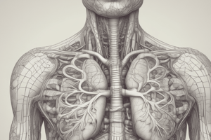
- The mediastinum is the median septum of the thorax between the two lungs.
- The ascending aorta begins at the aortic orifice and is approximately 2.5 cm in diameter.
- The arch of the aorta (aortic arch) is the curved continuation of the ascending aorta, beginning posterior to the 2nd right sternocostal joint at the level of the sternal angle.
Great Vessels
- The arch of the aorta ascends anterior to the right pulmonary artery and the bifurcation of the trachea, reaching its apex at the left side of the trachea and esophagus.
- The arch of the aorta ends by becoming the thoracic aorta posterior to the 2nd left sternocostal joint.
- The usual branches of the arch of the aorta are the brachiocephalic trunk, left common carotid artery, and left subclavian artery.
Pulmonary Fissures
- The left lung is separated into upper lobe and lower lobe by the oblique or major fissure.
- An additional transverse or minor fissure produces the middle lobe of the right lung.
- The right oblique fissure starts opposite the fourth thoracic spinous process and follows the line of the fifth rib, or a line just below, to end near the right sixth costochondral junction or just above in the fifth intercostal space.
Studying That Suits You
Use AI to generate personalized quizzes and flashcards to suit your learning preferences.


