Podcast
Questions and Answers
What anatomical reference point may be modified during tumoral expansion of the pituitary gland?
What anatomical reference point may be modified during tumoral expansion of the pituitary gland?
- Foramen magnum
- Anterior clinoidal process (correct)
- Sphenoid sinus
- Optic chiasm
Which imaging technique provides a two-dimensional projection of three-dimensional body parts?
Which imaging technique provides a two-dimensional projection of three-dimensional body parts?
- Skull X-ray (correct)
- Computed Tomography
- Magnetic Resonance Imaging
- Ultrasound
What is the main disadvantage of using X-rays for visualizing the pituitary gland?
What is the main disadvantage of using X-rays for visualizing the pituitary gland?
- They are unable to differentiate between soft tissues. (correct)
- They use minimal radiation.
- They are less sensitive than CT scans.
- They provide a three-dimensional view.
What year was CT originally introduced, and by whom?
What year was CT originally introduced, and by whom?
What appearance may the normal pituitary gland have on a CT scan?
What appearance may the normal pituitary gland have on a CT scan?
What factor relates the absorption of X-rays to the imaging of body structures?
What factor relates the absorption of X-rays to the imaging of body structures?
What is a common allergic reaction associated with CT scans?
What is a common allergic reaction associated with CT scans?
What is the diameter of the sella turcica in millimeters for its length?
What is the diameter of the sella turcica in millimeters for its length?
What is the normal range for urinary amylase?
What is the normal range for urinary amylase?
Which mode is primarily used for a thyroid scintigram?
Which mode is primarily used for a thyroid scintigram?
What characterizes the concentric rings formed in the Rosenbach reaction?
What characterizes the concentric rings formed in the Rosenbach reaction?
What is the primary function of contrast dye in imaging procedures?
What is the primary function of contrast dye in imaging procedures?
What is NOT a characteristic of MRI as described?
What is NOT a characteristic of MRI as described?
Which radioactive isotope is commonly used for thyroid imaging?
Which radioactive isotope is commonly used for thyroid imaging?
What is the primary purpose of using short half-life isotopes in nuclear medicine?
What is the primary purpose of using short half-life isotopes in nuclear medicine?
What is a limitation of MRI in detecting pituitary tumors?
What is a limitation of MRI in detecting pituitary tumors?
During the Thyroid Scintigraphy, how is the information ultimately processed?
During the Thyroid Scintigraphy, how is the information ultimately processed?
How does MRI differ from CT in terms of image acquisition?
How does MRI differ from CT in terms of image acquisition?
Which statement about the interaction of MRI with metallic objects is true?
Which statement about the interaction of MRI with metallic objects is true?
In the histogram mode, what effect do more pictures over time have on the pixels?
In the histogram mode, what effect do more pictures over time have on the pixels?
What is the initial step in performing the Rosenbach reaction for bile pigment identification?
What is the initial step in performing the Rosenbach reaction for bile pigment identification?
What is a prevalent issue faced by a percentage of healthy individuals on an MRI scan of the pituitary gland?
What is a prevalent issue faced by a percentage of healthy individuals on an MRI scan of the pituitary gland?
Which best explains why no harmful biological effects have been proven for MRI under standard conditions?
Which best explains why no harmful biological effects have been proven for MRI under standard conditions?
Which part of the pituitary gland is typically represented as whiter in MRI imaging?
Which part of the pituitary gland is typically represented as whiter in MRI imaging?
What is the typical scanning time after administration of I123 agents?
What is the typical scanning time after administration of I123 agents?
Which statement about thyroid nodules is correct?
Which statement about thyroid nodules is correct?
In the context of thyroid scintigraphy, what does the term 'cold nodule' indicate?
In the context of thyroid scintigraphy, what does the term 'cold nodule' indicate?
Graves' disease is associated with which of the following conditions?
Graves' disease is associated with which of the following conditions?
What risk percentage pertains to malignancy in a cold nodule?
What risk percentage pertains to malignancy in a cold nodule?
Which factor is implicated in the autoimmune nature of Graves' disease?
Which factor is implicated in the autoimmune nature of Graves' disease?
What major role does scintigraphy play in assessing a thyroid nodule?
What major role does scintigraphy play in assessing a thyroid nodule?
What percentage of solitary, hot nodules are typically benign?
What percentage of solitary, hot nodules are typically benign?
What is the primary disadvantage of using urine to determine amylase activity compared to plasma?
What is the primary disadvantage of using urine to determine amylase activity compared to plasma?
At what temperature should the test tubes be incubated during the amylase determination procedure?
At what temperature should the test tubes be incubated during the amylase determination procedure?
What does one Wohlgemuth Unit signify in terms of starch hydrolysis?
What does one Wohlgemuth Unit signify in terms of starch hydrolysis?
Which dilution factor corresponds to an undiluted urine that contains 64 Wohlgemuth Units if the test shows colorless at the sixth tube?
Which dilution factor corresponds to an undiluted urine that contains 64 Wohlgemuth Units if the test shows colorless at the sixth tube?
In the context of chronic pancreatitis with exocrine pancreatic insufficiency, how is the amylase level expected to be affected?
In the context of chronic pancreatitis with exocrine pancreatic insufficiency, how is the amylase level expected to be affected?
How does the hyperamylasemia function in the diagnosis of acute pancreatitis?
How does the hyperamylasemia function in the diagnosis of acute pancreatitis?
What is the initial step for creating dilutions for amylase determination?
What is the initial step for creating dilutions for amylase determination?
What color indicates the presence of undigested starch in the test tubes after the procedure?
What color indicates the presence of undigested starch in the test tubes after the procedure?
Flashcards
What causes the majority of pituitary abnormalities?
What causes the majority of pituitary abnormalities?
Pituitary tumors are the most common type of pituitary pathology. They often cause compression and erosion of the sphenoid bone.
What bony structure is affected during pituitary tumor expansion?
What bony structure is affected during pituitary tumor expansion?
The sella turcica is a bony structure that houses the pituitary gland. Its walls can be altered or disappear due to pituitary tumor growth.
What imaging technique can show the bony walls of the sella turcica?
What imaging technique can show the bony walls of the sella turcica?
X-rays are used to visualize the bony walls of the sella turcica.
How do x-rays work?
How do x-rays work?
Signup and view all the flashcards
What imaging technique provides a detailed cross-sectional view of the sella turcica?
What imaging technique provides a detailed cross-sectional view of the sella turcica?
Signup and view all the flashcards
How do different tissues appear in a CT scan?
How do different tissues appear in a CT scan?
Signup and view all the flashcards
What information does a CT scan provide about the pituitary gland?
What information does a CT scan provide about the pituitary gland?
Signup and view all the flashcards
What is a potential downside of using CT scans?
What is a potential downside of using CT scans?
Signup and view all the flashcards
Contrast dye
Contrast dye
Signup and view all the flashcards
CT Scan (Computed Tomography)
CT Scan (Computed Tomography)
Signup and view all the flashcards
MRI Scan (Magnetic Resonance Imaging)
MRI Scan (Magnetic Resonance Imaging)
Signup and view all the flashcards
Pituitary gland
Pituitary gland
Signup and view all the flashcards
Pituitary adenoma
Pituitary adenoma
Signup and view all the flashcards
Macroadenoma
Macroadenoma
Signup and view all the flashcards
Microadenoma
Microadenoma
Signup and view all the flashcards
Multiplanar imaging
Multiplanar imaging
Signup and view all the flashcards
Urine Amylase Test
Urine Amylase Test
Signup and view all the flashcards
Amylase
Amylase
Signup and view all the flashcards
Pancreatitis
Pancreatitis
Signup and view all the flashcards
Exocrine Pancreatic Insufficiency
Exocrine Pancreatic Insufficiency
Signup and view all the flashcards
Wohlgemuth Unit (WU)
Wohlgemuth Unit (WU)
Signup and view all the flashcards
Serial Dilution
Serial Dilution
Signup and view all the flashcards
Half-life
Half-life
Signup and view all the flashcards
Lugol's Solution
Lugol's Solution
Signup and view all the flashcards
Thyroid Scintigraphy
Thyroid Scintigraphy
Signup and view all the flashcards
Hot nodule
Hot nodule
Signup and view all the flashcards
Warm nodule
Warm nodule
Signup and view all the flashcards
Cold nodule
Cold nodule
Signup and view all the flashcards
Goiter
Goiter
Signup and view all the flashcards
Graves' disease
Graves' disease
Signup and view all the flashcards
TSH receptor antibodies
TSH receptor antibodies
Signup and view all the flashcards
Ectopic thyroid tissue
Ectopic thyroid tissue
Signup and view all the flashcards
24-hour Urinary Amylase Test
24-hour Urinary Amylase Test
Signup and view all the flashcards
Rosenbach Reaction
Rosenbach Reaction
Signup and view all the flashcards
Thyroid Scintigram
Thyroid Scintigram
Signup and view all the flashcards
Histogram Mode
Histogram Mode
Signup and view all the flashcards
Three Dimensional Reconstruction
Three Dimensional Reconstruction
Signup and view all the flashcards
Single Photon Emission Computed Tomography (SPECT)
Single Photon Emission Computed Tomography (SPECT)
Signup and view all the flashcards
Positron Emission Tomography (PET)
Positron Emission Tomography (PET)
Signup and view all the flashcards
Study Notes
Pituitary Gland Radiological Exploration
- Pituitary pathologies are primarily tumors (adenomas).
- Growing tumors compress bone, eroding and invading the sphenoid bone.
- Normal sella turcica is defined by anterior and posterior clinoid processes; these may disappear during tumor expansion.
- X-rays, discovered by Roentgen (1895), can visualize these injuries.
- Skull X-rays (lateral view): X-rays penetrate body structures, projecting shadows onto detectors.
- Bones appear in varying shades of gray; soft tissues appear black.
- Three-dimensional structures are projected onto two-dimensional film, losing information.
- X-rays can visualize sella turcica bony walls.
- Normal sella turcica dimensions: 15/12/19 mm (length/depth/width).
- Normal image variations form the basis for pituitary adenoma diagnosis and classification.
Computed Tomography (CT)
- Hounsfield introduced CT in 1971, earning a Nobel Prize in 1979.
- CT extends basic X-ray technology.
- X-ray tube and detectors rotate around patient (head to toe).
- X-ray absorption is proportional to tissue density.
- Density profile of a body slice (transverse or axial cut) is obtained.
- Detection system includes scintillating crystals and photodiodes.
- Computer reconstructs the image from raw scan data.
- Bone appears white; gases and liquids are black; tissues are gray.
- CT provides tomodensitometric analysis of sella turcica (pituitary tissue).
- Normal pituitary gland may have a non-homogeneous appearance (mixed light and dense areas).
- This heterogeneity relates to microscopic variations in the anterior and posterior lobes.
- Contrast dye enhances image contrast, facilitating tumor demarcation.
Magnetic Resonance Imaging (MRI)
- MRI uses hydrogen's magnetic properties with a large external magnetic field.
- Protons in body water align with the magnetic field and pulsed radio waves modify their orientation.
- Protons return to their alignment and emit characteristic radio waves.
- These radio waves are detected by antennas.
- Computer translates the wave patterns into detailed images.
- Contrast material can improve image quality.
- MRI images can be obtained in any plane.
- MRI does not use X-rays; no proven harmful effects.
- MRI is the best imaging technique to identify pituitary tumors (macro- or microadenomas).
- MRI may not detect microadenomas smaller than 3 mm.
- Relatively low cost, no electricity, no radiation.
Normal Head MRI
- Sagittal view through pituitary gland; large arrow indicates anterior portion, small arrow posterior.
- Coronal MRI showing posterior pituitary bright spot (ectopic) at hypothalamus base.
- Sagittal MRI view of infant pituitary gland exhibiting transected hypothalamic-pituitary stalk.
Urinary Amylase Determination
- Amylase is filtered by kidneys and expelled in urine.
- Acute pancreatitis causes elevated plasma amylase, which can be reflected in urine.
- Amylase activity in plasma varies widely and depends on inflammation severity and pancreatic function.
- Urine amylase levels show gradual reduction over 4-10 days, reaching normal levels.
- Wohlgemuth method establishes urine amylase activity, useful but with wider reference ranges compared to serum.
- Urine disadvantages include variable diuresis, making ranges broader than for serum.
- Chronic pancreatitis with exocrine insufficiency shows decreased urinary amylase.
Rosenbach Reaction (Bile Pigments Identification)
- Used to identify bile pigments.
- Filter paper, bile pigments solutions, concentrated nitric acid, sodium nitrite crystals required.
- Drop bile pigment solution onto filter paper, then nitric acid.
- Concentric rings form (yellow/red, violet/blue, green).
- Green rings indicate bilirubin presence.
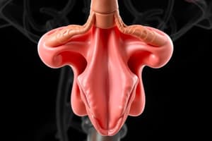
Thyroid Scintigram
- Nuclear medicine investigation (histogram mode, list mode, synchronized recordings, 3D reconstruction).
- Thyroid imaging can detect activity distribution using gamma cameras.
- Histogram mode uses pixels that become darker with increasing picture accumulation.
- Three-dimensional reconstruction (SPECT) processes multiple two-dimensional images to create 3D image.
- Positron emission tomography (PET) is more precise, using positron-emitting isotopes and recording simultaneous emissions, determining precise location of radiation source.
- Radiolabeled material is injected; gamma camera detects emission information and sends it to computer for image processing.
- Iodine 123 (I123), Iodine 131 (I131), Technetium (Tc 9m) pertechnetate, and Thallium 201 (Tl 201) isotopic agents are used for imaging.
- Scans are performed at specific intervals post-injection depending on the agent.
- Thyroid scintigrams provide information on thyroid gland size, shape, activity distribution (homogeneous or inhomogeneous), and nodule details.
Studying That Suits You
Use AI to generate personalized quizzes and flashcards to suit your learning preferences.