Podcast
Questions and Answers
What is the primary function of veins in the human circulatory system?
What is the primary function of veins in the human circulatory system?
Veins carry blood towards the heart.
Name the arteries that branch from the internal carotid artery.
Name the arteries that branch from the internal carotid artery.
The anterior and middle cerebral arteries, and the ophthalmic arteries.
What important function does the cerebral arterial circle perform?
What important function does the cerebral arterial circle perform?
It equalizes blood pressure in the brain and provides alternative blood delivery routes.
Identify the major veins involved in draining the head and neck.
Identify the major veins involved in draining the head and neck.
Which veins return blood directly to the heart from the lower body?
Which veins return blood directly to the heart from the lower body?
What arteries supply blood to the scalp and skull?
What arteries supply blood to the scalp and skull?
How do vertebral arteries contribute to cerebral blood supply?
How do vertebral arteries contribute to cerebral blood supply?
What are the primary tributaries of the facial veins?
What are the primary tributaries of the facial veins?
What effects do vasoconstriction and vasodilation have on peripheral resistance?
What effects do vasoconstriction and vasodilation have on peripheral resistance?
How does hypertensive blood pressure differ from hypotensive blood pressure?
How does hypertensive blood pressure differ from hypotensive blood pressure?
What is the primary function of the pulmonary circulation?
What is the primary function of the pulmonary circulation?
What does an increase in blood flow to skeletal muscles during exercise indicate?
What does an increase in blood flow to skeletal muscles during exercise indicate?
Explain the path of deoxygenated blood from the right ventricle to the lungs.
Explain the path of deoxygenated blood from the right ventricle to the lungs.
Describe the differences in blood pressure between pulmonary circulation and systemic circulation.
Describe the differences in blood pressure between pulmonary circulation and systemic circulation.
What causes orthostatic hypotension?
What causes orthostatic hypotension?
Identify the three main branches of the aortic arch.
Identify the three main branches of the aortic arch.
How does blood return to the right side of the heart from the lungs?
How does blood return to the right side of the heart from the lungs?
What is the significance of lower blood pressure in pulmonary capillaries?
What is the significance of lower blood pressure in pulmonary capillaries?
What does the increase in blood flow to the skin during exercise signify?
What does the increase in blood flow to the skin during exercise signify?
Define the roles of the superior and inferior vena cava.
Define the roles of the superior and inferior vena cava.
What is the role of the coronary sinus?
What is the role of the coronary sinus?
How does blood pressure in the aorta compare to that in the pulmonary circulation?
How does blood pressure in the aorta compare to that in the pulmonary circulation?
What happens to total blood flow when there is a steeper pressure gradient, assuming resistance remains unchanged?
What happens to total blood flow when there is a steeper pressure gradient, assuming resistance remains unchanged?
How does increased resistance in the liver, such as in cirrhosis, affect total blood flow?
How does increased resistance in the liver, such as in cirrhosis, affect total blood flow?
What is the primary reason for Arlene's light-headedness upon standing suddenly?
What is the primary reason for Arlene's light-headedness upon standing suddenly?
Which hormone is specifically not released during exercise, impacting regulation of blood pressure?
Which hormone is specifically not released during exercise, impacting regulation of blood pressure?
What are the three tunics found in most blood vessels?
What are the three tunics found in most blood vessels?
Why is an overweight individual advised to lose weight in the context of high blood pressure?
Why is an overweight individual advised to lose weight in the context of high blood pressure?
Compare the thickness of the tunica media between arteries and veins.
Compare the thickness of the tunica media between arteries and veins.
In terms of blood flow velocity, where is the blood flow the slowest?
In terms of blood flow velocity, where is the blood flow the slowest?
What are the primary branches of the common carotid artery and their significance?
What are the primary branches of the common carotid artery and their significance?
Describe the function of the dural venous sinuses in the cranial cavity.
Describe the function of the dural venous sinuses in the cranial cavity.
Identify the three main unpaired arteries that supply the gastrointestinal tract.
Identify the three main unpaired arteries that supply the gastrointestinal tract.
What is the significance of the hepatic portal system in circulation?
What is the significance of the hepatic portal system in circulation?
How does fetal circulation differ from postnatal circulation?
How does fetal circulation differ from postnatal circulation?
Explain the role of one-way valves in veins.
Explain the role of one-way valves in veins.
What types of capillaries exist and how do they differ?
What types of capillaries exist and how do they differ?
How do the branches of the celiac trunk contribute to abdominal organ supply?
How do the branches of the celiac trunk contribute to abdominal organ supply?
What major vessel drains blood from the thoracic cavity into the systemic circulation?
What major vessel drains blood from the thoracic cavity into the systemic circulation?
What is the primary venous drainage of the kidney?
What is the primary venous drainage of the kidney?
Describe the anastomoses between arteries in the abdominal region.
Describe the anastomoses between arteries in the abdominal region.
Identify the arteries that supply the diaphragm.
Identify the arteries that supply the diaphragm.
How does the structure of veins differ from that of arteries?
How does the structure of veins differ from that of arteries?
What is the function of the posterior tibial artery?
What is the function of the posterior tibial artery?
Explain the importance of the circle of Willis in cerebral circulation.
Explain the importance of the circle of Willis in cerebral circulation.
Flashcards are hidden until you start studying
Study Notes
Peripheral Resistance
- Peripheral resistance opposes blood flow in vessels and is influenced by vessel radius, length, and blood viscosity.
- Vasoconstriction elevates peripheral resistance and increases blood pressure, while vasodilation decreases both.
Blood Volume
- Increased fluid intake raises blood volume and blood pressure; decreased output leads to lower levels.
- Hypertension is chronic high blood pressure; hypotension is chronic low blood pressure, causing fatigue, dizziness, and fainting.
Clinical View
- Chronic hypertension may damage blood vessel walls, leading to atherosclerosis or arteriosclerosis and increasing heart workload, potentially causing congestive heart failure.
- Orthostatic hypotension is a sudden drop in blood pressure upon changing positions.
Blood Flow Distribution
- Total blood flow increases during exercise due to faster heartbeat and blood redistribution to active tissues.
- Key increases in blood flow during exercise include:
- Coronary circulation
- Skeletal muscles (increase from 1100 mL/min to 12,500 mL/min)
- Skin (increase from 400 mL/min to 1900 mL/min)
- Brain blood flow remains significant throughout.
Pulmonary Circulation Pathway
- Deoxygenated blood is pumped from the right ventricle into the pulmonary trunk, dividing into left and right pulmonary arteries toward the lungs.
- Gas exchange occurs in pulmonary capillaries, with venules draining into pulmonary veins, transporting oxygenated blood to the left atrium.
Blood Pressure in Pulmonary Circulation
- Systolic pressure in pulmonary arteries ranges from 15-25 mm Hg, lower than the aorta's systolic pressure.
- Nearly all returning blood to the right side of the heart is pumped to the lungs.
Features of the Pulmonary Circulation
- Lower blood pressure in pulmonary capillaries (about 10 mm Hg) compared to systemic capillaries (around 40 mm Hg) facilitates gas exchange.
- Pulmonary arteries have less elastic tissue and wider lumens, allowing for lower resistance and shorter distances to the heart.
Comparison of Pulmonary and Systemic Circulation
- Blood pressure fluctuations in pulmonary circulation are lower than those in systemic circulation.
- Arteries and veins in the pulmonary route are named based on adjacent regions or bones.
Arterial Supply to the Body
- Oxygenated blood is pumped from the left ventricle into the ascending aorta, which curves into the aortic arch.
- Major branches from the aortic arch include the brachiocephalic trunk, left common carotid artery, and left subclavian artery to supply head, neck, and upper limbs.
Venous Drainage
- Superior vena cava collects blood from the head, neck, upper limbs, while inferior vena cava collects from the lower limbs and abdomen.
- The coronary sinus drains blood from the heart myocardium.
Arterial Supply to the Head and Neck
- Common carotid arteries supply blood to the head and neck; subclavian arteries serve the upper limbs.
Venous Drainage of the Head and Neck
- Internal and external jugular veins drain blood from the head, neck, and are connected to brachiocephalic veins leading to superior vena cava.
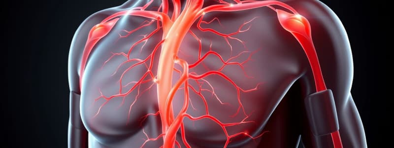
Human Veins and Arteries System
- The body has a complex network of veins (returning blood) and arteries (delivering blood).
- Major veins include internal and external jugular veins, brachiocephalic veins, and inferior vena cava.
Arterial Supply to the Head and Neck
- Arterial supply includes common carotid arteries, vertebral artery, and branches like the external carotid artery, which supply facial structures.
Cerebral Arterial Circle
- Also called the circle of Willis, this system ensures stable brain blood pressure and alternative blood flow routes in case of vessel blockage.
Venous Drainage
- Key pairs of veins include external jugular veins (from scalp and skull), anterior jugular veins (from face and neck), and facial veins (from face and tributaries).### Major Arteries and Veins of the Head and Neck
- Common carotid artery branches into internal and external carotid arteries.
- External carotid artery further branches into superficial temporal, maxillary, posterior auricular, occipital, facial, lingual, ascending pharyngeal, superior thyroid arteries, and thyrocervical trunk.
- Additional vital arteries include the brachiocephalic artery, vertebral artery, and subclavian artery.
- Major veins include vertebral, external jugular, internal jugular, superficial temporal, maxillary, posterior auricular, occipital, facial, lingual, superior thyroid, and right subclavian veins.
- Right brachiocephalic vein drains head and neck regions.
Dural Venous Sinuses
- Function to drain blood from the brain and collect excess cerebrospinal fluid.
- Blood from the dural sinuses primarily drains into the internal jugular vein.
Cerebral Arterial Circle
- Also known as the circle of Willis, an anastomosis of blood vessels surrounding the sphenoid bone's hypophyseal fossa.
Thoracic and Abdominal Wall Anatomy
- Major arteries supplying thoracic and abdominal walls include the internal thoracic artery, which branches into anterior intercostal arteries and musculophrenic artery, and transitions into the superior epigastric artery.
- Venous drainage in these areas is more complex than arterial supply, with key veins including inferior epigastric vein, supreme intercostal vein, and veins of the azygos system.
- Anastomoses play a significant role in maintaining blood supply, especially between inferior and superior epigastric arteries.
Arterial Supply to the Abdominal Organs
- Three major arteries arise from the abdominal aorta: celiac trunk, superior mesenteric artery, and inferior mesenteric artery.
- Celiac trunk supplies stomach, esophagus, spleen, pancreas, and liver through branches like left gastric, splenic, and common hepatic arteries.
- Superior mesenteric artery supplies the intestines, while inferior mesenteric artery supplies the distal colon.
Hepatic Portal System
- Composed of veins transporting blood from the gastrointestinal tract, spleen, and pancreas to the liver for processing.
- Blood flows through portal vein and is processed in the liver before drainage to the inferior vena cava.
Arterial and Venous Supply to the Limbs
- Upper limbs are mainly supplied by the subclavian artery, which branches into the axillary and brachial arteries.
- Lower limbs are supplied by the external iliac artery, later renamed the femoral artery beneath the inguinal ligament.
- Veins in upper limbs include cephalic, basilic, and brachial veins; lower limb veins include great and small saphenous veins draining into the femoral vein.
Fetal Circulation
- Umbilical vein carries oxygenated blood from the placenta; ductus venosus shunts blood from the liver to the inferior vena cava.
- Foramen ovale and ductus arteriosus bypass pulmonary circulation, facilitating efficient blood flow to vital organs during fetal development.
- Postnatal changes include closure of ductus venosus, foramen ovale, and umbilical vessels.
Blood Vessel Functionality
- Arteries transport blood away from the heart; veins return blood towards the heart, equipped with valves to prevent backflow.
- Capillaries serve as the primary site for nutrient and gas exchange due to their thin walls.
- Total cross-sectional area inversely affects blood flow velocity, with the slowest flow occurring in capillaries to enhance exchange efficiency.
Regulation of Blood Flow
- Blood pressure is influenced by cardiac output, vessel resistance, and blood volume, regulated through neural and hormonal mechanisms.
- Local blood flow is modified by tissue vascularization, myogenic responses, and local regulatory factors, ensuring adequate perfusion under various conditions.
Key Differences Between Fetal and Postnatal Circulation
- Fetal circulation relies on placental oxygen and nutrient transfer, whereas postnatal circulation demands lung oxygenation.
- Structural modifications occur at birth to establish independent circulation, including closure of fetal shunts and transformations of umbilical vessels.### Understanding Blood Flow and Blood Pressure
- Total blood flow increases with a steeper pressure gradient, assuming resistance remains constant.
- In cirrhosis of the liver, total blood flow decreases due to increased resistance while cardiac output is stable.
- Adequate perfusion of all tissues relies heavily on maintaining significant total blood flow.
- Capillary exchange remains unaffected despite variations in total blood flow.
- Increased colloid osmotic pressure in capillaries retains fluid in blood and decreases hydrostatic pressure.
- When colloid osmotic pressure decreases, fluid can accumulate in interstitial spaces, leading to edema.
- Blood flow velocity is slowest in veins compared to other blood vessels.
Regulation of Blood Pressure
- Light-headedness from sudden standing can result from baroreceptors in the carotid artery detecting reduced stretch, triggering an autonomic reflex to enhance cerebral blood flow.
- Hormones that regulate blood pressure include epinephrine, atrial natriuretic peptide, angiotensin II, and norepinephrine.
- Aortic bodies respond to increased blood pressure by initiating a chemoreceptor reflex that lowers blood pressure.
Blood Vessels
- Most blood vessels consist of three layers: tunica intima, tunica media, and tunica adventitia.
- Arteries possess many vascular anastomoses, especially around joints like the elbow and knee.
- Shorter blood vessel length correlates with decreased blood pressure.
- Weight loss in overweight individuals is recommended to alleviate compressive forces on thoracic organs, subsequently reducing resistance and blood pressure.
- The innermost layer of blood vessels, tunica intima, directly contacts the blood.
- Tunica media, the middle layer, is responsible for the contraction and relaxation of blood vessels.
- Tunica adventitia, the outer layer, provides structural support to blood vessels.
- Arteries transport blood away from the heart, whereas veins return it.
- Arteries feature a thicker tunica media and larger lumen compared to veins, allowing them to handle higher blood pressure.
Studying That Suits You
Use AI to generate personalized quizzes and flashcards to suit your learning preferences.


