Podcast
Questions and Answers
What structures are included in the Peripheral Nervous System (PNS)?
What structures are included in the Peripheral Nervous System (PNS)?
- Spinal cord, sensory receptors, and blood vessels
- Only sensory receptors and peripheral nerves
- Ganglia, motor endings, and brain
- Sensory receptors, peripheral nerves, associated ganglia, and motor endings (correct)
Which of the following connective tissue coverings surrounds the individual axons in a nerve?
Which of the following connective tissue coverings surrounds the individual axons in a nerve?
- Myoneurium
- Epineurium
- Perineurium
- Endoneurium (correct)
What type of nerve carries impulses to and from the central nervous system (CNS)?
What type of nerve carries impulses to and from the central nervous system (CNS)?
- Mixed nerves (correct)
- Sensory nerves
- Autonomic nerves
- Motor nerves
Which of the following statements correctly describes cranial nerves?
Which of the following statements correctly describes cranial nerves?
What is the primary function of Cranial Nerve I (Olfactory)?
What is the primary function of Cranial Nerve I (Olfactory)?
Which nerve arises from the retina of the eye?
Which nerve arises from the retina of the eye?
Where do the optic nerves converge?
Where do the optic nerves converge?
What characterizes the structure of a nerve?
What characterizes the structure of a nerve?
What type of nerve is mostly found within the human body?
What type of nerve is mostly found within the human body?
What is the primary route of afferent impulses for the sense of smell?
What is the primary route of afferent impulses for the sense of smell?
Which cranial nerve is responsible for motor functions to the parotid salivary gland?
Which cranial nerve is responsible for motor functions to the parotid salivary gland?
What unique feature distinguishes the vagus nerve from other cranial nerves?
What unique feature distinguishes the vagus nerve from other cranial nerves?
Which cranial nerve arises from both the medulla and the spinal cord?
Which cranial nerve arises from both the medulla and the spinal cord?
What function is primarily associated with Cranial Nerve XII?
What function is primarily associated with Cranial Nerve XII?
What type of fibers makes up most of the motor functions of the vagus nerve?
What type of fibers makes up most of the motor functions of the vagus nerve?
Which cranial nerve conducts taste sensations from the tongue?
Which cranial nerve conducts taste sensations from the tongue?
Where do the fibers of the accessory nerve exit the cranium?
Where do the fibers of the accessory nerve exit the cranium?
Which structure is primarily innervated by Cranial Nerve XI?
Which structure is primarily innervated by Cranial Nerve XI?
What is the main sensory function of the vagus nerve?
What is the main sensory function of the vagus nerve?
Which cranial nerve has both motor and sensory functions related to the throat?
Which cranial nerve has both motor and sensory functions related to the throat?
Which cranial nerve is responsible solely for carrying afferent impulses for vision?
Which cranial nerve is responsible solely for carrying afferent impulses for vision?
What is the primary function of Cranial Nerve III: Oculomotor?
What is the primary function of Cranial Nerve III: Oculomotor?
Where do the parasympathetic cell bodies of the Oculomotor nerve reside?
Where do the parasympathetic cell bodies of the Oculomotor nerve reside?
Which cranial nerve innervates the superior oblique muscle?
Which cranial nerve innervates the superior oblique muscle?
Which cranial nerve is composed of three divisions: ophthalmic (V1), maxillary (V2), and mandibular (V3)?
Which cranial nerve is composed of three divisions: ophthalmic (V1), maxillary (V2), and mandibular (V3)?
What primary function does Cranial Nerve VI: Abducens serve?
What primary function does Cranial Nerve VI: Abducens serve?
What sensory function is associated with Cranial Nerve VII: Facial?
What sensory function is associated with Cranial Nerve VII: Facial?
Which cranial nerve has both cochlear and vestibular divisions and is solely sensory?
Which cranial nerve has both cochlear and vestibular divisions and is solely sensory?
Which cranial nerve is primarily responsible for facial expression and transmitting autonomic impulses to glands?
Which cranial nerve is primarily responsible for facial expression and transmitting autonomic impulses to glands?
Cranial Nerve V: Trigeminal provides sensory impulses from which areas?
Cranial Nerve V: Trigeminal provides sensory impulses from which areas?
What is the primary type of nerve function associated with Cranial Nerve XI: Accessory?
What is the primary type of nerve function associated with Cranial Nerve XI: Accessory?
Which cranial nerve extends beyond the head and neck to innervate organs like the heart and lungs?
Which cranial nerve extends beyond the head and neck to innervate organs like the heart and lungs?
Which function is primarily associated with Cranial Nerve IX: Glossopharyngeal?
Which function is primarily associated with Cranial Nerve IX: Glossopharyngeal?
Which cranial nerve innervates both intrinsic and extrinsic muscles of the tongue?
Which cranial nerve innervates both intrinsic and extrinsic muscles of the tongue?
What exits the skull via the jugular foramen?
What exits the skull via the jugular foramen?
What distinguishes mixed nerves from sensory and motor nerves in the Peripheral Nervous System?
What distinguishes mixed nerves from sensory and motor nerves in the Peripheral Nervous System?
Which connective tissue layer wraps around groups of axons to form fascicles in a peripheral nerve?
Which connective tissue layer wraps around groups of axons to form fascicles in a peripheral nerve?
Which cranial nerve is primarily responsible for vision by transmitting information from the retina?
Which cranial nerve is primarily responsible for vision by transmitting information from the retina?
What is the primary role of cranial nerves that carry parasympathetic fibers?
What is the primary role of cranial nerves that carry parasympathetic fibers?
What term refers to the nerves that arise from the spinal column?
What term refers to the nerves that arise from the spinal column?
Which cranial nerve is responsible for conducting taste sensations from the anterior two-thirds of the tongue?
Which cranial nerve is responsible for conducting taste sensations from the anterior two-thirds of the tongue?
What is the correct order of structures for the nerve impulse pathway from the sensory receptor to the CNS?
What is the correct order of structures for the nerve impulse pathway from the sensory receptor to the CNS?
Which of the following statements about the structure of a nerve is accurate?
Which of the following statements about the structure of a nerve is accurate?
Which cranial nerve is primarily responsible for directing the eyeball and controlling lens shape?
Which cranial nerve is primarily responsible for directing the eyeball and controlling lens shape?
Which cranial nerve is solely sensory and involved in both equilibrium and hearing?
Which cranial nerve is solely sensory and involved in both equilibrium and hearing?
What is the primary motor function of Cranial Nerve VI?
What is the primary motor function of Cranial Nerve VI?
Which cranial nerve conveys sensory impulses from the face and supplies motor fibers for mastication?
Which cranial nerve conveys sensory impulses from the face and supplies motor fibers for mastication?
From which location do the fibers of Cranial Nerve IV emerge?
From which location do the fibers of Cranial Nerve IV emerge?
What autonomic functions are performed by Cranial Nerve VII?
What autonomic functions are performed by Cranial Nerve VII?
What is the pathway for the fibers of Cranial Nerve V: Trigeminal for its ophthalmic division?
What is the pathway for the fibers of Cranial Nerve V: Trigeminal for its ophthalmic division?
Which cranial nerve is responsible for taste sensations from the anterior two-thirds of the tongue?
Which cranial nerve is responsible for taste sensations from the anterior two-thirds of the tongue?
Which spinal nerves contribute to the formation of the lumbar plexus?
Which spinal nerves contribute to the formation of the lumbar plexus?
What is the primary nerve associated with the sacral plexus?
What is the primary nerve associated with the sacral plexus?
Which of the following statements accurately reflects the function of dermatomes?
Which of the following statements accurately reflects the function of dermatomes?
Which two nerves compose the sciatic nerve?
Which two nerves compose the sciatic nerve?
What is a major function of the lumbar plexus?
What is a major function of the lumbar plexus?
How are spinal nerves categorized?
How are spinal nerves categorized?
Which component of the spinal nerve contains motor fibers?
Which component of the spinal nerve contains motor fibers?
What is the primary function of nerve plexuses?
What is the primary function of nerve plexuses?
What is a consequence of damage to a single spinal segment?
What is a consequence of damage to a single spinal segment?
Which spinal nerves are classified under lumbar nerves?
Which spinal nerves are classified under lumbar nerves?
What type of fibers do dorsal roots contain?
What type of fibers do dorsal roots contain?
What is the role of the spinal nerves that arise from the sacral region?
What is the role of the spinal nerves that arise from the sacral region?
Which regions of the body get their nerve supply from the plexuses formed by the ventral rami?
Which regions of the body get their nerve supply from the plexuses formed by the ventral rami?
What is the primary function of the phrenic nerve?
What is the primary function of the phrenic nerve?
Which of the following does NOT arise from the brachial plexus?
Which of the following does NOT arise from the brachial plexus?
Which of the following is a primary component of the brachial plexus?
Which of the following is a primary component of the brachial plexus?
What muscles are primarily innervated by the radial nerve?
What muscles are primarily innervated by the radial nerve?
How many ventral rami contribute to the brachial plexus?
How many ventral rami contribute to the brachial plexus?
Which of the following nerves supplies the biceps brachii?
Which of the following nerves supplies the biceps brachii?
Which cervical nerves primarily form the cervical plexus?
Which cervical nerves primarily form the cervical plexus?
What is the major role of the intercostal nerves?
What is the major role of the intercostal nerves?
Flashcards
Peripheral Nervous System (PNS)
Peripheral Nervous System (PNS)
All neural structures outside the brain and spinal cord, including sensory receptors, nerves, ganglia, and motor endings.
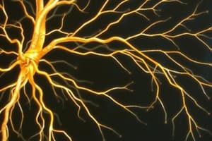
Nerve Structure
Nerve Structure
A cordlike organ of the PNS, composed of bundled peripheral axons surrounded by connective tissue layers.
Endoneurium
Endoneurium
Loose connective tissue surrounding individual axons in a nerve.
Perineurium
Perineurium
Signup and view all the flashcards
Epineurium
Epineurium
Signup and view all the flashcards
Sensory (Afferent) Nerve
Sensory (Afferent) Nerve
Signup and view all the flashcards
Motor (Efferent) Nerve
Motor (Efferent) Nerve
Signup and view all the flashcards
Mixed Nerve
Mixed Nerve
Signup and view all the flashcards
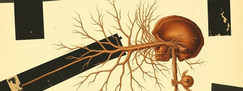
Cranial Nerves
Cranial Nerves
Signup and view all the flashcards
Olfactory Nerve (I)
Olfactory Nerve (I)
Signup and view all the flashcards
Optic Nerve (II)
Optic Nerve (II)
Signup and view all the flashcards
Oculomotor Nerve (III)
Oculomotor Nerve (III)
Signup and view all the flashcards
Trochlear Nerve (IV)
Trochlear Nerve (IV)
Signup and view all the flashcards
Trigeminal Nerve (V)
Trigeminal Nerve (V)
Signup and view all the flashcards
Abducens Nerve (VI)
Abducens Nerve (VI)
Signup and view all the flashcards
Facial Nerve (VII)
Facial Nerve (VII)
Signup and view all the flashcards
Vestibulocochlear Nerve (VIII)
Vestibulocochlear Nerve (VIII)
Signup and view all the flashcards
Glossopharyngeal Nerve (IX)
Glossopharyngeal Nerve (IX)
Signup and view all the flashcards
Vagus Nerve (X)
Vagus Nerve (X)
Signup and view all the flashcards
Accessory Nerve (XI)
Accessory Nerve (XI)
Signup and view all the flashcards
Hypoglossal Nerve (XII)
Hypoglossal Nerve (XII)
Signup and view all the flashcards
Spinal Nerves
Spinal Nerves
Signup and view all the flashcards
Study Notes
Peripheral Nervous System (PNS)
- The PNS includes all neural structures outside the brain and spinal cord.
- It includes sensory receptors, peripheral nerves, associated ganglia, and motor endings.
- The PNS provides links to and from the external environment.
Structure of a Nerve
- A nerve is a cordlike organ of the PNS, consisting of peripheral axons enclosed by connective tissue.
- Connective tissue coverings in a nerve:
- Endoneurium: loose connective tissue surrounding axons.
- Perineurium: coarse connective tissue bundling fibers into fascicles.
- Epineurium: tough fibrous sheath around a nerve.
Classification of Nerves
- Nerves are classified based on their function:
- Sensory or afferent: carry impulses to the CNS.
- Motor or efferent: carry impulses from the CNS.
- Mixed: carry sensory and motor fibers to and from the CNS; most common type of nerve.
Peripheral Nerves
- Mixed nerves carry both somatic and autonomic (visceral) impulses.
- Types of mixed nerves:
- Somatic afferent and somatic efferent: involved in voluntary movement and sensory information.
- Visceral afferent and visceral efferent: involved in involuntary bodily functions and sensory information from internal organs.
- Peripheral nerves originate from the brain or spinal column.
Cranial Nerves
- Twelve pairs of cranial nerves arise from the brain.
- Functions: sensory, motor, or both.
- Each nerve is identified by a number (I through XII) and a name.
- Four cranial nerves carry parasympathetic fibers that serve muscles and glands.
Cranial Nerve I: Olfactory
- Arises from the olfactory epithelium.
- Passes through the cribriform plate of the ethmoid bone.
- Fibers run through the olfactory bulb and terminate in the primary olfactory cortex.
- Sole function: carrying afferent impulses for the sense of smell.
Cranial Nerve II: Optic
- Arises from the retina of the eye.
- Optie nerves pass through the optic canals and converge at the optic chiasm, the X-shaped structure formed at the point below the brain where the two optic nerves cross over each other.
- They continue to the thalamus where they synapse.
- From there, the optic radiation fibers run to the visual cortex.
- Sole function: carrying afferent impulses for vision.
Cranial Nerve III: Oculomotor
- Fibers extend from the ventral midbrain, pass through the superior orbital fissure, and go to the extrinsic eye muscles.
- Functions:
- Raising the eyelid.
- Directing the eyeball.
- Constriction of the iris.
- Controlling lens shape.
- Parasympathetic cell bodies are in the ciliary ganglia.
Cranial Nerve IV: Trochlear
- Fibers emerge from the dorsal midbrain and enter the orbits via the superior orbital fissures; innervate the superior oblique muscle.
- Primarily a motor nerve that directs the eyeball.
Cranial Nerve V: Trigeminal
- Composed of three divisions: ophthalmic (V1), maxillary (V2), and mandibular (V3).
- Fibers run from the face to the pons via the:
- Superior orbital fissure (V1).
- Foramen rotundum (V2).
- Foramen ovale (V3).
- Conveying sensory impulses from various areas of the face (V1) and (V2), and supplies motor fibers (V3) for mastication.
Cranial Nerve VI: Abducens
- Fibers leave the inferior pons and enter the orbit via the superior orbital fissure.
- Primarily a motor nerve innervating the lateral rectus muscle, which allows for outward movement of the eye.
Cranial Nerve VII: Facial
- Fibers leave the pons, travel through the internal acoustic meatus, and emerge through the stylomastoid foramen to the lateral aspect of the face.
- Mixed nerve with five major branches.
- Motor functions:
- Facial expression.
- Transmittal of autonomic impulses to lacrimal and salivary glands.
- Sensory function: taste from the anterior two-thirds of the tongue.
Cranial Nerve VIII: Vestibulocochlear
- Fibers arise from the hearing and equilibrium apparatus of the inner ear, pass through the internal acoustic meatus, and enter the brainstem at the pons-medulla border.
- Two divisions: cochlear (hearing) and vestibular (balance).
- Solely sensory: equilibrium and hearing.
Cranial Nerve IX: Glossopharyngeal
- Fibers emerge from the medulla, leave the skull via the jugular foramen, and run to the throat.
- Mixed nerve with motor and sensory functions.
- Motor:
- Innervates part of the tongue and pharynx.
- Provides motor fibers to the parotid salivary gland.
- Sensory:
- Fibers conduct taste and general sensory impulses from the tongue and pharynx.
Cranial Nerve X: Vagus
- The only cranial nerve that extends beyond the head and neck.
- Fibers emerge from the medulla via the jugular foramen.
- Mixed nerve.
- Most motor fibers are parasympathetic fibers to the heart, lungs, and visceral organs.
- Sensory function: taste.
Cranial Nerve XI: Accessory
- Formed from a cranial root emerging from the medulla and a spinal root arising from the superior region of the spinal cord.
- The spinal root passes upward into the cranium via the foramen magnum.
- Leaves the cranium via the jugular foramen.
- Primarily a motor nerve:
- Supplies fibers to the larynx, pharynx, and soft palate.
- Innervates the trapezius and sternocleidomastoid, which move the head and neck.
Cranial Nerve XII: Hypoglossal
- Fibers arise from the medulla and exit the skull via the hypoglossal canal.
- Innervates both extrinsic and intrinsic muscles of the tongue, which contribute to swallowing and speech.
Peripheral Nervous System (PNS)
- The PNS includes all nerve structures outside of the brain and spinal cord.
- It is composed of sensory receptors, peripheral nerves, associated ganglia, and motor endings.
- The PNS transmits information to and from the external environment.
Structure of a Nerve
- A nerve is a cord-like organ of the PNS composed of peripheral axons enclosed in connective tissue.
- Connective tissue coverings include the endoneurium (surrounding axons), perineurium (bundling fibers into fascicles), and epineurium (tough fibrous sheath around the nerve).
Classification of Nerves
- Nerves are classified based on their functions:
- Sensory (afferent): Carry impulses to the CNS.
- Motor (efferent): Carry impulses from the CNS.
- Mixed: Carry both sensory and motor fibers, the most common type.
Peripheral Nerves
- Peripheral nerves are mixed nerves that carry both somatic and autonomic (visceral) impulses. There are four types: somatic afferent, somatic efferent, visceral afferent, and visceral efferent.
- Peripheral nerves originate from the brain or spinal column.
Cranial Nerves
- There are twelve pairs of cranial nerves arising from the brain.
- Each cranial nerve has a unique number (I-XII) and name.
- These nerves can have sensory, motor, or both sensory and motor functions.
- Four of the cranial nerves carry parasympathetic fibers that serve muscles and glands.
Cranial Nerve I: Olfactory
- This nerve originates from the olfactory epithelium and passes through the cribriform plate of the ethmoid bone.
- Its fibers travel through the olfactory bulb and terminate in the primary olfactory cortex.
- This nerve solely carries afferent impulses for the sense of smell.
Cranial Nerve II: Optic
- This nerve originates from the retina of the eye.
- The optic nerves pass through the optic canals and converge at the optic chiasm, where they cross over each other.
- They continue to the thalamus where they synapse, and then the optic radiation fibers run to the visual cortex.
- This nerve is solely responsible for carrying afferent impulses for vision.
Cranial Nerve III: Oculomotor
- Fibers extend from the ventral midbrain, passing through the superior orbital fissure to the extrinsic eye muscles.
- This nerve controls the raising of the eyelid, directing the eyeball, constricting the iris, and adjusting lens shape.
- Parasympathetic cell bodies are located in the ciliary ganglia.
Cranial Nerve IV: Trochlear
- Fibers emerge from the dorsal midbrain and enter the orbits via the superior orbital fissures, innervating the superior oblique muscle.
- Primarily a motor nerve that directs the eyeball.
Cranial Nerve V: Trigeminal
- Composed of three divisions: ophthalmic (V1), maxillary (V2), and mandibular (V3).
- Fibers run from the face to the pons via the superior orbital fissure (V1), the foramen rotundum (V2), and the foramen ovale (V3).
- This nerve is responsible for conveying sensory impulses from different areas of the face (V1, V2) and provides motor fibers (V3) for mastication.
Cranial Nerve VI: Abducens
- Fibers leave the inferior pons and enter the orbit via the superior orbital fissure.
- This is primarily a motor nerve that innervates the lateral rectus muscle.
Cranial Nerve VII: Facial
- Fibers leave the pons, travel through the internal acoustic meatus, and emerge through the stylomastoid foramen to the lateral aspect of the face.
- This nerve is a mixed nerve with five major branches.
- Motor functions include facial expression and transmitting autonomic impulses to lacrimal and salivary glands.
- Sensory function includes taste from the anterior two-thirds of the tongue.
Cranial Nerve VIII: Vestibulocochlear
- Fibers arise from the hearing and equilibrium apparatus of the inner ear, pass through the internal acoustic meatus, and enter the brainstem at the pons-medulla border.
- It has two divisions: cochlear (hearing) and vestibular (balance).
- Its sole functions are sensory, responsible for equilibrium and hearing.
Cranial Nerve IX: Glossopharyngeal
- Fibers emerge from the medulla, leave the skull via the jugular foramen, and run to the throat.
- This nerve is a mixed nerve with motor and sensory functions.
- Motor functions include innervating part of the tongue and pharynx, and providing motor fibers to the parotid salivary gland.
- Sensory functions include conducting taste and general sensory impulses from the tongue and pharynx.
Cranial Nerve X: Vagus
- This is the only cranial nerve that extends beyond the head and neck.
- Fibers emerge from the medulla via the jugular foramen.
- This nerve is a mixed nerve.
- Most motor fibers are parasympathetic fibers to the heart, lungs, and visceral organs.
- Its sensory function is in taste.
Cranial Nerve XI: Accessory
- This nerve is formed from a cranial root emerging from the medulla and a spinal root arising from the superior region of the spinal cord.
- The spinal root passes upward into the cranium via the foramen magnum.
- The accessory nerve leaves the cranium via the jugular foramen.
- Primarily a motor nerve, it supplies fibers to the larynx, pharynx, and soft palate.
- It also innervates the trapezius and sternocleidomastoid muscles, which move the head and neck.
Cranial Nerve XII: Hypoglossal
- Fibers arise from the medulla and exit the skull via the hypoglossal canal.
- This nerve innervates the extrinsic and intrinsic muscles of the tongue, important for swallowing and speech.
Spinal Nerves
- Thirty-one pairs of mixed nerves arise from the spinal cord and supply all parts of the body except the head
- They are named according to their point of issue
- 8 cervical (C1-C8)
- 12 thoracic (T1-T12)
- 5 Lumbar (L1-L5)
- 5 Sacral (S1-S5)
- 1 Coccygeal (C0)
- Each spinal nerve connects to the spinal cord via two medial roots
- Each root forms a series of rootlets that attach to the spinal cord
- Ventral roots arise from the anterior horn and contain motor (efferent) fibers
- Dorsal roots arise from sensory neurons in the dorsal root ganglion and contain sensory (afferent) fibers
Nerve Plexuses
- All ventral rami except T2-T12 form interlacing nerve networks called plexuses
- Plexuses are found in the cervical, brachial, lumbar, and sacral regions
- Each resulting branch of a plexus contains fibers from several spinal nerves
- Fibers travel to the periphery via several different routes
- Each muscle receives a nerve supply from more than one spinal nerve
- Damage to one spinal segment cannot completely paralyze a muscle
Spinal Nerve Innervation
- The back is innervated by dorsal rami via several branches
- The thorax is innervated by ventral rami T1-T12 as intercostal nerves
- Intercostal nerves supply muscles of the ribs, anterolateral thorax, and abdominal wall
Cervical Plexus
- The cervical plexus is formed by ventral rami of C1-C4
- Most branches are cutaneous nerves of the neck, ear, back of head, and shoulders
- The most important nerve of this plexus is the phrenic nerve
- The phrenic nerve is the major motor and sensory nerve of the diaphragm
Brachial Plexus
- Formed by C5-C8 and T1 (C4 and T2 may also contribute to this plexus)
- It gives rise to the nerves that innervate the upper limb
- There are four major branches of this plexus
- Roots – five ventral rami (C5-T1)
- Trunks – upper, middle, and lower, which form divisions
- Divisions – anterior and posterior serve the front and back of the limb
- Cords – lateral, medial, and posterior fiber bundles
Brachial Plexus: Nerves
- Axillary – innervates the deltoid and teres minor
- Musculocutaneous – sends fibers to the biceps brachii and brachialis
- Median – branches to most of the flexor muscles of arm
- Ulnar – supplies the flexor carpi ulnaris and part of the flexor digitorum profundus
- Radial – innervates essentially all extensor muscles
Lumbar Plexus
- Arises from L1-L4 and innervates the thigh, abdominal wall, and related muscles
- The major nerves are the femoral and the obturator
Sacral Plexus
- Arises from L4-S4 and serves the buttock, lower limb, pelvic structures, and the related sacral areas
- Major nerve is the sciatic, the longest and thickest nerve of the body
- The sciatic is actually composed of two nerves: the tibial and the common fibular nerves
Dermatomes
- A dermatome is the area of skin innervated by the cutaneous branches of a single spinal nerve
- All spinal nerves except C1 participate in dermatomes
Studying That Suits You
Use AI to generate personalized quizzes and flashcards to suit your learning preferences.