Podcast
Questions and Answers
Which of the following are components of the periodontium?
Which of the following are components of the periodontium?
- Periodontal ligament (correct)
- Gingival tissue (correct)
- Cementum (correct)
- Pulp
- Alveolar bone (correct)
A normal, intact periodontium will always show bone loss in furcation areas.
A normal, intact periodontium will always show bone loss in furcation areas.
False (B)
What is the average distance, in millimeters, between the cementoenamel junction (CEJ) and the alveolar bone crest (AC) in a healthy individual?
What is the average distance, in millimeters, between the cementoenamel junction (CEJ) and the alveolar bone crest (AC) in a healthy individual?
2
The inter-dental crestal bone is not directly connected to the lamina dura of adjacent teeth.
The inter-dental crestal bone is not directly connected to the lamina dura of adjacent teeth.
Periodontal diseases are a set of conditions characterized by an inflammatory host response that primarily affects the hard tissues around the teeth.
Periodontal diseases are a set of conditions characterized by an inflammatory host response that primarily affects the hard tissues around the teeth.
Once periodontitis has developed, alveolar bone loss can be reversed without intervention.
Once periodontitis has developed, alveolar bone loss can be reversed without intervention.
Radiographs can solely determine the presence of clinical periodontal health on a reduced periodontium.
Radiographs can solely determine the presence of clinical periodontal health on a reduced periodontium.
Which of the following clinical examinations can be used to assess the extent of periodontal disease?
Which of the following clinical examinations can be used to assess the extent of periodontal disease?
2D radiographs can provide a complete view of the bucco-lingual aspects of the interproximal bone.
2D radiographs can provide a complete view of the bucco-lingual aspects of the interproximal bone.
Incorporating digital image receivers, such as phosphor storage plates or solid-state detectors, is not necessary for minimizing radiation exposure.
Incorporating digital image receivers, such as phosphor storage plates or solid-state detectors, is not necessary for minimizing radiation exposure.
For patients with generalized periodontal pockets measuring less than 5 mm in depth, bitewing radiographs are not recommended.
For patients with generalized periodontal pockets measuring less than 5 mm in depth, bitewing radiographs are not recommended.
The bisecting-angle technique for acquiring periapical radiographs is preferred over the paralleling technique due to its superior geometric configuration.
The bisecting-angle technique for acquiring periapical radiographs is preferred over the paralleling technique due to its superior geometric configuration.
Panoramic radiography is not a low-dose radiation technique.
Panoramic radiography is not a low-dose radiation technique.
What is the major limitation of panoramic radiography that affects the reliability of linear measurements?
What is the major limitation of panoramic radiography that affects the reliability of linear measurements?
The upward angulation of the X-ray beam used in panoramic radiography can cause horizontal overlap and distortion, making it difficult to visualize fine structures like the periodontal ligament space.
The upward angulation of the X-ray beam used in panoramic radiography can cause horizontal overlap and distortion, making it difficult to visualize fine structures like the periodontal ligament space.
What are the primary limitations of 2D radiographs in periodontal diagnosis?
What are the primary limitations of 2D radiographs in periodontal diagnosis?
Early periodontal lesions are easily diagnosed accurately using radiographic imaging.
Early periodontal lesions are easily diagnosed accurately using radiographic imaging.
Radiographic images provide valuable information regarding the detection of periodontal pockets and tooth mobility, crucial aspects for periodontal diagnosis.
Radiographic images provide valuable information regarding the detection of periodontal pockets and tooth mobility, crucial aspects for periodontal diagnosis.
Changes in the internal aspect of the alveolar bone can only reflect a reduction in bone structure/trabeculation.
Changes in the internal aspect of the alveolar bone can only reflect a reduction in bone structure/trabeculation.
Horizontal bone loss is the most common pattern of bone loss in periodontal disease.
Horizontal bone loss is the most common pattern of bone loss in periodontal disease.
Vertical bone loss occurs solely due to a lack of horizontal bone loss.
Vertical bone loss occurs solely due to a lack of horizontal bone loss.
Vertical bone loss always appears as three walled defects.
Vertical bone loss always appears as three walled defects.
It is always possible to recognize vertical defects on a radiograph.
It is always possible to recognize vertical defects on a radiograph.
Periodontal furcation bone loss occurs only in single-rooted teeth.
Periodontal furcation bone loss occurs only in single-rooted teeth.
Peri-implant health is characterized by the absence of visual signs of inflammation and bleeding on gentle probing.
Peri-implant health is characterized by the absence of visual signs of inflammation and bleeding on gentle probing.
Peri-implant diseases are classified as either peri-implant mucositis or peri-implantitis, but not both.
Peri-implant diseases are classified as either peri-implant mucositis or peri-implantitis, but not both.
Peri-implant mucositis is an inflammatory condition that involves the loss of bone around the dental implant.
Peri-implant mucositis is an inflammatory condition that involves the loss of bone around the dental implant.
Peri-implant mucositis is irreversible and cannot be treated.
Peri-implant mucositis is irreversible and cannot be treated.
The thickness of keratinized mucosa around dental implants does not play a role in the development of peri-implant mucositis.
The thickness of keratinized mucosa around dental implants does not play a role in the development of peri-implant mucositis.
Periodontal abscesses are always associated with pain and tenderness in the gingiva.
Periodontal abscesses are always associated with pain and tenderness in the gingiva.
The most common finding in a periodontal abscess is a suppurative discharge on probing or sampling.
The most common finding in a periodontal abscess is a suppurative discharge on probing or sampling.
Radiographically, periodontal abscesses typically appear well-defined around the root of the affected tooth.
Radiographically, periodontal abscesses typically appear well-defined around the root of the affected tooth.
Endodontic and periodontal lesions are always distinct and never overlap.
Endodontic and periodontal lesions are always distinct and never overlap.
The presence of a sinus tract is always a sign of an endodontic lesion.
The presence of a sinus tract is always a sign of an endodontic lesion.
Radiographs are the primary method for diagnosing endodontic and periodontal lesions.
Radiographs are the primary method for diagnosing endodontic and periodontal lesions.
What are the four components of the periodontium?
What are the four components of the periodontium?
A normal, intact periodontium should show evidence of bone loss in furcation areas.
A normal, intact periodontium should show evidence of bone loss in furcation areas.
What is the average distance, in millimeters, from the most coronal portion of the alveolar bone crest (AC) to the cementoenamel junction (CEJ) in a healthy individual?
What is the average distance, in millimeters, from the most coronal portion of the alveolar bone crest (AC) to the cementoenamel junction (CEJ) in a healthy individual?
The inter-dental crestal bone is continuous with the lamina dura of the adjacent teeth in a healthy periodontium.
The inter-dental crestal bone is continuous with the lamina dura of the adjacent teeth in a healthy periodontium.
What is the name of the inflammatory host response that characterizes periodontal diseases?
What is the name of the inflammatory host response that characterizes periodontal diseases?
Clinical periodontal health can be determined using radiographs alone.
Clinical periodontal health can be determined using radiographs alone.
Which of these is NOT a clinical examination technique used in the assessment of periodontal conditions?
Which of these is NOT a clinical examination technique used in the assessment of periodontal conditions?
What type of radiographs help in evaluating the extent of bone loss around teeth, crucial for diagnosing the severity of periodontal disease?
What type of radiographs help in evaluating the extent of bone loss around teeth, crucial for diagnosing the severity of periodontal disease?
Which of these is NOT a benefit of using digital radiography in periodontal assessment?
Which of these is NOT a benefit of using digital radiography in periodontal assessment?
Which type of radiograph is considered most commonly used for assessing generalized periodontal pockets measuring < 5 mm in depth?
Which type of radiograph is considered most commonly used for assessing generalized periodontal pockets measuring < 5 mm in depth?
The paralleling technique for acquiring periapical radiographs is generally considered less accurate than the bisecting-angle technique.
The paralleling technique for acquiring periapical radiographs is generally considered less accurate than the bisecting-angle technique.
What is the main advantage of using panoramic radiographs for periodontal assessment?
What is the main advantage of using panoramic radiographs for periodontal assessment?
Panoramic radiography is the preferred imaging modality for periodontal assessment, especially when complemented with intraoral radiographs.
Panoramic radiography is the preferred imaging modality for periodontal assessment, especially when complemented with intraoral radiographs.
Which of the following is a limitation of using 2D projections for periodontal assessment?
Which of the following is a limitation of using 2D projections for periodontal assessment?
Radiographic assessment can provide a complete view of the bucco-lingual aspects of the interproximal bone.
Radiographic assessment can provide a complete view of the bucco-lingual aspects of the interproximal bone.
Radiographic images are always reliable in accurately assessing early stages of periodontal disease.
Radiographic images are always reliable in accurately assessing early stages of periodontal disease.
Which of these is NOT a limitation of radiographs, as they relate to periodontal diagnosis?
Which of these is NOT a limitation of radiographs, as they relate to periodontal diagnosis?
Which of the following is an initial radiographic change seen in periodontitis?
Which of the following is an initial radiographic change seen in periodontitis?
In severe cases of periodontal disease, the posterior regions of the alveolar bone crest may appear rounded off with an irregular and diffuse border between the lamina dura and alveolar crest.
In severe cases of periodontal disease, the posterior regions of the alveolar bone crest may appear rounded off with an irregular and diffuse border between the lamina dura and alveolar crest.
Changes to the internal aspect of the alveolar bone can reflect which of these conditions? (Select all that apply)
Changes to the internal aspect of the alveolar bone can reflect which of these conditions? (Select all that apply)
What is the most common pattern of bone loss in periodontal disease?
What is the most common pattern of bone loss in periodontal disease?
Vertical bone loss indicates the presence of vertical or angular osseous defects.
Vertical bone loss indicates the presence of vertical or angular osseous defects.
What is the term used to describe a vertical defect that is surrounded by three bony walls?
What is the term used to describe a vertical defect that is surrounded by three bony walls?
Vertical defects are always easily recognizable on a radiograph.
Vertical defects are always easily recognizable on a radiograph.
What type of bone loss occurs in the furcation area of multi-rooted teeth?
What type of bone loss occurs in the furcation area of multi-rooted teeth?
Match the grade of furcation involvement with its description:
Match the grade of furcation involvement with its description:
Peri-implant diseases are broadly divided into which two categories?
Peri-implant diseases are broadly divided into which two categories?
Peri-implant mucositis is a reversible condition.
Peri-implant mucositis is a reversible condition.
What is the minimum thickness of keratinized mucosa that may contribute to the development of peri-implant mucositis?
What is the minimum thickness of keratinized mucosa that may contribute to the development of peri-implant mucositis?
Peri-implant mucositis is primarily caused by plaque-associated inflammation.
Peri-implant mucositis is primarily caused by plaque-associated inflammation.
Which of the following is NOT a potential cause of peri-implant mucositis?
Which of the following is NOT a potential cause of peri-implant mucositis?
What are the common clinical features of a periodontal abscess? (Select all that apply)
What are the common clinical features of a periodontal abscess? (Select all that apply)
What is a common clinical finding associated with periodontal abscesses?
What is a common clinical finding associated with periodontal abscesses?
Which of the following can contribute to the formation of a periodontal abscess? (Select all that apply)
Which of the following can contribute to the formation of a periodontal abscess? (Select all that apply)
Severe anatomic alterations, such as invaginated teeth or dens evaginatus, can contribute to the formation of periodontal abscesses.
Severe anatomic alterations, such as invaginated teeth or dens evaginatus, can contribute to the formation of periodontal abscesses.
Vertical root fractures or cracked tooth syndrome extending through the root can lead to periodontal abscesses.
Vertical root fractures or cracked tooth syndrome extending through the root can lead to periodontal abscesses.
What is the radiographic appearance of a periodontal abscess?
What is the radiographic appearance of a periodontal abscess?
Match the type of lesion with its corresponding radiographic finding for periodontal disease:
Match the type of lesion with its corresponding radiographic finding for periodontal disease:
Flashcards
Create flashcards
Create flashcards
Develop cards for learning
10 flashcards
10 flashcards
Ten cards for learning
Term
Term
Key word or phrase
Definition
Definition
Signup and view all the flashcards
Hint
Hint
Signup and view all the flashcards
Memory tip
Memory tip
Signup and view all the flashcards
Key concepts
Key concepts
Signup and view all the flashcards
Simple explanations
Simple explanations
Signup and view all the flashcards
Skip junk
Skip junk
Signup and view all the flashcards
Flashcard format
Flashcard format
Signup and view all the flashcards
Periodontium
Periodontium
Signup and view all the flashcards
Lamina Dura
Lamina Dura
Signup and view all the flashcards
Alveolar Bone Crest
Alveolar Bone Crest
Signup and view all the flashcards
Cementoenamel Junction (CEJ)
Cementoenamel Junction (CEJ)
Signup and view all the flashcards
Periodontal Ligament (PDL)
Periodontal Ligament (PDL)
Signup and view all the flashcards
Interdental Crest
Interdental Crest
Signup and view all the flashcards
What does a healthy PDL space look like on an x-ray?
What does a healthy PDL space look like on an x-ray?
Signup and view all the flashcards
Periodontal Disease
Periodontal Disease
Signup and view all the flashcards
Periodontitis
Periodontitis
Signup and view all the flashcards
What is the key symptom of periodontitis?
What is the key symptom of periodontitis?
Signup and view all the flashcards
Clinical Attachment Loss (CAL)
Clinical Attachment Loss (CAL)
Signup and view all the flashcards
How is CAL measured?
How is CAL measured?
Signup and view all the flashcards
Gingival Recession
Gingival Recession
Signup and view all the flashcards
What can cause gingival recession?
What can cause gingival recession?
Signup and view all the flashcards
Attached Gingiva
Attached Gingiva
Signup and view all the flashcards
Tooth Mobility
Tooth Mobility
Signup and view all the flashcards
What can cause tooth mobility?
What can cause tooth mobility?
Signup and view all the flashcards
Role of 2D Radiographs in Periodontal Disease Diagnosis
Role of 2D Radiographs in Periodontal Disease Diagnosis
Signup and view all the flashcards
What can 2D radiographs reveal about the bone?
What can 2D radiographs reveal about the bone?
Signup and view all the flashcards
What hidden problems can 2D radiographs reveal?
What hidden problems can 2D radiographs reveal?
Signup and view all the flashcards
How do 2D radiographs aid in treatment planning?
How do 2D radiographs aid in treatment planning?
Signup and view all the flashcards
How can 2D radiographs monitor progress over time?
How can 2D radiographs monitor progress over time?
Signup and view all the flashcards
Intraoral Radiographs
Intraoral Radiographs
Signup and view all the flashcards
What are the advantages of intraoral radiographs?
What are the advantages of intraoral radiographs?
Signup and view all the flashcards
Periapical Radiograph
Periapical Radiograph
Signup and view all the flashcards
Bitewing Radiograph
Bitewing Radiograph
Signup and view all the flashcards
What are phosphor storage plates (PSP) and solid-state detectors?
What are phosphor storage plates (PSP) and solid-state detectors?
Signup and view all the flashcards
Rectangular Collimator
Rectangular Collimator
Signup and view all the flashcards
What is the benefit of rectangular collimators?
What is the benefit of rectangular collimators?
Signup and view all the flashcards
Study Notes
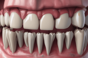
Periodontium Components
- The periodontium comprises cementum, periodontal ligament (PDL), alveolar bone, and gingival tissue.
Radiographic Features of Intact Periodontium
- Intact lamina dura, both laterally and at the alveolar crest.
- Absence of bone loss in furcation areas.
- Alveolar bone crest (AC) to cementoenamel junction (CEJ) distance averaging 2mm, though ranging from 1.0 to 3.0mm in healthy individuals.
- Continuous inter-dental crestal bone with adjacent teeth lamina dura.
- Thin, even width of the mesial and distal periodontal ligament spaces.
- Double PDL space appearance due to tooth shape.
Periodontal Diseases
- Inflammatory host response in periodontal tissues.
- Localized or generalized alterations in soft tissues.
- Loss of supporting bone.
- Potential for tooth loss.
- Periodontitis, by definition, involves alveolar bone loss due to inflammation.
Clinical Examination in Periodontal Assessment
- Increased periodontal probing.
- Bleeding sites identification.
- Gingival recession measurement.
- Tooth mobility assessment.
- Clinical attachment loss determination.
- Attached gingiva evaluation.
Role of 2D Imaging in Periodontal Disease
- Radiographs assess bone levels around teeth for diagnosing periodontal disease severity.
Detection of Pathology via Radiographs
- Reveal hidden issues like subgingival calculus and root fractures.
- Aid treatment planning by providing detailed bone and tooth structure information.
- Monitor disease progression and treatment effectiveness.
Anatomical Appearance Assessment
- Bone quantity assessment.
- Alveolar crests evaluation.
- Furcation areas analysis.
- Periodontal ligament space measurement.
- Root length and crown-to-root ratio assessment.
- Maxillary sinus positioning relative to periodontal deformities.
Local Irritating Factors
- Calculus identification.
- Poorly contoured restorations.
- Overextended restorations and open interproximal contacts.
Pathologic Considerations
- Caries assessment
- Periapical lesion identification
- Root resorption evaluation
Radiographic Modalities
- Intraoral radiographs (periapical and bitewing) are frequently used.
- Radiographic recommendations for patients with pockets <5mm often include bitewing radiographs.
- Panoramic radiographs offer a comprehensive view of the dentition.
Limitations of 2D Radiographs
- 2D view of a 3D structure may hide bony details.
- Relationships between hard and soft tissues aren't fully displayed.
- Incomplete view of bucco-lingual aspects of the interproximal bone.
- Early lesions may be difficult to diagnose radiographically, as changes become obvious only after significant bone loss (>30%-50%).
- Tendency to underestimate bone loss in early stages and overestimate it in severe stages.
- No information on periodontal pockets or tooth mobility.
Initial Radiographic Changes in Periodontitis
- Erosion of interproximal alveolar bone crests.
- Anterior regions may exhibit blunting of alveolar crests and reduced alveolar bone height.
- Posterior regions often show rounded, irregular borders between lamina dura and alveolar crest.
Studying That Suits You
Use AI to generate personalized quizzes and flashcards to suit your learning preferences.