Podcast
Questions and Answers
What is the orientation of the heart's apex?
What is the orientation of the heart's apex?
- Pointing downwards and backwards
- Pointing upwards and backwards
- Pointing sideways
- Pointing downwards and forwards (correct)
In terms of anatomical positioning, which end of the heart is referred to as the base?
In terms of anatomical positioning, which end of the heart is referred to as the base?
- The bottom end that faces down
- The side facing the lungs
- The blunt end that faces backwards (correct)
- The pointed end that faces forwards
Which of the following best describes the shape of the heart as initially perceived?
Which of the following best describes the shape of the heart as initially perceived?
- Heart-shaped with the apex facing downwards (correct)
- A perfect sphere
- An elongated oval
- Heart-shaped with the apex facing upwards
What is the main function of the cardiac skeleton?
What is the main function of the cardiac skeleton?
Which layer is NOT part of the heart wall?
Which layer is NOT part of the heart wall?
Early in development, how was the heart originally positioned?
Early in development, how was the heart originally positioned?
What angle does the apex of the heart face relative to the body's midline?
What angle does the apex of the heart face relative to the body's midline?
Which statement correctly describes the consequences of the heart's orientation?
Which statement correctly describes the consequences of the heart's orientation?
What is the location of the apex of the heart?
What is the location of the apex of the heart?
What structure is primarily found at the thick end of the heart wedge?
What structure is primarily found at the thick end of the heart wedge?
Which surface of the heart is in contact with the diaphragm?
Which surface of the heart is in contact with the diaphragm?
Which margin of the heart is referred to as the obtuse margin?
Which margin of the heart is referred to as the obtuse margin?
What is the direction of contraction for the atrial muscles?
What is the direction of contraction for the atrial muscles?
What is the primary characteristic of the acute margin of the heart?
What is the primary characteristic of the acute margin of the heart?
During which phase do the atrioventricular valves open?
During which phase do the atrioventricular valves open?
What ensures the nearly complete emptying of blood from the ventricles?
What ensures the nearly complete emptying of blood from the ventricles?
Which side of the heart is primarily associated with the pulmonary surface?
Which side of the heart is primarily associated with the pulmonary surface?
What occurs during ventricular systole?
What occurs during ventricular systole?
The heart muscle fibers are primarily attached to what structure?
The heart muscle fibers are primarily attached to what structure?
What happens to the outflow valves when the AV valves are open?
What happens to the outflow valves when the AV valves are open?
What is the term for the relaxation phase of the ventricles?
What is the term for the relaxation phase of the ventricles?
What occurs to the heart's structure during the 4th week of intrauterine life?
What occurs to the heart's structure during the 4th week of intrauterine life?
Which statement best describes the position of the heart’s right and left sides after rotation?
Which statement best describes the position of the heart’s right and left sides after rotation?
What is the primary function of the right heart in the circulatory system?
What is the primary function of the right heart in the circulatory system?
Where does the left atrium receive blood from?
Where does the left atrium receive blood from?
Why do the names of the heart's parts reflect the fetal position?
Why do the names of the heart's parts reflect the fetal position?
In which area does the right coronary artery arise?
In which area does the right coronary artery arise?
How does blood flow from the atria to the ventricles?
How does blood flow from the atria to the ventricles?
What does the term 'pulmona' signify in Latin concerning the heart's function?
What does the term 'pulmona' signify in Latin concerning the heart's function?
What is the primary function of the fluid within the pericardial cavity?
What is the primary function of the fluid within the pericardial cavity?
Which layer of pericardium directly covers the heart's surface?
Which layer of pericardium directly covers the heart's surface?
What is the total volume of fluid typically found in the pericardial cavity?
What is the total volume of fluid typically found in the pericardial cavity?
Which structure acts as a barrier separating the heart chambers from the myocardium?
Which structure acts as a barrier separating the heart chambers from the myocardium?
Which layer is classified as the outermost layer of the pericardium?
Which layer is classified as the outermost layer of the pericardium?
What is the function of the epicardium?
What is the function of the epicardium?
Which layer is referred to as the serous pericardium?
Which layer is referred to as the serous pericardium?
What type of tissue is found beneath the visceral pericardium?
What type of tissue is found beneath the visceral pericardium?
Flashcards are hidden until you start studying
Study Notes
Pericardial Cavity and Layers
- The pericardial cavity lies between the parietal and visceral layers of the pericardium.
- Contains 10-50 mL of serous fluid, reducing friction from heartbeats.
- Principal function of the pericardium is to minimize friction during heart movement.
- Multiple layers of pericardium include the fibrous pericardium, parietal pericardium, and visceral pericardium (epicardium).
Heart Wall Structure
- The heart wall consists of several layers:
- Epicardium (visceral pericardium)
- Myocardium (thick cardiac muscle)
- Endocardium (thin connective tissue and epithelium lining heart chambers).
- The epicardium may contain varying amounts of fat known as epicardial fat.
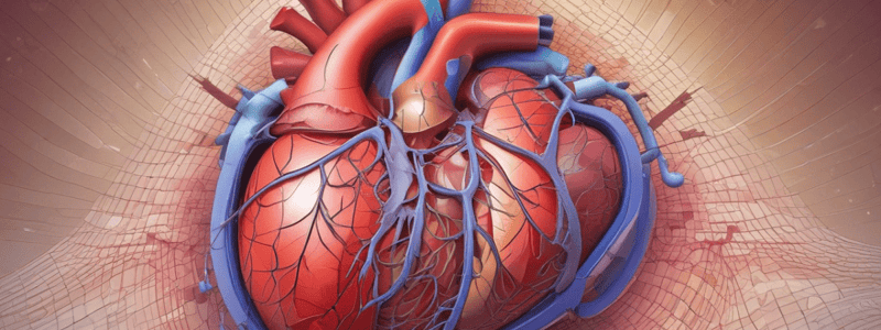
Orientation and Structure of the Heart
- The heart has a pointed end called the apex, which tilts downwards and forwards at a 70-degree angle.
- The base of the heart is blunt and faces backward.
- The anatomical position of the heart includes right and left sides, anterior and posterior surfaces.
Development and Rotation of the Heart
- The fetal heart positions with the apex pointing downwards and base upwards, but rotates to the left around the 4th week of intrauterine life.
- After rotation, the anterior surface becomes the superior surface; the right side becomes more anterior and the left side more posterior.
Heart Chambers
- Composed of four chambers:
- Right atrium and right ventricle (deoxygenated blood)
- Left atrium and left ventricle (oxygenated blood).
- Blood flows from atria to ventricles via gravity and contraction; ventricles pump blood into pulmonary and systemic circulation.
Cardiac Skeleton
- Provides support for heart muscle fibers and surrounds heart valves.
- Muscle fibers contract in opposite directions: atrial muscle downwards, ventricles upwards.
- Allows for effective contraction and maintains structural integrity.
Heart Valves
- Atrioventricular (AV) valves open during ventricular diastole (ventricles relaxed).
- Ventricles contract during ventricular systole, closing AV valves and opening outflow (semilunar) valves.
- Key valves include left (mitral) and right (tricuspid) atrioventricular valves, pulmonary and aortic semilunar valves.
Heart Orientation and Surface Features
- Heart resembles a wedge with the apex on the left side.
- Acute margin formed by the lower part of the right ventricle in contact with the diaphragm.
- The base, located at the thicker end of the wedge, is where atria receive blood.
Surface Features
- Anterior surface is superior, while the posterior surface (diaphragmatic surface) is inferior.
- Right atrium principally corresponds to the right pulmonary surface, and left ventricle to the left pulmonary surface.
- The obtuse margin of the heart is a feature of the left ventricle's curved side adjacent to the anterior surface.
Summary
- The structural complexity and arrangement of the heart ensure efficient blood flow and support vital cardiac functions, with distinct layers, chambers, orientations, and crucial roles of the cardiac skeleton and valves in circulation.
Studying That Suits You
Use AI to generate personalized quizzes and flashcards to suit your learning preferences.