Podcast
Questions and Answers
What is the primary role of pericardial fluid?
What is the primary role of pericardial fluid?
- To protect the heart from infections
- To supply nutrients to the heart
- To act as a lubricant for heart movement (correct)
- To regulate heart rate
What clinical condition is associated with inflammation of the pericardium?
What clinical condition is associated with inflammation of the pericardium?
- Pericarditis (correct)
- Myocardial infarction
- Cardiac tamponade
- Coronary artery disease
What symptom is most commonly associated with pericarditis?
What symptom is most commonly associated with pericarditis?
- Lightning pain in the arms
- Shortness of breath
- Continuous central chest pain (correct)
- Palpitations
What is a possible complication of pericardial effusion?
What is a possible complication of pericardial effusion?
Where is fluid aspiration performed to relieve cardiac tamponade?
Where is fluid aspiration performed to relieve cardiac tamponade?
Which chamber forms the anterior border of the heart?
Which chamber forms the anterior border of the heart?
What is the base of the heart primarily composed of?
What is the base of the heart primarily composed of?
What is the main function of the ventricles in the heart?
What is the main function of the ventricles in the heart?
Which structures form the right border of the heart?
Which structures form the right border of the heart?
What is the primary function of the portal vein?
What is the primary function of the portal vein?
Which lymphatic structure drains the right side of the upper body?
Which lymphatic structure drains the right side of the upper body?
What happens to lymph nodes during infections?
What happens to lymph nodes during infections?
Which of the following processes characterizes the function of the lymphatic system?
Which of the following processes characterizes the function of the lymphatic system?
What is the enlargement of the tonsils due to infection called?
What is the enlargement of the tonsils due to infection called?
What is the largest single mass of lymphoid tissue in the body?
What is the largest single mass of lymphoid tissue in the body?
Which chamber of the heart is responsible for receiving deoxygenated blood from the lower part of the body?
Which chamber of the heart is responsible for receiving deoxygenated blood from the lower part of the body?
What guards the opening between the right atrium and the right ventricle?
What guards the opening between the right atrium and the right ventricle?
Which structure is responsible for bringing oxygenated blood from the lungs to the left atrium?
Which structure is responsible for bringing oxygenated blood from the lungs to the left atrium?
What is the function of the left ventricle in the circulatory system?
What is the function of the left ventricle in the circulatory system?
What separates the right and left atria of the heart?
What separates the right and left atria of the heart?
Which chamber of the heart has the thickest walls?
Which chamber of the heart has the thickest walls?
What is the function of veins in the circulatory system?
What is the function of veins in the circulatory system?
What type of valves are located at the opening of the pulmonary artery?
What type of valves are located at the opening of the pulmonary artery?
Which structure is the largest artery in the body?
Which structure is the largest artery in the body?
Which veins bring blood from the upper part of the body to the right atrium?
Which veins bring blood from the upper part of the body to the right atrium?
Where does the ascending aorta originate?
Where does the ascending aorta originate?
What is the role of the bicuspid valve?
What is the role of the bicuspid valve?
What prevents the backflow of blood in veins?
What prevents the backflow of blood in veins?
What does the pulmonary trunk convey?
What does the pulmonary trunk convey?
Which feature helps separate the right and left ventricles?
Which feature helps separate the right and left ventricles?
What branches from the descending thoracic aorta?
What branches from the descending thoracic aorta?
Which part of the aorta lies behind the manubrium sterni?
Which part of the aorta lies behind the manubrium sterni?
What is the continuation of the subclavian artery in the arm?
What is the continuation of the subclavian artery in the arm?
What does the systemic circulation involve?
What does the systemic circulation involve?
Which artery is located at the back of the knee?
Which artery is located at the back of the knee?
What initiates the connection between the heart and the aorta?
What initiates the connection between the heart and the aorta?
Which vessel is primarily used to measure blood pressure?
Which vessel is primarily used to measure blood pressure?
Which of the following is NOT a part of the descending aorta?
Which of the following is NOT a part of the descending aorta?
Where is the pulse typically taken in the upper limb?
Where is the pulse typically taken in the upper limb?
What does the internal iliac artery primarily supply?
What does the internal iliac artery primarily supply?
Which vein collects blood from the upper half of the body?
Which vein collects blood from the upper half of the body?
Which vein is commonly used for intravenous injections in the upper limb?
Which vein is commonly used for intravenous injections in the upper limb?
What is the first artery branching from the external iliac artery as it enters the thigh?
What is the first artery branching from the external iliac artery as it enters the thigh?
From which vein can blood also be drawn besides the median cubital vein?
From which vein can blood also be drawn besides the median cubital vein?
What region does the inferior vena cava drain blood from?
What region does the inferior vena cava drain blood from?
Flashcards are hidden until you start studying
Study Notes
Pericardial Fluid and Pericarditis
- Pericardial fluid lubricates the heart, allowing for frictionless movement.
- Pericarditis is inflammation of the pericardium.
- Pericarditis can cause chest pain, friction rub between pericardium layers, and pericardial effusion.
- Pericardial effusion is an excess of fluid in the pericardial cavity, potentially compressing the atria and hindering heart filling during diastole.
- Cardiac tamponade is compression of the heart by excessive pericardial fluid, leading to reduced cardiac output.
- Fluid aspiration from the pericardial cavity is used to relieve cardiac tamponade, performed with a needle at the left infrasternal angle, through the cardiac notch of the left lung.
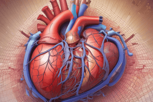
Heart Anatomy and Chambers
- The heart is a muscular organ that pumps blood throughout the body via the circulatory system.
- The heart contains four chambers:
- Two atria (inflow chambers).
- Two ventricles (outflow chambers).
- The heart's inferior (diaphragmatic) surface sits on the diaphragm, composed mainly of the left ventricle and a small portion of the right ventricle.
- The heart's anterior (sternocostal) surface faces inferiorly, resting on the diaphragm and separated from the heart's base by the coronary sinus.
- The heart's base (posterior surface) is quadrilateral, directed posteriorly, consisting of the left atrium, a small portion of the right atrium, and the proximal parts of the great veins (superior and inferior venae cavae and pulmonary veins).
Heart Borders
- The heart's right border is formed by the right atrium.
- The heart's left border is formed by the left auricle and left ventricle.
- The heart's inferior/lower border is formed by the right ventricle and a small portion of the left ventricle near the apex.
Heart Chambers and Valves
- The heart's atria are separated by the interatrial septum, and the ventricles are separated by the interventricular septum.
- The right atrium receives deoxygenated blood from the body through three veins:
- Superior vena cava (SVC): brings blood from the upper body.
- Inferior vena cava (IVC): brings blood from the lower body.
- Coronary sinus: brings blood from the heart.
- The right atrium empties into the right ventricle through an opening guarded by the tricuspid valve.
- The right ventricle receives blood from the right atrium via the tricuspid opening and pumps it to the lungs through the pulmonary trunk.
- The pulmonary artery opening is guarded by three semilunar valves known as the pulmonary valve.
- The left atrium receives oxygenated blood from the lungs via four pulmonary veins.
- The left atrium sends blood to the left ventricle through the bicuspid opening, guarded by the bicuspid or mitral valve.
- The left ventricle receives blood from the left atrium through the bicuspid opening and pumps it to the whole body through the aorta.
- The left ventricle's walls are thicker due to the pressure required to pump blood throughout the body.
- The aortic opening is guarded by the semilunar aortic valves.
Blood Circulation
- Blood in the body circulates in two circuits:
- Pulmonary circulation: between the lungs and the heart.
- Systemic circulation: between the heart and the rest of the body.
Arteries and Veins
- Arteries carry blood away from the heart.
- The pulmonary artery carries blood to the lungs.
- The aorta is the largest artery in the body, originating from the left ventricle.
- The aorta has three parts:
- Ascending aorta.
- Arch of aorta.
- Descending aorta:
- Thoracic part.
- Abdominal part.
Ascending Thoracic Aorta
- The ascending aorta originates at the base of the left ventricle, lying behind the right half of the sternum at the level of the sternal angle, where it continues as the arch of the aorta.
- The root of the ascending aorta has bulges known as the sinuses of the aorta.
- The ascending aorta's branches include:
- The right coronary artery.
- The left coronary artery.
Arch of the Aorta
- The arch of the aorta lies behind the manubrium sterni, arching upward, backward, and to the left in front of the trachea.
- It passes downward to the left of the trachea and continues as the descending thoracic aorta at the level of the sternal angle.
Descending Thoracic Aorta
- The descending thoracic aorta lies in the posterior mediastinum, beginning as a continuation of the arch of the aorta at the level of the lower border of the 4th thoracic vertebra on its left side (opposite the sternal angle).
- It passes behind the diaphragm (through the aortic opening) at the level of the 12th vertebra.
- Its branches include:
- Posterior intercostal arteries.
- Subcostal arteries.
- Pericardial, esophageal, and bronchial arteries.
Clinical Notes on Aorta
- Aneurysm and coarctation of the aorta are potential clinical conditions.
Pulmonary Trunk
- The pulmonary trunk carries deoxygenated blood from the right ventricle of the heart to the lungs.
- It runs upwards, backwards, and to the left.
- It is approximately 2 inches long.
- It terminates into the right and left pulmonary arteries.
Arteries of the Upper Limb
- The subclavian artery supplies the upper limb.
- The subclavian artery changes name according to its location:
- Axillary artery in the armpit.
- Brachial artery in the arm, which divides into:
- Radial artery.
- Ulnar artery.
Clinical Points on Upper Limb Arteries
- Blood pressure is measured from the brachial artery.
- Pulse is taken by pressing the radial artery at the lateral side of the wrist.
- Intravenous injections are commonly administered into the median cubital vein.
Arteries of the Lower Limb
- The external iliac artery continues into the thigh:
- Femoral artery in the front of the thigh.
- Popliteal artery at the back of the knee.
- Anterior tibial artery in the front of the leg.
- Posterior tibial artery at the back of the leg.
Clinical Point on Lower Limb Arteries
- The femoral artery pulse can be felt in the front of the thigh.
Arteries of the Pelvis
- The internal iliac artery supplies the pelvic organs, walls, and muscles.
Veins of the Body
- Veins carry blood back to the heart.
- Small veins join to form larger veins that enter the heart.
- Veins have valves to prevent blood backflow.
- The smallest veins are called venules.
Superior Vena Cava
- Veins from the upper half of the body join to form the Superior vena cava (SVC).
- Tributaries (veins draining into) the Superior vena cava include:
- Veins of the head and neck.
- Veins of the upper limb.
- Thoracic veins.
Veins of the Upper Limb
- Veins of the upper limb all join to form the subclavian veins, which enter the SVC.
- Superficial veins are visible on the dorsum of the hand, and one is present in front of the elbow called the median cubital vein.
Clinical Points on Upper Limb Veins
- Intravenous (IV) injections are often administered into the median cubital vein.
- Blood can be drawn from this vein.
- Injections can also be given into veins on the dorsum of the hand.
Inferior Vena Cava
- Veins from the lower half of the body drain into the Inferior vena cava (IVC).
- Tributaries (veins draining into) the inferior vena cava include:
- Veins of the abdomen.
- Veins of the pelvis.
- Veins of the lower limb.
Veins of the Abdomen
- Veins of the abdomen (renal veins, hepatic veins, etc.) open into the IVC.
- The abdomen contains a special vein called the portal vein, which drains the digestive tract and then enters the liver.
Veins of the Pelvis
- Veins of the pelvis form the internal iliac vein, which joins the external iliac vein to form the common iliac vein.
- The two common iliac veins join to form the inferior vena cava (IVC).
Portal Circulation
- Veins carrying nutrition-rich blood from the gastrointestinal tract enter the portal vein.
- The portal vein breaks up into capillaries again in the liver.
- Blood from the liver then flows to the IVC.
Lymphatic System
- Fluid that passes out of capillaries into tissues is called tissue fluid/lymph.
- Tissue fluid either enters veins or is carried by the lymphatic system.
Components of the Lymphatic System
- Lymphatic vessels: carry some tissue fluid (lymph) before it enters large veins.
- Lymphoid organs: include lymph nodes, thymus, tonsils, and spleen.
Thoracic Duct
- The thoracic duct originates in the abdomen and passes through the posterior part of the thorax to the neck, where it opens into the subclavian vein on the left side.
- It drains the left side of the upper half of the body and all of the lower body (both lower limbs, the entire pelvis and abdomen, and the left side of the thorax, left upper limb, and left side of the head and neck).
Right Lymphatic Duct
- The right lymphatic duct is smaller, draining the right side of the thorax, the right upper limb, and the right side of the head and neck.
- It opens into the right subclavian vein.
Lymph
- Tissue fluid that flows through lymphatic vessels is called lymph.
Lymph Nodes
- Lymph nodes enlarge in infections and cancers.
- Enlarged lymph nodes indicate the area drained by those nodes is affected.
Tonsils
- Tonsils are lymphoid organs present in the oropharynx.
- They prevent infectious agents from entering the respiratory and digestive tracts.
- Enlargement of the tonsils due to infection is called tonsillitis.
Spleen
- The spleen is reddish and the largest single mass of lymphoid tissue in the body.
Studying That Suits You
Use AI to generate personalized quizzes and flashcards to suit your learning preferences.