Podcast
Questions and Answers
What is a primary function of the iliac crest?
What is a primary function of the iliac crest?
- Protects the spinal cord from injury
- Attachment site for back muscles only
- Forms the anterior border of the pelvic inlet
- Key landmark and attachment site for muscles and ligaments (correct)
Which of the following ligaments is the strongest and most stabilizing ligament of the sacroiliac (SI) joint?
Which of the following ligaments is the strongest and most stabilizing ligament of the sacroiliac (SI) joint?
- Interosseous sacroiliac ligament (correct)
- Posterior sacroiliac ligament
- Superior pubic ligament
- Anterior sacroiliac ligament
The sacrospinous ligament transforms the greater sciatic notch into which foramen?
The sacrospinous ligament transforms the greater sciatic notch into which foramen?
- Obturator foramen
- Iliolumbar foramen
- Lesser sciatic foramen
- Greater sciatic foramen (correct)
Which of the following structures form the lateral border of the pelvic inlet?
Which of the following structures form the lateral border of the pelvic inlet?
Which characteristic of the female pelvic outlet is important for childbirth?
Which characteristic of the female pelvic outlet is important for childbirth?
Which condition can arise from compression or trauma involving the sacrospinous and sacrotuberous ligaments?
Which condition can arise from compression or trauma involving the sacrospinous and sacrotuberous ligaments?
What is the primary articulation involved in the lumbosacral joint?
What is the primary articulation involved in the lumbosacral joint?
What is the function of the nucleus pulposus within the intervertebral disc of the lumbosacral joint?
What is the function of the nucleus pulposus within the intervertebral disc of the lumbosacral joint?
Which type of joint combines to form the unique structure of the sacroiliac (SI) joint?
Which type of joint combines to form the unique structure of the sacroiliac (SI) joint?
Which ligaments directly stabilize the sacroiliac (SI) joint by limiting excessive movement in both directions?
Which ligaments directly stabilize the sacroiliac (SI) joint by limiting excessive movement in both directions?
Branches from which arteries supply blood to the pubic symphysis?
Branches from which arteries supply blood to the pubic symphysis?
What hormone influences the slight expansion of the pubic symphysis during pregnancy?
What hormone influences the slight expansion of the pubic symphysis during pregnancy?
Through which structure do the abdominal aorta and inferior vena cava pass as they enter the pelvic region?
Through which structure do the abdominal aorta and inferior vena cava pass as they enter the pelvic region?
Which structure passes through the lesser sciatic foramen to re-enter the perineum?
Which structure passes through the lesser sciatic foramen to re-enter the perineum?
Which of the following muscles is NOT part of the anatomical walls of the pelvic cavity?
Which of the following muscles is NOT part of the anatomical walls of the pelvic cavity?
The levator ani muscle group, which supports the pelvic organs, includes which of the following muscles?
The levator ani muscle group, which supports the pelvic organs, includes which of the following muscles?
Which muscle aids in fecal continence by forming a loop around the rectum?
Which muscle aids in fecal continence by forming a loop around the rectum?
Which muscle is responsible for supporting the prostate and maintaining urinary continence in males?
Which muscle is responsible for supporting the prostate and maintaining urinary continence in males?
Which of the following connective tissue structures provides stability and support to pelvic organs, bones, and blood vessels?
Which of the following connective tissue structures provides stability and support to pelvic organs, bones, and blood vessels?
Which ligament directly connects the sacrum to the ischial tuberosity?
Which ligament directly connects the sacrum to the ischial tuberosity?
Which of the following ligaments primarily supports the uterus?
Which of the following ligaments primarily supports the uterus?
The rectovesical pouch is located between which two structures in males?
The rectovesical pouch is located between which two structures in males?
In females, which peritoneal pouch is located between the uterus and the rectum?
In females, which peritoneal pouch is located between the uterus and the rectum?
Which of the following best describes the position of the bladder relative to the uterus in a sagittal section of the female pelvis?
Which of the following best describes the position of the bladder relative to the uterus in a sagittal section of the female pelvis?
In males, which structures does the deep perineal pouch contain?
In males, which structures does the deep perineal pouch contain?
Which of the following structures transitions into the anal canal as it passes through the pelvic diaphragm?
Which of the following structures transitions into the anal canal as it passes through the pelvic diaphragm?
From which artery does the internal pudendal artery originate?
From which artery does the internal pudendal artery originate?
What is the origin of the testicular arteries?
What is the origin of the testicular arteries?
Where does the right testicular vein drain?
Where does the right testicular vein drain?
Which component within the spermatic cord is responsible for cooling arterial blood?
Which component within the spermatic cord is responsible for cooling arterial blood?
Which cells within the seminiferous tubules are responsible for spermatogenesis?
Which cells within the seminiferous tubules are responsible for spermatogenesis?
What lies in the anterior relations of the seminal vesicle?
What lies in the anterior relations of the seminal vesicle?
Which component of prostatic secretion is an enzyme that liquefies coagulated semen?
Which component of prostatic secretion is an enzyme that liquefies coagulated semen?
Which zone of the prostate is most commonly affected by benign prostatic hyperplasia (BPH)?
Which zone of the prostate is most commonly affected by benign prostatic hyperplasia (BPH)?
Which of the following structures prevents compression of the urethra during an erection?
Which of the following structures prevents compression of the urethra during an erection?
From what structure is the cremaster muscle derived?
From what structure is the cremaster muscle derived?
Which nerves directly regulate the contraction of the ductus deferens during ejaculation?
Which nerves directly regulate the contraction of the ductus deferens during ejaculation?
In the uterine arteries, which artery has anastomotic connections?
In the uterine arteries, which artery has anastomotic connections?
Which of the following structures is found within the suspensory ligament of the ovary?
Which of the following structures is found within the suspensory ligament of the ovary?
Which feature distinguishes the male pelvis from the female pelvis in terms of general structure?
Which feature distinguishes the male pelvis from the female pelvis in terms of general structure?
During pregnancy, the hormone relaxin has the MOST direct effect on which pelvic structure?
During pregnancy, the hormone relaxin has the MOST direct effect on which pelvic structure?
Which movement is primarily facilitated by the flexion and extension capabilities of the lumbosacral joint?
Which movement is primarily facilitated by the flexion and extension capabilities of the lumbosacral joint?
What structural component of the intervertebral disc directly contributes to the lumbosacral joint's ability to withstand compressive forces during movement?
What structural component of the intervertebral disc directly contributes to the lumbosacral joint's ability to withstand compressive forces during movement?
Which combination of joint types BEST describes the unique structure of the sacroiliac joint?
Which combination of joint types BEST describes the unique structure of the sacroiliac joint?
What is the primary role of the obturator nerve as it passes through the obturator canal?
What is the primary role of the obturator nerve as it passes through the obturator canal?
Which structure passes through the greater sciatic foramen and is MOST at risk of compression due to its size and location?
Which structure passes through the greater sciatic foramen and is MOST at risk of compression due to its size and location?
Which set of structures passes through the lesser sciatic foramen?
Which set of structures passes through the lesser sciatic foramen?
Which anatomical feature is MOST important for fecal continence?
Which anatomical feature is MOST important for fecal continence?
Which primary function is uniquely attributed to the puboprostaticus muscle in males?
Which primary function is uniquely attributed to the puboprostaticus muscle in males?
Which action is performed by the iliococcygeus muscle?
Which action is performed by the iliococcygeus muscle?
Which type of issue might arise from damage to the sacrospinous ligament?
Which type of issue might arise from damage to the sacrospinous ligament?
How does the rectouterine pouch's anatomical position MOST significantly contribute to its clinical relevance?
How does the rectouterine pouch's anatomical position MOST significantly contribute to its clinical relevance?
Which anatomical relationship is MOST critical when assessing a transverse section of the female pelvis?
Which anatomical relationship is MOST critical when assessing a transverse section of the female pelvis?
Which structure runs through the deep perineal pouch in males?
Which structure runs through the deep perineal pouch in males?
The primary function of the internal pudendal artery to the perineum, external genitalia, and anal sphincters is carried by which major artery in the pelvis?
The primary function of the internal pudendal artery to the perineum, external genitalia, and anal sphincters is carried by which major artery in the pelvis?
What is the MOST significant functional adaptation observed in the female pelvis compared to the male pelvis?
What is the MOST significant functional adaptation observed in the female pelvis compared to the male pelvis?
Which physiological response is MOST directly facilitated by the hormone relaxin during pregnancy?
Which physiological response is MOST directly facilitated by the hormone relaxin during pregnancy?
Which component of the lumbosacral joint is MOST directly responsible for resisting compressive forces experienced during heavy lifting?
Which component of the lumbosacral joint is MOST directly responsible for resisting compressive forces experienced during heavy lifting?
Which statement BEST describes the dual structural nature of the sacroiliac (SI) joint and its functional implications?
Which statement BEST describes the dual structural nature of the sacroiliac (SI) joint and its functional implications?
Compression of which nerve as it exits the greater sciatic foramen is MOST likely to cause pain radiating down the posterior aspect of the lower limb?
Compression of which nerve as it exits the greater sciatic foramen is MOST likely to cause pain radiating down the posterior aspect of the lower limb?
What is the MAIN functional role of the pubococcygeus muscle during increased intra-abdominal pressure, such as during coughing or sneezing?
What is the MAIN functional role of the pubococcygeus muscle during increased intra-abdominal pressure, such as during coughing or sneezing?
Which anatomical feature is MOST important in preventing stress urinary incontinence in females?
Which anatomical feature is MOST important in preventing stress urinary incontinence in females?
In addition to the pudendal nerve, the structures that pass through the ischioanal fossa are supplied by which artery?
In addition to the pudendal nerve, the structures that pass through the ischioanal fossa are supplied by which artery?
Which statement BEST reflects the clinical significance of the rectouterine pouch relating to pelvic infections?
Which statement BEST reflects the clinical significance of the rectouterine pouch relating to pelvic infections?
Which artery lies within the suspensory ligament of the ovary, contributing to its clinical relevance?
Which artery lies within the suspensory ligament of the ovary, contributing to its clinical relevance?
What is the primary mechanism by which the bulbospongiosus muscle supports erectile function in males?
What is the primary mechanism by which the bulbospongiosus muscle supports erectile function in males?
Which muscle is the MOST likely site of origin for infections leading to ischioanal abscesses?
Which muscle is the MOST likely site of origin for infections leading to ischioanal abscesses?
What type of cells within the testes are DIRECTLY responsible for the synthesis of testosterone?
What type of cells within the testes are DIRECTLY responsible for the synthesis of testosterone?
What is indicated by an elevated PSA level in males?
What is indicated by an elevated PSA level in males?
The absence of what secretion in the seminal fluid would MOST directly impair sperm's function?
The absence of what secretion in the seminal fluid would MOST directly impair sperm's function?
What is the primary role of the tunica albuginea in the male testes?
What is the primary role of the tunica albuginea in the male testes?
An ultrasound examination of the scrotum reveals dilated veins within the spermatic cord. Which condition is MOST likely indicated by the examination findings?
An ultrasound examination of the scrotum reveals dilated veins within the spermatic cord. Which condition is MOST likely indicated by the examination findings?
How does the venous drainage differ between the right and left testicles, and what implications does this difference have?
How does the venous drainage differ between the right and left testicles, and what implications does this difference have?
Which statement BEST describes the functions of the seminal vesicles?
Which statement BEST describes the functions of the seminal vesicles?
Which anatomic relation is critical in gynecology in determining the location for uterine removal?
Which anatomic relation is critical in gynecology in determining the location for uterine removal?
Flashcards
What is the bony structure of the Pelvis?
What is the bony structure of the Pelvis?
Connects spine to lower limbs, transferring weight and providing muscle/ligament attachment.
Bones forming each hip bone
Bones forming each hip bone
Ilium, ischium, and pubis
Ilium
Ilium
Largest, uppermost part of the hip bone with the iliac crest and ASIS.
Ischium
Ischium
Signup and view all the flashcards
Pubis
Pubis
Signup and view all the flashcards
Sacrum
Sacrum
Signup and view all the flashcards
Coccyx
Coccyx
Signup and view all the flashcards
Key Sacroiliac Ligaments
Key Sacroiliac Ligaments
Signup and view all the flashcards
Key Sacrum Ligaments
Key Sacrum Ligaments
Signup and view all the flashcards
Key Pubic Ligaments
Key Pubic Ligaments
Signup and view all the flashcards
Iliolumbar Ligament
Iliolumbar Ligament
Signup and view all the flashcards
Pelvic Inlet
Pelvic Inlet
Signup and view all the flashcards
Pelvic Inlet Boundaries
Pelvic Inlet Boundaries
Signup and view all the flashcards
Pelvic Outlet
Pelvic Outlet
Signup and view all the flashcards
Lumbosacral Joint role
Lumbosacral Joint role
Signup and view all the flashcards
The Lumbosacral Joint
The Lumbosacral Joint
Signup and view all the flashcards
Intervertebral Disc
Intervertebral Disc
Signup and view all the flashcards
Facet Joints
Facet Joints
Signup and view all the flashcards
Iliolumbar Ligament Role
Iliolumbar Ligament Role
Signup and view all the flashcards
Sacroiliac Joint Function
Sacroiliac Joint Function
Signup and view all the flashcards
Sacroiliac Joint movement
Sacroiliac Joint movement
Signup and view all the flashcards
Sacroiliac Joint makeup
Sacroiliac Joint makeup
Signup and view all the flashcards
SI Joints transferring weight
SI Joints transferring weight
Signup and view all the flashcards
SI Joints ensuring balance
SI Joints ensuring balance
Signup and view all the flashcards
The Pubic Symphysis
The Pubic Symphysis
Signup and view all the flashcards
Study Notes
- The study notes below are a summary of the provided document.
Introduction
- This document is a study guide for the pelvis and perineum covering anatomy, structure, function, and clinical significance.
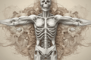
Skeletal Components of the Pelvis
- The pelvis connects the spine to the lower limbs acting as attachments for muscles and ligaments for locomotion.
- It is comprised of Hip Bone (os coxae or innominate bones), ilium, ischium, pubis, sacrum, and the coccyx.
- Hip bones are formed by the fusion of the ilium, ischium, and pubis during development.
- The iliac crest, a key landmark, includes the anterior superior iliac spine for muscle attachment and clinical assessments.
- The ischial tuberosity bears the body's weight when sitting and is a site for hamstring muscles to join.
- The two pubic bones meet at the inter cartilageinous pubic symphysis, it allows slight movement and it reinforced by ligaments.
- The sacrum is a triangular bone at the spine base, formed by five fused sections and it connects to hip bones via sacroiliac joints for weight equilibrium.
- The coccyx, or tailbone, has four fused vertebrae, and functions as muscle and ligament attachments of the pelvic floor.
- These bones makeup the pelvic girdle and work by supporting the body, protect pelvic structures, and promote movement.
Ligamentous Components of the Pelvis
- Ligaments are key stabilisers of the pelvis, promoting joint support and bone connection.
- Sacroiliac Ligaments stabilise the sacroiliac joints joining sacrum and ilium.
- The anterior Sacroiliac ligament assists in the front section of the sacroiliac joint.
- The posterior Sacroiliac ligament reinforces the rear of the joint.
- The interosseous Sacroiliac ligament is contained deep in with the joint, it is the most supportive ligament in that zone.
- The sacrospinous ligament extends out from the sacrum to ischial spine, it transforms major sciatic indentation to the greater sciatic opening, this is a passway for nerves and blood vessels.
- The sacrotuberous ligament stretches out from the sacrum to the ischial tuberosity, it helps to form the lesser sciatic foramen, it controls sacral revolution and reinforces the pelvis.
- The lumbar (L5) vertebrae is secured to the iliac crest, reinforcing the spinal linkage with the hips.
- Pubic Symphysis- the fibrocartilaginous joint located between the pubic bones is reinforced by public ligaments.
- The superior Pubic ligament strengthens the highest area of the pubic symphysis.
- The inferior Pubic ligament offers assistance across the lower borderline of the pubic symphysis.
Pelvic Inlet
- The pelvic inlet marks the border across greater with the abdominal viscera and lesser with the pelvic organs.
- Anterior border is the pubic crest.
- Lateral boundary by the pectineal line and also the arcuate line of the ilium.
- Posterior is the sacral point.
Pelvic Outlet
- The pelvic outlet makes up the inferior opening across the smallerpelvic area.
- It offers pathways, these include the rectum, vaginal passage , in females only, and channels in the urinary tract and it is involved in maintaining the body core and during delivery.
- The pelvis forms a diamond shape controlled via the placement of bones and ligamnets.
- Females have a broader pelvis ideal for baby delivery.
- Males posses a narrower shape with a narrower outlet.
Clinical Significance of Pelvis
- Abnormal or contracted pelvis is identified during pre natal examinations for birthing guidance.
- Fractures to the pelvis from trauma can cause damage to organs, blood vessels and nerves.
- Injuries to the public ligaments causes pelvic organ prolapse, along with urinary and bowel related complications.
- The spinal Ilia articulation bears loads and provides weight shifting from the axial spine along the lower body.
- Sciatica from sacral pressure along the sacrotuberous spine may cause pressure on nerves or vessels.
- Forcep or cesarean intervention may be required when delivery obstructions occur in vaginal births.
Mechanical Differences Between Male and Female Pelvis
- Male : Narrower broader bone heavier built skeleton.
- Male has a heart shaped pelvic inlet.
- Female: Wider rounder lighter bone structure.
- Female has a oval inlet shape. wider arch of the pubis .
- Female has a flexible coccyx.
- Wider greater sciatic alignment.
Female During Pregnancy
- Relaxin hormone loosens joints and allows flexibilities, most notably the public symphysis for child birth passage.
- Lumbar curve is increased to balance loads.
- The sacrum back tilts open up the pathway for passage.
- Fetal load places increased load on supportive muscles.
Lumbosacral Joint
- The articulation between the lumbar spine and first sacral vertebrae.
- Includes both cartilage and moving components for mobility and stability as well as weight stabilisation.
- Key point is the intervertebral disc is between each vertebrea.
- Key supports are the annulus, a Fibrosus, and shock distributing properties from nucleus pulposus.
- Synovial gliding actions come from the zygapophoseal alignment.
- Articular cartilage aids with smooth gliding motions.
- Illiolumbar ligaments give support from excessive movement potential.
- Some movements: bending, rotating and tilting.
- Transfer upper weight from lumbar down too sacrum, to the pelvis, down to femur bones.
- Shock reduction from activity.
- The L5 and S1 are reinforces by the ligaments disc and gliding facet joints in the spine, allowing safe controlled loading.
Sarcoiliac Joint
- Uniquely has both cartilaginous joint and moving sections: The sacroiliac joint joins the sacrum with the ilium bone providing rotation and stabilization.
- Synovial fluid filled with cartilage pads.
- Fibrous support by interlocking articulations with a multitude of ligaments.
- It is an important joint transferring the spines weight to the hips.
- Mobility is limited, but required rotation between parts .
- Some shock capacity during loading .
- Aids to manage centre position.
Pubic Symphysis
- Has a fibro cartilage structure allowing flexibility and movement and pelvic stability.
- Made up of secondary cartilaginous joint.
- The parts are linked with sections of fibrocartage, a wedge-shaped shock pad.
- Boney articulations connect surfaces with a thin lining of hyaline-cartilage with fibers connecting.
- There are superior and inferior ligament parts with a degree of movement allowance, approximately one to two mm.
- Nerves are proprioreceptors for sensing the body's self position, these include inguinal and pudendal locations.
- Is a shock absorbent buffer.
- Some shifts for movement, small though they maybe.
- Expands during pregnancy allowing the joints some give with relaxin hormones.
Openings
- Allow pathways into the perineum and allow nerves and arteries an free flow .
- Pelvic inlet is with the abdominals.
- Vessels are of the abdominal aorta origin.
- Ureters and muscles have passage throughout .
- Pelvic outlets allow the pudendal vasculature out, also muscles from membrane wall.
- Greater sciatic allows many major structures through.
- Lesser sciatic for pudendal.
Anatomical Walls of the Pelvic Cavity
- Shaped like a funnel inside the pelvic are, containing connective and other soft tissues.
- The anterior wall is for the public bone articulations, connecting components of the public symphysis.
- Back walls are for protection, and load transmission through the lower segment..
- The sides have bones like hips ischium sections connected with obturator sections, these reinforce core stability .
- Inferior wall is lined with tissue membranes.
- A strong base floor must support with muscles.
Pelvic Diaphragm
- Made of filament and ligaments to support the core anatomy of the pelvis.
- The parts of the Levariani control the anus passage.
- The posterior diaphragm aids with core stability and pelvic positioning.
- Sex specific parts are controlled by muscle tissues, and anal, vagina control mechanisms.
Pelvic Ligaments
- Sacrospinous: runs Sacrum to coccyx bone to ischial locations
- Sacrotuberous: runs sacrum ischial tuberosities.
- Illiolumbar : L5 linkage and iliac sections and some nerve branching.
- Anterior helps stabilises of a joint alignment.
- The uterosacral sections assists females.
- Critical for support, sacral integrity and organ management.
Peritoneal Pouches
- The abdomen cavities are of spaces in between parts and anatomical arrangements.
- Pockets are created from reflections of a membrane covering abdominals regions.
- They are often of clinical significance, resulting in build up, fluids in those zones .
- Sex specifics make some areas exclusive.
- Pouch is deep between the female anatomy
- Anterior vagina regions and the uterus.
- The rear section, douglas section, that has a fold running though .
- It is typical for fluids in female to be there as its low and more common for fluid build up.
- Infection and endometriosis may result.
- needle implementation is easy through this.
Pelvic Viscera
- Include organs for digestive, reproductive and urinary.
- Sagittal planes is as viewed from sides.
- Bladder to recum in male
- Bladder the uterus then out to rectum..
- frontal displays the bladder.
- Prostate seminal then deferens
- Female has uterus with vagina.
- Transversley viewing is for the body regions and the bladder.
- uterus, vagina then the vaginal canal.
Perineum
- Regions under the core diaphragm, this includes.
- bladder is in smaller area
- The urinary passages in men is in prostate with a deep.
- Penile positions.
- Females are shorter passages , directly in membrane openings and vagina.
- Lower region digestive section transition to the anal passage with internal components.
- The male system have testes, and cord parts, to vasculature cords.
- Both seminal vesical, and prostates.
- Females have an expansion going out of of the cervix to passageway to the vagina outer regions.
- A series of passages for menstruation and child brith are in sequence .
- The lower regions include vaginal exterior openings, these are attached with to the core passages of reproduction.
The Iliac Artery
- Vessels to parts in core. It runs to areas like pelvic , gluteal and more lower extremity.
- Origins are at key connection at L5/ s1 where common artery separates into the iliac parts.
- The arterial supplies the bladder, vaginally region for females too,.
- and rectum, , prostate ..
- Nerves in lower sections.
- Thigh is at gluteals sections.
Pelvis and Perineum Venous
- The pelvic area vessels have a interconnected network that transports blood cells to the core organ systems and support areas .
Pelvis Lymphatics
- Vessels for immune purpose within the pelvic cavity.
- Nodes connect to internal and external regions.
- Key nodes connect to iliac channels.
- Pathways connect both pelvic and abdominal vasculature tracts.
- Perineum has its own unique superficial and deep connection for filtration purposes.
Sacral
- Nerve alignment that provides motor and sensory to the pelvis , perineum gluteaus , leg and foot area .
- L4 and L5 provide rami that intersect vertebral bodies.
- This allows connection within piriformis muscle and vertebral cavities.
- This supply's the glutes, outer skin, back of leg ,and lower area .
- Sciatica as its called.
- Provides innervation to the external genitalia region and helps with core support regions.
- Motor parts control the outer most regions of the anus and helps posture .
- Pelvic helps regulate bladder or digestion.
Studying That Suits You
Use AI to generate personalized quizzes and flashcards to suit your learning preferences.