Podcast
Questions and Answers
What are the three bones that make up the innominate bone in the pelvis?
What are the three bones that make up the innominate bone in the pelvis?
- Pubis, sacrum, and coccyx
- Ilium, ischium, and pubis (correct)
- Lumbar vertebrae, ilium, and ilium
- Sternum, ilium, and ischium
Why is the triradiate cartilage significant in a pediatric pelvis?
Why is the triradiate cartilage significant in a pediatric pelvis?
- It is a protective structure for the acetabulum.
- It indicates muscle attachment points.
- It indicates the joining of the ilium, ischium, and pubis. (correct)
- It shows the joint between the sacrum and coccyx.
What role does the iliac crest play in physical therapy exams?
What role does the iliac crest play in physical therapy exams?
- It serves as an attachment site for muscles. (correct)
- It provides a landmark for hip joint stress testing.
- It protects the triradiate cartilage.
- It aids in stabilizing the coccyx.
Which two landmarks are known as the ASIS and AIIS?
Which two landmarks are known as the ASIS and AIIS?
What can shape the acetabulum in a pediatric hip joint?
What can shape the acetabulum in a pediatric hip joint?
What anatomical feature separates the abdominal cavity from the pelvic cavity?
What anatomical feature separates the abdominal cavity from the pelvic cavity?
Which of the following correctly describes the true pelvis?
Which of the following correctly describes the true pelvis?
Which statement accurately describes a characteristic of the female pelvis?
Which statement accurately describes a characteristic of the female pelvis?
In anatomical position, which structures align in a flat plane?
In anatomical position, which structures align in a flat plane?
What differentiates the false pelvis from the true pelvis?
What differentiates the false pelvis from the true pelvis?
What is the primary function of the acetabulum in the pelvic region?
What is the primary function of the acetabulum in the pelvic region?
Which landmark serves as an attachment for the sacrospinous ligament?
Which landmark serves as an attachment for the sacrospinous ligament?
What is the significance of the greater sciatic notch?
What is the significance of the greater sciatic notch?
Which feature indicates the location where sacral nerve roots exit?
Which feature indicates the location where sacral nerve roots exit?
What does the term 'ramus' refer to in the context of the ischium?
What does the term 'ramus' refer to in the context of the ischium?
What unique feature do female hip joints have compared to male hip joints?
What unique feature do female hip joints have compared to male hip joints?
Which component of the acetabulum is covered with hyaline cartilage?
Which component of the acetabulum is covered with hyaline cartilage?
What is the purpose of the acetabular labrum?
What is the purpose of the acetabular labrum?
Which ligament connects to the head of the femur and carries an artery supplying blood to it?
Which ligament connects to the head of the femur and carries an artery supplying blood to it?
What are the retinacula in the hip joint primarily responsible for?
What are the retinacula in the hip joint primarily responsible for?
What condition can result from the disruption of the retinacula and capsule, such as with a dislocated hip?
What condition can result from the disruption of the retinacula and capsule, such as with a dislocated hip?
Which ligament is responsible for limiting hip abduction?
Which ligament is responsible for limiting hip abduction?
How does the ischiofemoral ligament contribute to hip joint stability?
How does the ischiofemoral ligament contribute to hip joint stability?
What limits hip extension according to the ligaments discussed?
What limits hip extension according to the ligaments discussed?
What is the significance of fiber direction in hip joint ligaments?
What is the significance of fiber direction in hip joint ligaments?
Flashcards are hidden until you start studying
Study Notes
The Bony Pelvis
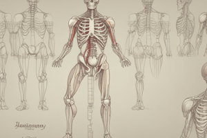
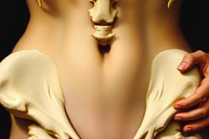
- The bony pelvis is comprised of three major components: the innominate or pelvic bone (ilium, ischium, and pubis), the sacrum, and the coccyx.
- In pediatric patients, the ilium, ischium, and pubis are not fully fused, and this is visible as triradiate cartilage.
- Important landmarks on the ilium include the iliac crest, iliac fossa, anterior superior iliac spine (ASIS), and anterior inferior iliac spine (AIIS).
- The acetabulum is a socket on the innominate bone that articulates with the head of the femur to form the hip joint. It features a bony rim and a fossa.
- The ischial spine is an attachment for several gluteal muscles and the sacrospinous ligament.
- Other notable landmarks of the pubic bone are: the pectineal line, the body of the pubis, and the symphyseal surface.
- The ischium extends from the inferior pubic ramus to the ischial tuberosity, a critical attachment point for many muscles.
- Key landmarks on the sacrum include the auricular surface for the sacroiliac joint, the base of the sacrum (functioning as a vertebral body), and the sacral promontory.
- The sacrum also has anterior and posterior sacral foramina, where sacral nerve roots exit the spinal canal.
- The sacral hiatus is the opening at the base of the spinal canal.
- The ala or wings are broad, wide lateral structures of the sacrum.
- The pelvic cavity is separated from the abdominal cavity by the superior pelvic aperture, which is defined by the ala of the sacrum, the sacral promontory, the arcuate line, and the pectineal line.
- The inferior pelvic aperture is formed by the pubic arch, ischial tuberosities, ligaments, and the tip of the coccyx, and helps establish the boundary for the pelvic floor muscles.
- The true pelvis, sometimes referred to as the lesser pelvis, is located below the superior pelvic aperture and contains pelvic organs.
- The false pelvis, also known as the greater pelvis, lies above the superior pelvic aperture and forms part of the abdominal cavity.
- The anatomical position of the pelvis is described as the innominate in the anatomical position, where the ASIS and pubic symphysis are in the same plane.
Sex Differences in the Pelvis
- The female pelvis is wider than the male pelvis and exhibits specific adaptations for childbirth.
- Features of the female pelvis include a wider sacrum, a less curved sacrum, wider ASIS, wider ischial tuberosities, wider superior and inferior pelvic apertures, a shallower false pelvis, and an anatomical position where the ASIS, AIIS, and pubic symphysis are in a plane.
The Hip Joint
- The hip joint is a synovial ball and socket joint between the head of the femur and the acetabulum of the pelvic bone.
- The acetabulum is created by the fusion of bone from the ilium, pubis, and ischium.
- The lunate surface, covered by hyaline cartilage, is the articulating surface of the acetabulum.
- The acetabular labrum, a ring of fibrocartilage, deepens the acetabulum and helps stabilize the joint.
- The acetabular notch, located inferiorly, is bridged by the transverse ligament of the acetabulum.
- The ligamentum teres (ligament of the head of the femur) is another ligament found within the acetabular notch, carrying a small artery that contributes to the blood supply of the head of the femur.
- The entire spherical surface of the head of the femur is covered with hyaline cartilage.
- There is a small indentation on the head of the femur called the fovea capitis, where the ligamentum teres attaches.
- Ligaments of the hip joint are thickenings within the joint capsule.
- The joint capsule extends from the acetabular labrum to the intertrochanteric line and crest on the femur, encompassing the head and neck of the femur.
Hip Movements
- Hip flexion, extension, abduction, adduction, internal/external rotation, and horizontal abduction/adduction
- Movements can be open chain (femur moving on stable pelvis) or closed chain (pelvis moving on stable femur)
- Open chain: Examples: kicking, marching, swinging leg backwards
- Closed chain: Examples: anterior pelvic tilt, posterior pelvic tilt, lateral tilt (hip hiking)
Hip Joint Capsule
- Contains retinacula, carrying blood vessels to the femur head
- Disruptions to the capsule can negatively impact blood supply to the femur head
- Iliofemoral and pubofemoral ligaments located anteriorly, limiting hip extension and abduction
- Ischiofemoral ligament located posteriorly, wrapping around and limiting hip extension and contributing to joint stability
Hip Joint Blood Supply
- Three main sources: Lateral and medial circumflex arteries (branches of profunda femoris artery), foveal artery (branch of obturator artery), and a branch of the inferior gluteal artery
- Disruption of blood supply can negatively impact hip joint function
Hip Joint Innervation
- Key nerves include the femoral, obturator, superior gluteal, and nerve to quadratus femoris
- Nerves provide innervation to the joint and the muscles that cross it
- Hip joint pain may refer to the groin, medial thigh, or anterior lateral thigh depending on the nerve affected
Gluteal Region Surface Anatomy
- Key landmarks: iliac crest, PSIS, ASIS, sacrum, ischeal tuberosities, greater trochanter, natal/intergluteal cleft, gluteal fold
Gluteal Region Fascia
- Superficial fascia (hypodermis) contains a significant amount of fat
- Deep fascia is called the fascia lata, thickening to form the iliotibial tract
- Iliotibial tract is not a separate structure, just a thickening of fascia
- Gluteus maximus and tensor fascia lata attach to the iliotibial tract
Gluteal Muscles
- Two main layers: superficial and deep
- Superficial muscles: gluteus maximus, gluteus medius, gluteus minimus, and tensor fascia lata
Gluteus Maximus
- Strong hip extensor
- Stabilizes the pelvis during walking and standing
- Contributes to femur external rotation
- Originates on the posterior aspect of the ilium, posterior to the posterior gluteal line
- Inserts into the iliotibial tract (IT band) and the gluteal tuberosity on the proximal femur
- Innervated by the inferior gluteal nerve, primary nerve roots are S1 and S2 with some L5
- Action: Primary hip extensor, assists in walking, running, jumping, going upstairs, and lateral rotation of the hip.
Superficial Gluteal Muscles
-
Tensor Fascia Lata (TFL):
- Anterior attachment to the ASIS and the anterior portion of the iliac crest
- Inserts into the IT band
- Action: Abducts the thigh/hip, contributes to medial rotation.
- Innervated by the superior gluteal nerve (L5, S1)
-
Gluteus Medius:
- Attaches to the external or posterior surface of the ilium between the anterior and posterior gluteal lines
- Inserts into the greater trochanter
- Action: Primary hip abductor, stabilizes the femur during squatting.
- Innervated by the superior gluteal nerve (L5, S1)
-
Gluteus Minimus:
- Attaches to the posterior or external surface of the ilium between the anterior and inferior gluteal lines
- Inserts into the greater trochanter
- Action: Primary hip abductor, stabilizes the pelvis during single-limb stance (Trendelenburg gait).
- Innervated by the superior gluteal nerve (L5, S1)
Deep Gluteal Muscles ("Deep Six" or "PGOGOQ")
-
Piriformis:
- Originates on the anterior surface of the sacrum
- Inserts into the greater trochanter
- Action: Laterally rotates the thigh, stabilizes the hip.
- Innervated by the nerve to piriformis (L5, S1)
-
Superior Gemellus:
- Originates on the ischial spine
- Inserts into the greater trochanter
- Action: Laterally rotates the thigh, stabilizes the hip.
- Innervated by the nerve to obturator internus (L5, S1, S2)
-
Obturator Internus:
- Originates on the internal surface of the obturator membrane
- Inserts into the greater trochanter
- Action: Laterally rotates the thigh, stabilizes the hip.
- Innervated by the nerve to obturator internus (L5, S1, S2)
-
Inferior Gemellus:
- Originates on the ischial tuberosity
- Inserts into the greater trochanter
- Action: Laterally rotates the thigh, stabilizes the hip.
- Innervated by the nerve to quadratus femoris (L5, S1)
-
Obturator Externus:
- Originates on the external surface of the obturator membrane
- Inserts into the greater trochanter
- Action: Laterally rotates the thigh, stabilizes the hip.
- Innervated by the obturator nerve (L3, L4)
-
Quadratus Femoris:
- Originates on the ischial tuberosity
- Inserts into the intertrochanteric crest and greater trochanter
- Action: Laterally rotates the thigh, stabilizes the hip.
- Innervated by the nerve to quadratus femoris (L5, S1)
Bursa in the Gluteal Region
-
Trochanteric Bursa:
- Separates the insertion of the gluteus maximus and IT band from the greater trochanter.
-
Ischial Bursa:
- Separates the gluteus maximus from the ischial tuberosity
-
Gluteofemoral Bursa:
- Separates the IT band from quadriceps muscles.
Gluteal Region Neurovascular Supply
- Superior and Inferior Gluteal Arteries:
- Branches off the internal iliac artery
- Supply blood to the gluteal region
- Exit the pelvic cavity through the greater sciatic foramen
Structures Passing Through the Greater Sciatic Foramen
- Muscles:
- Piriformis: A muscle that passes through the greater sciatic foramen
- Blood Vessels:
- Superior Gluteal Artery and Vein: These emerge superior to the piriformis muscle and supply the gluteus medius, gluteus minimus, and tensor fasciae latae muscles.
- Inferior Gluteal Artery and Vein: These emerge inferior to the piriformis muscle and supply deeper gluteal muscles.
- Internal Pudendal Artery and Vein: These pass through the greater sciatic foramen but do not supply the gluteal region. They continue to the perineal region, providing blood supply.
- Nerves:
- Superior Gluteal Nerve: Emerges superior to piriformis, supplies gluteus medius, gluteus minimus, and tensor fasciae latae.
- Inferior Gluteal Nerve: Emerges inferior to piriformis, supplies gluteus maximus.
- Sciatic Nerve: A large nerve that emerges inferior to piriformis and continues into the thigh.
- Posterior Femoral Cutaneous Nerve: A thick nerve that also continues into the thigh.
- Nerve to Obturator Internus.
- Nerve to Quadratus Femoris.
- Pudendal Nerve.
Gluteal Region Clinical Considerations
- Piriformis Syndrome: Compression of the sciatic nerve by the piriformis muscle. Causes sciatica (radiating nerve pain).
- Hip Abductors: Gluteus medius and gluteus minimus play a significant role in hip stability, particularly during stance phase (standing on one leg). Weakness in these muscles can lead to hip drop.
- Trochanteric Bursitis: Inflammation of the bursa located near the greater trochanter, causing pain in the lateral hip.
- Femoroacetabular Impingement (FAI): Pinching between the femoral head and the acetabulum, often associated with athletic populations.
Deep Gluteal Muscles
- Piriformis: A large muscle that originates from the anterior surface of the sacrum and inserts into the greater trochanter.
- Superior Gemellus: A small muscle associated with the obturator internus.
- Obturator Internus: A muscle located on the inside of the pelvis.
- Inferior Gemellus: Another small muscle that is part of the same group as the obturator internus.
- Quadratus Femoris: This muscle sits inferior to the obturator internus and is located at the back of the hip joint.
- Obturator Externus: This muscle sits underneath the other deep gluteal muscles.
Studying That Suits You
Use AI to generate personalized quizzes and flashcards to suit your learning preferences.


