Podcast
Questions and Answers
Which structures define the medial extent of the parotid bed?
Which structures define the medial extent of the parotid bed?
- Posterior belly of the digastric and muscles attached to the styloid process (correct)
- Ramus of the mandible and styloid process
- External acoustic meatus and mastoid process
- Superoanterior border of the sternocleidomastoid
What anatomical structures are embedded within the substance of the parotid gland?
What anatomical structures are embedded within the substance of the parotid gland?
- Masseter muscle and zygomatic arch
- Facial nerve and parotid duct
- External carotid artery and retromandibular vein (correct)
- Internal carotid artery and internal jugular vein
Which cranial nerve enters the deep region of the parotid gland and exits as multiple branches?
Which cranial nerve enters the deep region of the parotid gland and exits as multiple branches?
- Glossopharyngeal nerve (CN IX)
- Trigeminal nerve (CN V)
- Vagus nerve (CN X)
- Facial nerve (CN VII) (correct)
The parotid duct pierces which muscle as it passes into the oral cavity?
The parotid duct pierces which muscle as it passes into the oral cavity?
Which of the following is NOT a component of the masticator space?
Which of the following is NOT a component of the masticator space?
Which structure serves as the inferior attachment for the temporalis muscle?
Which structure serves as the inferior attachment for the temporalis muscle?
What bony landmark is the roof of the infratemporal fossa?
What bony landmark is the roof of the infratemporal fossa?
Which ligament divides the infratemporal fossa from the parotid gland region?
Which ligament divides the infratemporal fossa from the parotid gland region?
What is the primary action of the masseter muscle?
What is the primary action of the masseter muscle?
What action is uniquely attributed to the lateral pterygoid muscle among the muscles of mastication?
What action is uniquely attributed to the lateral pterygoid muscle among the muscles of mastication?
The pterygoid plexus of veins drains structures supplied by which artery?
The pterygoid plexus of veins drains structures supplied by which artery?
Damage to the middle meningeal artery is most likely to result in what?
Damage to the middle meningeal artery is most likely to result in what?
Which nerve is sensory to the mucosa and skin of the cheek, emerging between the heads of the lateral pterygoid muscle?
Which nerve is sensory to the mucosa and skin of the cheek, emerging between the heads of the lateral pterygoid muscle?
What type of fibers does the chorda tympani carry, which then hitchhike on the lingual nerve?
What type of fibers does the chorda tympani carry, which then hitchhike on the lingual nerve?
What nerve is often inadvertently blocked during an inferior alveolar nerve block, leading to numbness of the tongue?
What nerve is often inadvertently blocked during an inferior alveolar nerve block, leading to numbness of the tongue?
Through which foramen does the mandibular nerve (V3) enter the infratemporal fossa (ITF)?
Through which foramen does the mandibular nerve (V3) enter the infratemporal fossa (ITF)?
The posterior superior alveolar artery supplies blood to which structure?
The posterior superior alveolar artery supplies blood to which structure?
Postganglionic parasympathetic fibers destined for the parotid gland hitchhike on which nerve?
Postganglionic parasympathetic fibers destined for the parotid gland hitchhike on which nerve?
What is the origin of the preganglionic parasympathetic fibers providing innervation to the parotid gland?
What is the origin of the preganglionic parasympathetic fibers providing innervation to the parotid gland?
A patient presents with an extradural hematoma that has resulted from a fracture of the temporal bone. Which artery is MOST likely damaged in this scenario?
A patient presents with an extradural hematoma that has resulted from a fracture of the temporal bone. Which artery is MOST likely damaged in this scenario?
Flashcards
What is the parotid bed?
What is the parotid bed?
Space between the ramus of the mandible, external acoustic meatus, and mastoid and styloid processes of the temporal bone.
What is the deep cervical layer of investing fascia?
What is the deep cervical layer of investing fascia?
A tough fibrous capsule that encloses the parotid gland, extending up from the neck.
What vessels are in the parotid gland?
What vessels are in the parotid gland?
The external carotid artery and retromandibular vein are embedded within this organ.
What is the masticator region/space?
What is the masticator region/space?
Signup and view all the flashcards
What is the temporal fossa?
What is the temporal fossa?
Signup and view all the flashcards
What forms the superior (roof) boundary of the infratemporal fossa?
What forms the superior (roof) boundary of the infratemporal fossa?
Signup and view all the flashcards
What forms the lateral boundary of the infratemporal fossa?
What forms the lateral boundary of the infratemporal fossa?
Signup and view all the flashcards
What is the stylomandibular ligament?
What is the stylomandibular ligament?
Signup and view all the flashcards
What does the pterygomaxillary fissure represent?
What does the pterygomaxillary fissure represent?
Signup and view all the flashcards
Mandibular nerve (V3)
Mandibular nerve (V3)
Signup and view all the flashcards
What is the function of the buccal nerve?
What is the function of the buccal nerve?
Signup and view all the flashcards
What is the pterygoid portion of the maxillary artery?
What is the pterygoid portion of the maxillary artery?
Signup and view all the flashcards
What does the Auriculotemporal n. supply?
What does the Auriculotemporal n. supply?
Signup and view all the flashcards
What does the lingual nerve supply?
What does the lingual nerve supply?
Signup and view all the flashcards
What does the mental nerve supply?
What does the mental nerve supply?
Signup and view all the flashcards
What is an inferior alveolar nerve block?
What is an inferior alveolar nerve block?
Signup and view all the flashcards
What is the inferior salivatory nucleus of the medulla?
What is the inferior salivatory nucleus of the medulla?
Signup and view all the flashcards
Study Notes
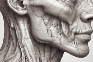
Parotid Bed Definition
- The parotid bed is the space between the ramus of the mandible, external acoustic meatus, and mastoid and styloid processes of the temporal bone.
- It extends medially to the posterior belly of the digastric and other muscles attached to the styloid process.
- Inferiorly, it is defined by the superoanterior border of the sternocleidomastoid.
- It is an irregular shaped area occupied by the parotid gland, which molds into the space and assists in filling out the contour of the jaw.
Parotid Gland
- It is enclosed by the deep cervical layer of investing fascia extending up from the neck.
- The superficial aspect extends superficially over the masseter muscle to the zygomatic arch.
- There is a deep (medial) extension to the medial wall of the region.
- The external carotid artery and retromandibular vein are embedded within the gland.
- The facial nerve (CN VII) enters the deep region of the gland and ascends to exit the gland as 5+ ramifying branches that are motor to the muscles of facial expression.
- The parotid (Stenson's) duct exits the anterior aspect of the superficial portion of the gland, passing superficial to the masseter and diving into the buccal fat pad. The duct pierces the buccinator muscle and opens in the oral cavity opposite the second upper maxillary molar. An extension of the parotid gland often extends out along the duct.
Infratemporal Fossa (ITF)
- The ITF may be the most important anatomical area for dental practitioners.
- Many important blood vessels, nerves and muscles are located here.
- Local anesthetic is often deposited in this area.
- It is part of the larger masticator region/space that includes: a portion of the parotid bed lateral to the mandibular ramus, temporal fossa, and the infratemporal fossa itself.
Temporal Fossa
- Contents are essentially the temporalis muscle.
- Superior attachment: superior and inferior temporal lines.
- Inferior attachment: coronoid process of the mandible.
- Actions: elevation, retraction, and translation.
Boundaries of the Infratemporal Fossa
- Superior (roof): Inferior surface of the greater wing of sphenoid and part of squamous portion of the temporal bone.
- Inferior: No structure, formed by an imaginary line drawn inward from the occlusal plane of the mandible.
- Lateral: Medial portion of ramus of mandible.
- Medial: Lateral surface of lateral pterygoid plate (part of sphenoid bone).
- Anterior: Posterior surface of maxilla.
- Posterior: Not bony, stylomandibular ligament divides the infratemporal fossa from the region of the parotid gland.
- The roof is separated from the temporal fossa above by the infratemporal crest.
Accessing the Infratemporal Fossa
- To gain access, the ramus of the mandible needs to be removed. Superficial to the ramus is the masseter muscle (the second muscle of mastication).
Muscles of Mastication in the Infratemporal Fossa
- After removing the ramus of the mandible, the infratemporal fossa is exposed
- The remaining two muscles of mastication are revealed.
- The medial pterygoid muscle is superficial.
- The lateral pterygoid muscle is upper and lightly deeper.
- The medial pterygoid muscle has two heads: a larger deep head and a smaller superficial head.
Medial Pterygoid Muscle Attachments and Actions
- Superficial head:
- Superior: maxillary tuberosity.
- Inferior: deep side angle of mandible.
- Deep head:
- Superior: medial side, lateral pterygoid plate.
- Inferior: deep side, angle of mandible.
- Actions: elevation, protraction, and translation.
Lateral Pterygoid Muscle
- Has two heads:
- The superior head arises from the greater wing of the sphenoid bone.
- The inferior head arises from the lateral surface of the lateral pterygoid plate of the sphenoid.
- Both heads pass posterolaterally to insert on the neck of the mandible, as well as the capsule and articular disc of the temporomandibular joint (TMJ).
Muscles of Mastication Functional Summary
- Masseter: elevation.
- Temporalis: elevation, retraction.
- Medial pterygoid: elevation, protraction.
- Lateral pterygoid: protraction.
- Suprahyoids: depression.
- Protraction is accomplished by the medial and lateral pterygoids working together on both sides.
- Side-to-side chewing movements are accomplished by unilateral contraction of the medial and lateral pterygoids.
- The lateral pterygoid muscles function in opening the mouth.
- The suprahyoid muscles (digastrics, geniohyoid, mylohyoid, stylohyoid) are accessory muscles used to open the mouth.
Temporomandibular Joint (TMJ)
- The TMJ is a modified hinge joint divided into two cavities by an (intra) articular disc, creating a superior and inferior joint cavity.
- Anterior gliding occurs between the disc and the mandibular fossa at the superior joint cavity.
- Rotation between the mandibular condyle and the disc occurs at the inferior joint cavity.
Superficial Infratemporal Fossa
- The pterygoid plexus of veins is a network of veins in the ITF that drains structures supplied by the maxillary artery
- The veins of the pterygoid plexus drain to the maxillary vein
- The maxillary vein joins with the superficial temporal vein to form the retromandibular vein.
Nerves in the Infratemporal Fossa
- The superficial dissection of the infratemporal fossa will mostly contain the initial part of the maxillary artery and a number of nerves.
- The buccal nerve (sensory branch of the anterior division of the mandibular nerve) appears from between the two heads of the lateral pterygoid muscle.
- From under the inferior aspect of the inferior head of the lateral pterygoid muscle are the lingual nerve and the inferior alveolar nerve.
- From lateral to medial are the inferior alveolar nerve, the lingual nerve, and finally the buccal nerve.
Maxillary Artery
- This is one of two terminal branches of the external carotid artery.
- It takes a twisted course through the infratemporal fossa and can be found either superficial or deep to the lateral pterygoid muscle.
- The maxillary artery can be divided into 3 parts: mandibular, pterygoid, and pterygopalatine.
- The mandibular portion extends between the medial surface of the mandible and the sphenomandibular ligament.
- The most important clinical branches are the middle meningeal and inferior alveolar arteries.
- The mylohyoid branch from the inferior alveolar artery supplies the mylohyoid muscle.
- The pterygoid portion passes either superficial or deep to the lateral pterygoid muscle.
- Numerous branches to the muscles of mastication emanate from this portion
- The buccal artery travels with the buccal nerve through the buccinator muscle.
- While the buccal nerve does not innervate the buccinator, the buccal artery supplies it with blood.
- The pterygopalatine portion passes through the pterygomaxillary fissure to enter the ptergopalatine fossa.
- It has branches that run with the maxillary nerve (CN V2).
- The posterior superior alveolar artery arises just before the maxillary artery enters the fissure
- It supplies blood to the maxillary molars and premolars, buccal gingiva, and regions of the wall of the maxillary sinus.
- The middle meningeal artery passes through the roof of the intratemporal fossa via the foramen spinosum.
- It lies between the dura and the bone of the calvarium to supply the dura with blood.
- The region of the temporal bone (squamous portion) is thin and impact to the bone creates sharp broken edges that can tear the artery and result in an extradural hematoma.
Pterygomaxillary Fissure
- Represents the lateral opening of the pterygopalatine fossa.
Mandibular Nerve (V3)
- The mandibular nerve enters the ITF through the foramen ovale.
- Under cover of the lateral pterygoid muscle, it quickly divides into an anterior and posterior division.
Branches of the Anterior Division of Mandibular Nerve (V3)
- Buccal nerve (long buccal n.):
- Sensory to mucosa and skin of the cheek and gingival buccal to mandibular molars.
- It does NOT supply the buccinator muscle
- It emerges between the superior and inferior heads of the lateral pterygoid muscle.
- Motor branches to the muscles of mastication (except the nerve to the medial pterygoid, which comes off the stem of V3).
Branches of the Posterior Division of Mandibular Nerve (V3)
- Auriculotemporal nerve:
- Splits around the middle meningeal artery and heads toward the TMJ.
- Sensory nerve that carries sensation from the external auditory meatus, lateral portion of tympanic membrane, temporomandibular joint, and skin over temporal surface head.
- Postganglionic parasympathetic fibers travel on the auriculotemporal nerve from the otic ganglion to the parotid gland.
- Lingual nerve:
- Runs almost parallel with the inferior alveolar nerve, but anterior to it.
- Sensory nerve that carries sensation from anterior 2/3 of tongue, mucosa on floor of mouth and lingual gingival.
- Receives the chorda tympani nerve (a branch of facial nerve) which carries parasympathetic fibers and taste fibers.
- Preganglionic parasympathetic fibers hitchhike on the lingual nerve to the submandibular ganglion where they synapse.
- Postganglionic fibers then travel to the submandibular gland, the sublingual gland and minor salivary glands of floor of mouth.
- Inferior Alveolar nerve:
- Has both motor and sensory fibers.
- Emerges from under the lateral pterygoid muscle along with the lingual nerve.
- Runs between the mandible and the medial pterygoid muscle until it enters the mandible at the mandibular foramen.
- Before entering the foramen the inferior alveolar nerve gives off a small motor branch to the nerve to the mylohyoid which provides motor innervation to both the mylohyoid muscle and the anterior belly digastric muscle.
- The interosseous portion sends branches to supply the pulps of mandibular teeth.
- The mental nerve is a branch of the inferior alveolar nerve that passes out of the mental foramen to provide sensory innervation to the skin and mucous membrane of lower lip, skin of chin and buccal gingival of mandibular premolars and incisors.
- The incisive nerve continues within the bone anterior to the mental foramen to supply the pulps of the mandibular anterior teeth.
Clinical Correlation for Inferior Alveolar Nerve Block
- A common local anesthetic injection in dentistry is the inferior alveolar nerve block.
- A block of this nerve provides anesthesia to the mandibular teeth on the side of the injection, the buccal gingiva anterior to premolars, and the lower lip and chin.
- Because the lingual nerve is close to the inferior alveolar nerve, the lingual nerve is also blocked during an inferior alveolar nerve block.
- The tongue, lingual gingival and floor of mouth become numb as well.
Autonomic Supply to the Parotid Gland
- The cell bodies for the preganglionic parasympathetic innervation are located in the inferior salivatory nucleus of the medulla.
- These fibers exit the skull in CN IX through the jugular foramen.
- The fibers travel to the inferior ganglion of CN IX and exit via the tympanic branch that contributes to the tympanic plexus (preganglionic parasympathetics, postganglionic sympathetics form the carotid plexus and general sensory fibers from sensory root of IX) in the middle ear cavity.
- The preganglionic parasympathetic fibers leave the tympanic plexus via the lesser petrosal nerve and pass through the foramen ovale to enter the ITF where they synapse at the otic ganglion.
- The postganglionic parasympathetic fibers then hitch a ride with the auriculotemporal nerve to the parotid gland.
Autonomic Supply to the Submandibular and Sublingual Glands
- Review the cranial nerve wiring diagrams on CN V3 showing the chorda tympani hitchhiking along the lingual nerve in the infratemporal fossa to the submandibular and sublingual glands.
Studying That Suits You
Use AI to generate personalized quizzes and flashcards to suit your learning preferences.