Podcast
Questions and Answers
What is one of the common symptoms of otitis media?
What is one of the common symptoms of otitis media?
- Persistent cough
- Fluctuating vision
- Ear pain (correct)
- Severe headaches
What structure separates the two nasal cavities?
What structure separates the two nasal cavities?
- Septum (correct)
- Nasal turbinates
- Nasal bridge
- Alveoli
Which part of the nose is responsible for trapping dirt and particles?
Which part of the nose is responsible for trapping dirt and particles?
- Hair and cilia (correct)
- Nerve cells
- Sinuses
- Nasal cavities
Which anatomical feature of the ear is primarily involved in balance?
Which anatomical feature of the ear is primarily involved in balance?
What is one function of the sinuses in relation to the nasal cavities?
What is one function of the sinuses in relation to the nasal cavities?
What is a common examination technique used to assess ear health?
What is a common examination technique used to assess ear health?
Which of the following statements about the parts of the nose is incorrect?
Which of the following statements about the parts of the nose is incorrect?
Which of the following symptoms is NOT typically associated with a middle ear infection?
Which of the following symptoms is NOT typically associated with a middle ear infection?
What is the primary function of the auricle or pinna in the ear?
What is the primary function of the auricle or pinna in the ear?
Which part of the ear separates the external ear from the middle ear?
Which part of the ear separates the external ear from the middle ear?
What structure connects the middle ear with the nasopharynx?
What structure connects the middle ear with the nasopharynx?
Which structure in the inner ear is responsible for receiving vibrations from the middle ear?
Which structure in the inner ear is responsible for receiving vibrations from the middle ear?
During an ear examination, what should a healthcare provider observe for in the external ear?
During an ear examination, what should a healthcare provider observe for in the external ear?
What symptom might indicate otitis externa during ear examination?
What symptom might indicate otitis externa during ear examination?
What are common symptoms to inquire about during a health assessment of the ear?
What are common symptoms to inquire about during a health assessment of the ear?
Which of the following parts of the ear is NOT considered part of the inner ear?
Which of the following parts of the ear is NOT considered part of the inner ear?
What appearance should the tympanic membrane have during examination?
What appearance should the tympanic membrane have during examination?
Which of the following can interfere with the ear's ability to conduct sound waves?
Which of the following can interfere with the ear's ability to conduct sound waves?
What signs and symptoms of otitis media might an adult present?
What signs and symptoms of otitis media might an adult present?
Which of the following describes acute otitis media?
Which of the following describes acute otitis media?
What may cause damage to the bony structures of the middle ear?
What may cause damage to the bony structures of the middle ear?
What observation should be made when inspecting the ear canal?
What observation should be made when inspecting the ear canal?
What can lead to otitis media according to the content?
What can lead to otitis media according to the content?
Which of the following is NOT a common symptom of earache?
Which of the following is NOT a common symptom of earache?
Flashcards are hidden until you start studying
Study Notes
Ear Examination
- Inspect the ear canal for discharge, redness, odor, nodules, or cysts.
- Perform otoscopic examination to assess tympanic membrane and middle ear structures.
- Normal tympanic membrane appears pearl gray, glistening, and transparent.
- Look for abnormalities such as bulging, retraction, bleeding, lesions, or perforations.
Common Ear Disorders
- Earache: Indicative of external or middle ear disorders; often linked to infections and can cause hearing loss or otorrhea.
- Hearing Loss: May be due to cerumen impaction, foreign bodies, polyps, or otitis media, which thickens fluid in the middle ear.
- Otosclerosis: Hardening of middle ear bones affecting sound transmission.
- Trauma: Can disrupt the bony chain in the middle ear.
Otitis Media
- Inflammation of the middle ear, stemming from eustachian tube blockage.
- Can be classified as suppurative (with infection) or secretory (without infection) and can be acute or chronic.
- Acute Otitis Media: Characterized by rapid onset, infected fluid, and short duration.
- Symptoms: Ear pain, especially when lying down, tugging at an ear, trouble sleeping, increased fussiness in children.
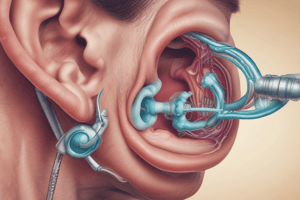
Anatomy of the Ear
- External Ear: Comprises elastic cartilage, includes the auricle (pinna) and auditory canal; collects and transmits sound.
- Middle Ear: Separated from the external ear by the tympanic membrane; contains malleus, incus, and stapes bones; connected to the nasopharynx via the eustachian tube for pressure equalization.
- Inner Ear: Fluid-filled structures (bony labyrinth) including the vestibule, semicircular canals, and cochlea; responsible for sending sound vibrations to the brain.
Health Assessment
- Gather subjective data: hearing troubles, use of hearing aids, symptoms like tinnitus or ear drainage, dizziness or balance issues.
- Conduct external observations: examine ear position, symmetry, lesions, drainage, or tenderness.
- Common Symptoms to Note: Ear pain, trouble hearing, fluid drainage, fever, loss of balance, headache, loss of appetite.
Anatomy of the Nose
- Lower two-thirds consist of flexible cartilage; upper third composed of rigid bone.
- Merges internally with the pharynx, divided into nasopharynx, oropharynx, and laryngopharynx.
Parts of the Nose
- Bone: Forms the hard upper bridge of the nose.
- Hair and Cilia: Trap dirt and particles, facilitating removal through sneezing or wiping.
- Lateral Walls: Cartilage outer walls covered in skin, forming nasal cavities and nostrils.
- Nasal Cavities: Two hollow spaces lined with mucous membranes for air flow.
- Nerve Cells: Responsible for the sense of smell through brain communication.
- Nostrils (Nares): Openings to nasal cavities located on the face.
- Septum: Bone and cartilage structure separating nasal cavities.
- Sinuses: Four pairs of air-filled pockets connected to nasal cavities, produce moisture-retaining mucus.
- Turbinates (Conchae): Three pairs located along nasal cavity sides; aid in warming and moistening inhaled air.
Studying That Suits You
Use AI to generate personalized quizzes and flashcards to suit your learning preferences.