Podcast
Questions and Answers
What is the function of the marginal gingiva?
What is the function of the marginal gingiva?
- To produce saliva
- To cover the alveolar processes of the jaws
- To form the soft tissue wall of the gingival sulcus (correct)
- To support the teeth
What is the gingival sulcus?
What is the gingival sulcus?
- A shallow crevice or space around the tooth neck (correct)
- A deep crevice or space around the tooth pulp
- A deep crevice or space around the tooth crown
- A shallow crevice or space around the tooth root
What is the width of the marginal gingiva?
What is the width of the marginal gingiva?
- About 10 mm
- About 0.5 mm
- About 1 mm (correct)
- About 5 mm
What is the attached gingiva?
What is the attached gingiva?
What determines the shape of the interdental gingiva?
What determines the shape of the interdental gingiva?
What is the histological composition of the gingiva?
What is the histological composition of the gingiva?
What is the normal depth of the gingival sulcus?
What is the normal depth of the gingival sulcus?
What happens to the width of the attached gingiva with age?
What happens to the width of the attached gingiva with age?
What is the structureless layer between the epithelium and lamina propria?
What is the structureless layer between the epithelium and lamina propria?
What is the function of the basal lamina?
What is the function of the basal lamina?
What type of collagen is found in the lamina densa?
What type of collagen is found in the lamina densa?
What is the thickness of the oral epithelium?
What is the thickness of the oral epithelium?
What is characteristic of the sulcular epithelium?
What is characteristic of the sulcular epithelium?
What is the thickness of the junctional epithelium?
What is the thickness of the junctional epithelium?
What is expressed in the junctional epithelium?
What is expressed in the junctional epithelium?
What is the result of the irregularity of the basement membrane?
What is the result of the irregularity of the basement membrane?
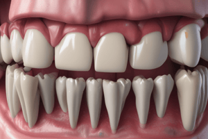
What is the normal color of the gingiva in adults?
What is the normal color of the gingiva in adults?
What is the shape of the gingival margin in adults?
What is the shape of the gingival margin in adults?
What is the consistency of the gingiva in adults?
What is the consistency of the gingiva in adults?
What is the surface texture of the gingiva in adults?
What is the surface texture of the gingiva in adults?
What is the normal depth of the gingival sulcus in newly erupted teeth?
What is the normal depth of the gingival sulcus in newly erupted teeth?
At what age is stippling typically present in the gingiva?
At what age is stippling typically present in the gingiva?
What percentage of connective tissue is formed by collagen fibers?
What percentage of connective tissue is formed by collagen fibers?
What is the main function of gingival fibers?
What is the main function of gingival fibers?
What is the main component of the ground substance in connective tissue?
What is the main component of the ground substance in connective tissue?
What type of fibers provide tensile strength to the lamina propria?
What type of fibers provide tensile strength to the lamina propria?
What is the origin of the dentogingival group of gingival fibers?
What is the origin of the dentogingival group of gingival fibers?
What is the main source of blood supply to the gingiva?
What is the main source of blood supply to the gingiva?
What is the main cell type responsible for forming collagen and elastic fibers in connective tissue?
What is the main cell type responsible for forming collagen and elastic fibers in connective tissue?
What is the main function of the reticular layer of connective tissue?
What is the main function of the reticular layer of connective tissue?
What is the main cell type in the oral epithelium?
What is the main cell type in the oral epithelium?
Where does the proliferation of keratinocytes most frequently occur?
Where does the proliferation of keratinocytes most frequently occur?
Which type of cell found in the basal layer functions as tactile receptors?
Which type of cell found in the basal layer functions as tactile receptors?
Which process involves the progressive flattening of the cell and the disappearance of the nucleus?
Which process involves the progressive flattening of the cell and the disappearance of the nucleus?
Which layers are Langerhans cells located in?
Which layers are Langerhans cells located in?
What is the turnover time for the gingiva in experimental animals?
What is the turnover time for the gingiva in experimental animals?
What is the primary function of gingival epithelium?
What is the primary function of gingival epithelium?
Which cells synthesize melanin?
Which cells synthesize melanin?
Flashcards are hidden until you start studying
Study Notes
Marginal Gingiva
- Forms the free edge of the gingiva
- Connects the attached gingiva to the tooth
- Width: 1-2 mm
Gingival Sulcus
- Space between the tooth and the free gingiva
- Filled with crevicular fluid
- Normal depth: 0-3 mm
- Depth increases with inflammation
Attached Gingiva
- Part of the gingiva that is firmly attached to the underlying bone
- Width decreases with age due to gingival recession
Interdental Gingiva
- Shape determined by the shape of the interproximal surfaces of adjacent teeth
Histological Composition of the Gingiva
- Epithelium: Oral epithelium and junctional epithelium
- Lamina propria: Connective tissue layer
Lamina Propria
- Consists of collagen fibers and ground substance
- Main cell type: Fibroblast
- Composed of reticular and papillary layers
Oral Epithelium
- Thickness: 0.2 - 0.5 mm
- Four layers: stratum basale, stratum spinosum, stratum granulosum, and stratum corneum
- Langerhans cells located in the stratum spinosum and stratum granulosum
Junctional Epithelium
- Thickness: 15-30 μm
- Epithelium attaches to the tooth via hemidesmosomes
- Rich in basal lamina: Connects epithelium to underlying connective tissue
- Expressed proteins include: Integrins, cadherins, and desmosomes
- Irregularity of the basement membrane: Results in a greater surface area for attachment
Sulcular Epithelium
- Characterized by its non-keratinized nature
Gingival Color
- Normal color: Pink
- Affected by vascularity, pigmentation, and thickness of the epithelium
Gingival Margin
- Shape: Crescentic in adults
Gingival Consistency
- Firm in adults
Gingival Texture
- Orange-peel-like stippling in adults
Gingival Development
- Stippling emerges around 6 years of age
- Normal sulcus depth in newly erupted teeth: 0-1.5 mm
Collagen Fibers
- 90% of connective tissue
- Main function: Provide support and stability to the gingiva
Ground Substance
- Main component: Glycosaminoglycans
Dentogingival Fibers
- Origin: Cemento-enamel junction
- Insert into: Lamina propria of the gingiva
Blood Supply
- Main source: Superior and inferior alveolar arteries
Fibroblasts
- Main cell type responsible for collagen and elastic fibers in connective tissue
Reticular Layer of Connective Tissue
- Located: Deepest layer of the lamina propria
- Contains: Larger blood vessels, nerves, and collagen fibers
Keratinocytes
- Main cell type in oral epithelium
- Proliferate most frequently in the stratum basale
Merkel Cells
- Tactile receptors located in the stratum basale
Keratinization
- Process involves: Progressive flattening of the cell and disappearance of the nucleus
Gingival Turnover Time
- 2-3 weeks in experimental animals
Gingival Epithelium
- Primary function: Barrier to infection and injury
Melanocytes
- Synthesize melanin: Responsible for gingival pigmentation
Studying That Suits You
Use AI to generate personalized quizzes and flashcards to suit your learning preferences.