Podcast
Questions and Answers
Which component accounts for half of the assessments in the OPT506 course?
Which component accounts for half of the assessments in the OPT506 course?
- Oral presentation
- Group project
- Practical exam (correct)
- Written exam (correct)
What is the primary focus of the OPT506 course?
What is the primary focus of the OPT506 course?
- General eye examination procedures
- Non-vision related health practices
- Specialist optometry skills (correct)
- General health assessments
What is the maximum duration of a practical session in the OPT506 course?
What is the maximum duration of a practical session in the OPT506 course?
- 120 minutes
- 50 minutes (correct)
- 60 minutes
- 30 minutes
Which of the following is NOT a topic covered in the OPT506 lectures?
Which of the following is NOT a topic covered in the OPT506 lectures?
Which item is essential for attendance in the practical sessions?
Which item is essential for attendance in the practical sessions?
How often are lectures scheduled in the OPT506 course?
How often are lectures scheduled in the OPT506 course?
Which of the following is a requirement for practical session attire?
Which of the following is a requirement for practical session attire?
What is the purpose of using narrow slit illumination in the examination process?
What is the purpose of using narrow slit illumination in the examination process?
Which phenomenon is mentioned as being similar to the principles of the conical beam technique?
Which phenomenon is mentioned as being similar to the principles of the conical beam technique?
What tool is advised to be turned off after use during practical sessions?
What tool is advised to be turned off after use during practical sessions?
In the retro-illumination technique, what is primarily observed?
In the retro-illumination technique, what is primarily observed?
What angle is recommended for the conical beam technique during examination?
What angle is recommended for the conical beam technique during examination?
What does the slit's height for the conical beam technique typically range between?
What does the slit's height for the conical beam technique typically range between?
What is the primary advantage of using a slit lamp for assessing the anterior segment of the eye?
What is the primary advantage of using a slit lamp for assessing the anterior segment of the eye?
Which structures can be examined with high magnification (20-25x) using a slit lamp?
Which structures can be examined with high magnification (20-25x) using a slit lamp?
What aspect of a slit lamp's observation system is crucial for achieving the desired magnification?
What aspect of a slit lamp's observation system is crucial for achieving the desired magnification?
Which of the following correctly identifies the components of the slit lamp that support patient positioning?
Which of the following correctly identifies the components of the slit lamp that support patient positioning?
In slit lamp bio-microscopy, what feature of the illumination system is specifically controlled to enhance evaluation?
In slit lamp bio-microscopy, what feature of the illumination system is specifically controlled to enhance evaluation?
What is the primary function of the diffuser filter in a slit lamp?
What is the primary function of the diffuser filter in a slit lamp?
Which filter is specifically used to enhance the contrast of fluorescein during examination?
Which filter is specifically used to enhance the contrast of fluorescein during examination?
Which procedure is NOT typically associated with the use of a slit lamp?
Which procedure is NOT typically associated with the use of a slit lamp?
What is one of the main purposes of assessing contact lenses during a slit lamp examination?
What is one of the main purposes of assessing contact lenses during a slit lamp examination?
What is an essential practice to ensure safety and hygiene before using a slit lamp?
What is an essential practice to ensure safety and hygiene before using a slit lamp?
What percentage of the overall assessment is dedicated to the practical exam in the OPT506 module?
What percentage of the overall assessment is dedicated to the practical exam in the OPT506 module?
Which of the following skills is NOT part of the competencies expected by the General Optical Council regarding visual impairment?
Which of the following skills is NOT part of the competencies expected by the General Optical Council regarding visual impairment?
Which technique is specifically assessed in Station 1 of the practical exam?
Which technique is specifically assessed in Station 1 of the practical exam?
Which intended learning outcome involves selecting optical or non-optical aids for patients?
Which intended learning outcome involves selecting optical or non-optical aids for patients?
What type of assessment occurs during the ISCE portion of the practical exam?
What type of assessment occurs during the ISCE portion of the practical exam?
Which of the following is NOT a component of evaluating and selecting contact lenses?
Which of the following is NOT a component of evaluating and selecting contact lenses?
Which outcome relates specifically to the use of slit lamp and its accessory equipment?
Which outcome relates specifically to the use of slit lamp and its accessory equipment?
Which of the following assessments is included in the practical exam for the previous year?
Which of the following assessments is included in the practical exam for the previous year?
What is the primary method for examining all corneal layers, excluding the endothelium?
What is the primary method for examining all corneal layers, excluding the endothelium?
Which illumination technique is specifically recommended for assessing corneal oedema and pathological changes?
Which illumination technique is specifically recommended for assessing corneal oedema and pathological changes?
When performing direct retro illumination, which initial magnification is suggested?
When performing direct retro illumination, which initial magnification is suggested?
Which of the following objects would most likely be examined using indirect retro illumination?
Which of the following objects would most likely be examined using indirect retro illumination?
What observation technique is used for examining keratic precipitates and other debris on the corneal endothelium?
What observation technique is used for examining keratic precipitates and other debris on the corneal endothelium?
What is the angle setting recommended for indirect retro illumination?
What is the angle setting recommended for indirect retro illumination?
What is the purpose of using the Van Herick’s technique?
What is the purpose of using the Van Herick’s technique?
Which condition is NOT likely to be observed using direct retro illumination?
Which condition is NOT likely to be observed using direct retro illumination?
Flashcards
What is Specialist Optometry Skills?
What is Specialist Optometry Skills?
A course designed to develop specialized optometry skills beyond general practice.
What does the OPT506 course cover?
What does the OPT506 course cover?
Includes topics like contact lens assessment, fitting, and management for various lens types (soft, rigid, etc.), low vision. It also covers optics and CL material.
How is OPT506 structured?
How is OPT506 structured?
The course is split into a combination of lectures and practical sessions, with assessments including a written and practical exam.
What is Slit Lamp Biomicroscopy?
What is Slit Lamp Biomicroscopy?
Signup and view all the flashcards
What is the purpose of Slit Lamp Biomicroscopy ?
What is the purpose of Slit Lamp Biomicroscopy ?
Signup and view all the flashcards
What are the important ground rules for OPT506?
What are the important ground rules for OPT506?
Signup and view all the flashcards
What is the goal of the OPT506 course?
What is the goal of the OPT506 course?
Signup and view all the flashcards
Slit Lamp Biomicroscopy
Slit Lamp Biomicroscopy
Signup and view all the flashcards
Competent Slit Lamp Use
Competent Slit Lamp Use
Signup and view all the flashcards
Slit Lamp Functions
Slit Lamp Functions
Signup and view all the flashcards
Slit Lamp Accessory Equipment
Slit Lamp Accessory Equipment
Signup and view all the flashcards
Slit Lamp Examination
Slit Lamp Examination
Signup and view all the flashcards
Cornea
Cornea
Signup and view all the flashcards
Conjunctiva
Conjunctiva
Signup and view all the flashcards
Iris
Iris
Signup and view all the flashcards
Diffuser filter
Diffuser filter
Signup and view all the flashcards
Wratten 12 filter
Wratten 12 filter
Signup and view all the flashcards
Gonioscopy
Gonioscopy
Signup and view all the flashcards
Keratometry
Keratometry
Signup and view all the flashcards
Pachymetry
Pachymetry
Signup and view all the flashcards
What is a slit lamp?
What is a slit lamp?
Signup and view all the flashcards
Why is the slit lamp's adjustable beam important?
Why is the slit lamp's adjustable beam important?
Signup and view all the flashcards
What is stereoscopic vision in the slit lamp?
What is stereoscopic vision in the slit lamp?
Signup and view all the flashcards
How does the slit lamp magnification help?
How does the slit lamp magnification help?
Signup and view all the flashcards
What can you see with the slit lamp?
What can you see with the slit lamp?
Signup and view all the flashcards
Direct Illumination
Direct Illumination
Signup and view all the flashcards
Diffuse Illumination
Diffuse Illumination
Signup and view all the flashcards
Slit Illumination
Slit Illumination
Signup and view all the flashcards
Conical Beam Illumination
Conical Beam Illumination
Signup and view all the flashcards
Retro-illumination
Retro-illumination
Signup and view all the flashcards
Parallelepiped Illumination
Parallelepiped Illumination
Signup and view all the flashcards
Direct Retro-illumination
Direct Retro-illumination
Signup and view all the flashcards
Indirect Retro-illumination
Indirect Retro-illumination
Signup and view all the flashcards
Direct Retro Illumination: Purpose
Direct Retro Illumination: Purpose
Signup and view all the flashcards
Indirect Retro illumination: Purpose
Indirect Retro illumination: Purpose
Signup and view all the flashcards
Assessing the Anterior Chamber Angle
Assessing the Anterior Chamber Angle
Signup and view all the flashcards
Optic Section
Optic Section
Signup and view all the flashcards
Study Notes
Recording Information
- The lecture will be recorded and available via Panopto on the module DLE pages.
- Questions asked during the lecture may appear on the recording.
- Students can ask the lecturer to pause the recording if they do not wish their question to be recorded.
Module Information
- The module is OPT506 Specialist Optometry Skills.
- The module focuses on introducing Slit lamp biomicroscopy.
- The module is a full-year course (terms 1 and 2).
- The module includes 20 credits (200 hours of work).
- Lectures: 30 hours
- Practical sessions: 48 hours
- Independent study: 122 hours
Assessments
- 50% Written exam on May 13, 2025.
- The exam includes 50 multiple-choice questions.
- 50% Practical exam on December 3, 2024.
- The exam consists of 3 OSCE stations and 1 ISCE station.
- Station 1: Slit lamp techniques
- Station 2: Keratometry and baseline measurements
- Station 3: RGP fit assessment(passive)
- ISCE (May 27-28, 2025): SCL Aftercare routine
Ground Rules
- Be respectful.
- Be ready.
- Be safe.
Specialist Optometry Skills Breakdown
- Optometry is a general skill.
- Specialist skills include:
- Binocular vision
- Pediatric vision
- Contact Lens
- Low Vision
Lecture Structure
- 1 lecture per week (2 hours each)
- Lecture outlines are uploaded 48 hours before lecture.
- Lecture slides are uploaded after the lecture.
- Lecture content:
- L1 - L5: Initial CL assessment
- L6 - L9: Rigid Gas Permeable Lenses (RGP)
- L10 - L11: Optics and CL material
- L12 - L16: Soft Contact Lens (SCL)
- L17 - L18: Low Vision
- L19 - L20: CL care and complications
Practical Sessions
- 1 practical session per week (2 hours each).
- Briefing/Demo/Q&A duration: 50 minutes.
- Group 1: 11:05-11:15am; 11:15am – 12:05pm
- Group 2: 9:00-9:10am ; 9:10 - 10:00am; 10:00am - 10:50am
- Students should arrive 10 minutes early.
- Set up the bay if you are first in your pair- don't wait.
Practical Session Guidelines
- All sessions have demo videos
- Hand hygiene is critical!
- Dress code is smart casual, with a name tag.
- No long nails or nail varnish.
- Clinical workbook and pen are required.
- Optometry equipment and stationery are to be placed in locker code 2244.
- No cell phones in the practical sessions.
- Turn off equipment after use (such as keratometer, SL, bay lights).
- Log out of PC before leaving the session.
- Leave the session bay tidy.
Intended Learning Outcomes
- Use a variety of instruments and techniques to assess the anterior eye.
- Understand the manufacturing process of contact lenses.
- Evaluate and select suitable contact lenses for various refractive issues.
- Perform appropriate contact lens aftercare.
- Understand magnification and alternative methods for managing visual impairment.
- Assess and select appropriate optical and non-optical aids.
- Develop a management plan for contact lens wearers.
Slit Lamp Biomicroscopy 1
- Slit lamp biomicroscopy is a critical skill.
- Understanding the anatomy of the anterior eye is key.
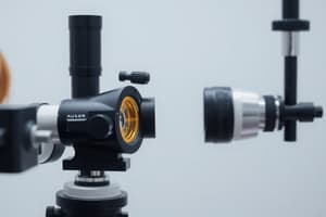
- The slit lamp is a crucial tool for assessing the anterior segment of the eye.
- It delivers excellent image quality and provides a stereoscopic view.
- Flexible illumination and magnification enhance its versatility.
Slit Lamp Components
- Patient support (chin rest and forehead rest)
- Illumination system
- Observation system
Observation System Components
- Eyepiece
- Magnification control
Magnification Levels
- Levels of magnification are critical in ophthalmology procedures:
- Low (6x): overview of the eye (lids, conjunctiva, cornea, limbus)
- Medium (10-16x): detailed examination of the anterior structures.
- High (20-25x): deeper examination of corneal layers (epithelium, stroma, endothelium)
- Very High (30-40x): detailed examination, identifying vacuoles, microcysts and dystrophies.
Slit Beam Height Setting
- The height of the slit beam can be adjusted using a dial on the illumination arm.
- A mm gauge display can be used for setting the correct height.
Diffuser Filters
- The diffuser filter is essential to prevent a focused beam from damaging the eye.
- Ground glass filters are often implemented to achieve diffuse illumination.
Slit Lamp Filters
- Heat reduction filter
- Neutral density filter
- Cobalt blue + Wratten filter
- Red free filter
Wratten 12 Filter
- A yellow filter used in conjunction with the observation system to enhance fluorescein's contrast.
- Commonly hand-held, though some slit lamps have inbuilt options.
Slit Lamp Uses
- Pachymetry
- Gonioscopy
- Fundoscopy
- Laser photocoagulation
- Contact tonometry
- Anterior eye assessment
Contact Lens Fitting Routine
- History & symptoms
- Baseline measurements: key step to determine the initial status.
- Keratometry: Measurement of corneal curvature. Vital for contact lens selection.
- Anterior eye assessment
- Selection of lens: based on the initial data.
- Insertion of lens: placing the lens onto the eye.
- Fit assessment: evaluating if the lens fits well to the eye. Crucial to ensuring a comfortable wearing experience.
- Over refraction: further adjustment of the spectacle or contact lens prescription.
- Prescribe/refit: finalising the contact lens prescription.
Slit Lamp and Contact Lens Fitting Procedures
- Patient suitability assessment
- Recording baseline measurements
- Monitoring the contact lens fitting process, including scheduled and unscheduled visits.
- Assessing the contact lens' condition, including surface properties and wetting.
- Assessing ocular integrity.
Things to Remember
- Wipe down the slit lamp.
- Maintain effective hand hygiene.
- The focusing of fully corrected emmetropic individuals should be within +/-0.50D.
Slit Lamp Examination Techniques Overview (Techniques and Purpose)
- Direct illumination: Basic overview, fit assessment
- Diffuse illumination: General overview
- Parallelepiped: Detailed corneal observation, no endothelium assessment needed
- Optic section: Detailed corneal observation
- Indirect illumination: Detailed endothelium assessment, tear film, lens surface -Specular reflection: Endothelium, tear film, crystalline lens surface -Sclerotic scatter: Corneal surface -Conical beam: Anterior chamber -Retro-illumination: Corneal oedema and pathological changes. -Van Herick's technique: Anterior chamber angle assessment
Diffuse Illumination Setup
- Angle: 30-45° between the microscope and illumination.
- Slit Width: Widest setting.
- Filter: Diffusing filter.
- Magnification: Low to medium.
- Illumination: Medium to high.
Diffuse Illumination Observation
- Good overall view of the eye (lids, lashes, conjunctiva, sclera, redness, iris, pupil, gross pathology, and media opacities).
- No fine details.
- Useful for a general survey.
Parallelepiped Setup
- Angle: 40-60° from the straight ahead position.
- Slit: Narrow, 1-2mm width.
- Filter: No filter initially.
- Magnification: 16x.
- Illumination: Medium.
- Microscope directly in front of the cornea.
Parallelepiped Observation
- Detect and examine corneal structures or defects.
- Wider beam illumination is necessary to evaluate the depth and extent of corneal scarring or foreign bodies.
- Corneal nerve features, appearing as fine white threads.
Optic Section Setup
- Angle: 45-60°
- Slit: Narrowest possible (careful to avoid turning the illumination off).
- Magnification: Medium to Maximum.
- Illumination: Maximum.
Optic Section Observation
- Localize: Nerve fibers, blood vessels, infiltrates, cataracts.
- Assess: Corneal thickness, thinning, foreign bodies, opacities (percentage of thickness).
- Visualize: Wide slices of the stroma.
Decoupling - Indirect Observation
- Separate rotation points for the illumination and observation systems.
Illumination Techniques
- Sclerotic scatter: -Beam focused on the limbus. -Angle(45-60°). -Slit: Narrow slit of 0.5 mm. -Filter: No filter -Magnification: 10-16x. -Illumination: Highest setting.
- Conical beam: -Angle(45-60°) -Slit: Parallelepiped, 1-2mm height. -Filter: No filter initially. -Magnification: Medium(10x) to maximum. -Illumination: Maximum -Focusing beam between the cornea and anterior lens surface and dark zone between cornea and anterior lens observed. -Useful for assessing anterior chamber transparency, floating cells and flare in anterior uveitis
Retro Illumination Overview
- Techniques specifically used for corneal edema and pathological changes.
- Used to assess vessels and neovascularizations.
Direct Retro Illumination Setup
- Angle: 30-50° for iris/lens; 0 - 10° for retina.
- Slit: Parallelepiped max height; parallelepiped, pupil size.
- Magnification: 16x initially, and increase
- Illumination: Medium-high; Low-medium setting
Direct Retro Illumination Observation
- Assess objects close to the cornea.
- Identify infiltrations, small scars, corneal vessels, microcysts, and vacuoles.
Indirect Retro Illumination Observation
- Infiltrations, small scars, corneal vessels, micro cysts, and vacuoles.
- Corneal features seen against a dark background.
Anterior Chamber Angle Assessment
- Assessment of ACA
- Assessment of ACD
- Van Herrick's grading system for assessing the anterior chamber angle (ACA).
- Grades 0 -4 based on the relative width of the anterior chamber and visible corneal structures (with the relationship between cornea and iris)
Studying That Suits You
Use AI to generate personalized quizzes and flashcards to suit your learning preferences.