Podcast
Questions and Answers
What are the two essential molecular features required for the diagnosis of oligodendroglioma, IDH mutant and 1p/19q codeleted, according to the CNS WHO 2021 definition?
What are the two essential molecular features required for the diagnosis of oligodendroglioma, IDH mutant and 1p/19q codeleted, according to the CNS WHO 2021 definition?
The two essential molecular features are the presence of an IDH1 or IDH2 mutation and codeletion of chromosome arms 1p and 19q. Both of these molecular alterations must be present for a definitive diagnosis.
Describe the typical microscopic appearance of oligodendroglioma cells, and explain the artifact that contributes to their characteristic 'fried egg' appearance.
Describe the typical microscopic appearance of oligodendroglioma cells, and explain the artifact that contributes to their characteristic 'fried egg' appearance.
Oligodendroglioma cells typically appear as closely packed cells with small, round, monotonous nuclei. The 'fried egg' appearance is due to perinuclear clearing, which is a formalin fixation artifact and not seen in frozen sections or smear preparations.
What is 'chicken wire vasculature' and why is it considered a characteristic feature of oligodendrogliomas?
What is 'chicken wire vasculature' and why is it considered a characteristic feature of oligodendrogliomas?
'Chicken wire vasculature' refers to the network of thin-walled, branching blood vessels seen in oligodendrogliomas. It is considered a characteristic feature because of its distinctive appearance under microscopy, aiding in histological diagnosis.
Explain the role of IDH mutations in the pathophysiology of oligodendroglioma, including the metabolic and epigenetic consequences.
Explain the role of IDH mutations in the pathophysiology of oligodendroglioma, including the metabolic and epigenetic consequences.
What is the most common presenting symptom in patients diagnosed with oligodendroglioma, and why is this symptom frequently associated with this type of brain tumor?
What is the most common presenting symptom in patients diagnosed with oligodendroglioma, and why is this symptom frequently associated with this type of brain tumor?
Differentiate between WHO grade 2 and grade 3 oligodendroglioma, IDH mutant and 1p/19q codeleted, based on histological features.
Differentiate between WHO grade 2 and grade 3 oligodendroglioma, IDH mutant and 1p/19q codeleted, based on histological features.
Describe the typical findings on MRI for oligodendroglioma, including characteristics related to contrast enhancement and diffusion restriction.
Describe the typical findings on MRI for oligodendroglioma, including characteristics related to contrast enhancement and diffusion restriction.
List three favorable prognostic factors and two unfavorable prognostic factors for patients with oligodendroglioma, IDH mutant and 1p/19q codeleted.
List three favorable prognostic factors and two unfavorable prognostic factors for patients with oligodendroglioma, IDH mutant and 1p/19q codeleted.
What is the standard initial treatment approach for oligodendroglioma, and what adjuvant therapies are typically considered?
What is the standard initial treatment approach for oligodendroglioma, and what adjuvant therapies are typically considered?
Explain why immunohistochemistry for IDH1 R132H is a useful diagnostic tool for oligodendroglioma, and what are its limitations?
Explain why immunohistochemistry for IDH1 R132H is a useful diagnostic tool for oligodendroglioma, and what are its limitations?
Describe the significance of 1p/19q codeletion in the context of oligodendroglioma diagnosis and prognosis.
Describe the significance of 1p/19q codeletion in the context of oligodendroglioma diagnosis and prognosis.
Name two molecular alterations, besides IDH mutation and 1p/19q codeletion, that are frequently found in oligodendrogliomas and their potential roles in tumorigenesis.
Name two molecular alterations, besides IDH mutation and 1p/19q codeletion, that are frequently found in oligodendrogliomas and their potential roles in tumorigenesis.
How does the staining pattern of p53 and ATRX help in differentiating oligodendroglioma, IDH mutant and 1p/19q codeleted from astrocytoma, IDH mutant?
How does the staining pattern of p53 and ATRX help in differentiating oligodendroglioma, IDH mutant and 1p/19q codeleted from astrocytoma, IDH mutant?
In the differential diagnosis of oligodendroglioma, list two tumor types with 'oligo-like' morphology and explain how they are distinguished from true oligodendrogliomas.
In the differential diagnosis of oligodendroglioma, list two tumor types with 'oligo-like' morphology and explain how they are distinguished from true oligodendrogliomas.
What is the role of MGMT promoter methylation in oligodendroglioma, and how might it influence treatment strategies?
What is the role of MGMT promoter methylation in oligodendroglioma, and how might it influence treatment strategies?
Explain the concept of 'secondary structures of Scherer' in the context of oligodendroglioma and provide examples.
Explain the concept of 'secondary structures of Scherer' in the context of oligodendroglioma and provide examples.
What are microcalcifications (calcospherites) and why are they considered a characteristic histological feature of oligodendrogliomas?
What are microcalcifications (calcospherites) and why are they considered a characteristic histological feature of oligodendrogliomas?
Describe the typical age group and location predilection for oligodendroglioma, IDH mutant and 1p/19q codeleted, within the central nervous system.
Describe the typical age group and location predilection for oligodendroglioma, IDH mutant and 1p/19q codeleted, within the central nervous system.
How is droplet digital PCR (ddPCR) used in the diagnosis of oligodendroglioma, and what advantage does it offer over Sanger sequencing?
How is droplet digital PCR (ddPCR) used in the diagnosis of oligodendroglioma, and what advantage does it offer over Sanger sequencing?
Explain how magnetic resonance spectroscopy (MRS) can be used as a non-invasive method to suggest the presence of an IDH mutation in suspected oligodendrogliomas.
Explain how magnetic resonance spectroscopy (MRS) can be used as a non-invasive method to suggest the presence of an IDH mutation in suspected oligodendrogliomas.
Flashcards
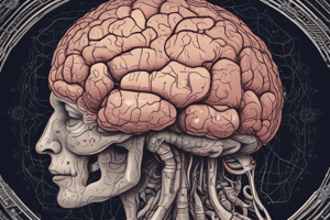
Oligodendroglioma Definition
Oligodendroglioma Definition
Diffusely infiltrating glioma with IDH1 or IDH2 mutation and codeletion of chromosome arms 1p and 19q.
Oligodendroglioma Morphology
Oligodendroglioma Morphology
Round monotonous nuclei, perinuclear halos, chicken wire vasculature, microcalcifications, and microcysts.
Oligodendroglioma Epidemiology
Oligodendroglioma Epidemiology
Peak incidence in fourth and fifth decades; slight male predominance; rare in infants and children.
Oligodendroglioma Common Locations
Oligodendroglioma Common Locations
Signup and view all the flashcards
Oligodendroglioma Pathophysiology
Oligodendroglioma Pathophysiology
Signup and view all the flashcards
Oligodendroglioma Clinical Features
Oligodendroglioma Clinical Features
Signup and view all the flashcards
Oligodendroglioma Grading
Oligodendroglioma Grading
Signup and view all the flashcards
Oligodendroglioma Diagnosis Methods
Oligodendroglioma Diagnosis Methods
Signup and view all the flashcards
Oligodendroglioma Radiology
Oligodendroglioma Radiology
Signup and view all the flashcards
Favorable Prognostic Factors
Favorable Prognostic Factors
Signup and view all the flashcards
Unfavorable Prognostic Factors
Unfavorable Prognostic Factors
Signup and view all the flashcards
Oligodendroglioma Treatment
Oligodendroglioma Treatment
Signup and view all the flashcards
Histologic Features
Histologic Features
Signup and view all the flashcards
Grade 3 Histologic Features
Grade 3 Histologic Features
Signup and view all the flashcards
Positive Stains
Positive Stains
Signup and view all the flashcards
Key Molecular Features
Key Molecular Features
Signup and view all the flashcards
Other Molecular Alterations
Other Molecular Alterations
Signup and view all the flashcards
Distinguishing from Astrocytoma
Distinguishing from Astrocytoma
Signup and view all the flashcards
Other Oligo-like tumors
Other Oligo-like tumors
Signup and view all the flashcards
Epigenetic Change
Epigenetic Change
Signup and view all the flashcards
Study Notes
Oligodendroglioma, IDH Mutant and 1p/19q Codeleted
- Defined as a diffusely infiltrating glioma with IDH1 or IDH2 mutation and codeletion of chromosome arms 1p and 19q.
- Classified as CNS WHO grade 2 or 3.
Essential Features
- Requires both IDH1 or IDH2 mutation and 1p/19q whole arm codeletion for diagnosis.
- Exhibits morphology similar to nonneoplastic oligodendrocytes, including round, monotonous nuclei and perinuclear halos.
- Characterized by chicken wire vasculature, microcalcifications, and microcysts.
- Astrocytic differentiation does not rule out diagnosis if molecular features are present, including small gemistocytes, especially in grade 3 tumors.
Additional Considerations
- The presence of atypical features such as multinucleated giant cells or sarcomatous features doesn't preclude diagnosis if molecular features are present.
ICD Coding
- 9450/3: Oligodendroglioma, NOS (Not Otherwise Specified).
- 9451/3: Anaplastic oligodendroglioma.
Epidemiology
- Incidence in the United States stands at 0.23 cases per 100,000 population.
- CNS WHO grade 3 oligodendroglioma incidence is 0.11.
- 0.9% of all brain tumors are oligodendroglioma WHO grade 2 and 0.4% are oligodendroglioma WHO grade 3.
- Peak incidence occurs in the fourth and fifth decades of life.
- Rare in infants and children.
- Slight male predominance.
Sites
- Typically involves white and gray matter.
- Can occur anywhere in the neuraxis, but most commonly in the frontal lobes (59%), temporal lobes (14%), and parietal lobes (10%).
- Rarely found in midline structures, brainstem, cerebellum, or spinal cord.
Pathophysiology
- The cell of origin for oligodendroglioma is unknown.
- IDH mutation is likely the initiating event, preceding 1p/19q codeletion.
- IDH mutations lead to increased production of 2-hydroxyglutarate (2HG).
- Elevated 2HG inhibits histone demethylation, causing a hypermethylation phenotype (G-CIMP).
Etiology
- Generally sporadic without significant known risk factors.
- Rare familial instances and genetic alterations are associated with increased risk.
Clinical Features
- Approximately 67% of patients present with seizures.
- Other symptoms include headache, focal neurologic deficits, or cognitive/mental status changes, depending on the tumor location.
Grading
- WHO grade 2 tumors lack anaplastic features.
- WHO grade 3 tumors exhibit prominent anaplastic features like necrosis, microvascular proliferation, or brisk mitotic activity.
- CDKN2A homozygous deletion may indicate CNS WHO grade 3.
Diagnosis
- Diagnosed via MRI, followed by stereotactic brain biopsy or surgical resection.
- Methods to detect IDH gene mutation: Immunohistochemistry for IDH1 R132H, Sanger sequencing, ddPCR, Next generation sequencing.
- Methods to detect 1p / 19q codeletion: FISH, array comparative genomic hybridization, PCR.
Radiology Description
- CT scans show mixed density (hypodense and isodense) in cortex or subcortical white matter, with high attenuation areas from calcifications.
- MRI shows heterogeneous appearance on T1 and T2 weighted imaging, poorly defined borders, and cystic changes.
- Contrast enhancement is present in < 20% of WHO grade 2 and > 70% of WHO grade 3 tumors.
Prognostic Factors
Favorable features:
- Younger age at diagnosis.
- Frontal lobe location.
- Presentation with seizures.
- Macroscopically complete surgical resection.
- CNS WHO grade 2 histology.
- Higher postoperative Karnofsky score.
Unfavorable features:
- Contrast enhancement on MRI.
- CNS WHO grade 3 histology.
- CDKN2A / CDKN2B homozygous deletion.
- Median overall survival is 11.6 years, with a 10 year survival rate of 51 - 63%.
- Local recurrence and malignant transformation are common.
Gross Description
- Variably well defined, gray-pink mass.
- May have a gelatinous consistency due to mucoid change.
- Cystic degeneration, calcifications, hemorrhage, or necrosis may be present.
Frozen Section Description
- Moderately cellular, diffusely infiltrating neoplasm.
- Mild to moderate nuclear atypia.
- Round nuclei with speckled chromatin.
- Calcifications, perineuronal satellitosis, or perivascular accumulation may be seen.
- Perinuclear halos are not seen on frozen sections or smear preparations.
- Anaplastic features may be seen in WHO grade 3 tumors.
Microscopic (Histologic) Description
- Closely packed cells with small, round, monotonous nuclei.
- Perinuclear clearing (fried egg appearance) is a formalin fixation artifact.
- Network of thin walled, branching blood vessels (chicken wire vasculature).
- Microcalcifications (calcospherites) are characteristic.
- Perineural, perivascular, or subpial aggregates of tumor cells (secondary structures of Scherer).
- Occasional mitoses and moderate nuclear atypia are consistent with grade 2.
- Well differentiated / fibrillary astrocytic morphology may be present.
Features of CNS WHO grade 3:
- Microvascular proliferation.
- Necrosis.
- Brisk mitotic activity.
Positive Stains
- IDH1 (R132H) is positive in > 90% of oligodendrogliomas.
- Olig2, GFAP.
- ATRX (retained; wildtype pattern).
- p53 (weak staining in rare cells; wildtype staining pattern).
- Ki67: Grade 2 tumors typically show < 5% positive nuclei, while Grade 3 tumors generally show > 10%.
- The trio of IDH1 (R132H), ATRX, and p53 distinguishes oligodendroglioma from IDH mutant astrocytoma.
Molecular / Cytogenetics Description
- Requires demonstration of both IDH1 or IDH2 mutation and 1p/19q codeletion for diagnosis.
- Incomplete or partial deletions are not compatible with oligodendroglioma diagnosis.
- Common molecular alterations include TERT promotor mutation, CIC mutation, FUBP1 mutations, and NOTCH1 mutation.
- Molecular alterations associated with tumor progression: increased copy number alterations, CDKN2A / CDKN2B deletions, PIK3CA mutation, TCF12 mutation, MYC amplification.
- Epigenetic changes: Glioma CpG island methylator phenotype (G-CIMP), MGMT promotor methylation.
Sample Pathology Report
- Includes integrated diagnosis, histologic diagnosis, CNS WHO grade, and molecular information (IDH1 mutation, 1p / 19q codeletion, TERT promotor mutation).
Differential Diagnosis
- Astrocytoma, IDH mutant: Lacks 1p/19q codeletion and TERT promotor mutations, harbors p53 or ATRX mutations.
- Other tumors with oligo-like morphology: Dysembryoplastic neuroepithelial tumor (DNET), central neurocytoma, polymorphous low grade neuroepithelial tumor of the young (PLNTY), clear cell ependymoma, metastatic clear cell carcinomas.
- Macrophage rich lesions: Stain positive with macrophage markers.
Studying That Suits You
Use AI to generate personalized quizzes and flashcards to suit your learning preferences.