Podcast
Questions and Answers
What characteristic allows acid-fast bacteria to retain the primary stain in a Gram stain procedure?
What characteristic allows acid-fast bacteria to retain the primary stain in a Gram stain procedure?
- The development of a capsule around the cell
- The presence of a thick peptidoglycan layer
- The formation of endospores
- The ability to resist decolorization by acid-alcohol (correct)
What type of bacteria is N.gonorrhoeae classified as?
What type of bacteria is N.gonorrhoeae classified as?
- Gram-positive cocci
- Acid-fast rods
- Gram-negative bacilli
- Gram-negative diplococci (correct)
What methodology enhances the contrast of samples in transmission electron microscopy?
What methodology enhances the contrast of samples in transmission electron microscopy?
- Embedding in paraffin wax
- Staining with iodine solution
- Applying fluorescent dyes
- Using heavy metals such as lead (correct)
The presence of acid-fast rods in a sample suggests which possible condition?
The presence of acid-fast rods in a sample suggests which possible condition?
What do the dots visible in the electron microscopy images of a spirochete represent?
What do the dots visible in the electron microscopy images of a spirochete represent?
What principle does scanning acoustic microscopy (SAM) rely on?
What principle does scanning acoustic microscopy (SAM) rely on?
What type of microscopy provides a three-dimensional view of surfaces?
What type of microscopy provides a three-dimensional view of surfaces?
What is the primary purpose of staining microorganisms?
What is the primary purpose of staining microorganisms?
How is bacteria affected by a basic dye during staining?
How is bacteria affected by a basic dye during staining?
Which component is NOT part of the Gram stain procedure?
Which component is NOT part of the Gram stain procedure?
What is the resolving power of a transmission electron microscope (TEM)?
What is the resolving power of a transmission electron microscope (TEM)?
In what situation would you use a negative stain?
In what situation would you use a negative stain?
Which microscopy technique uses a beam of electrons for imaging?
Which microscopy technique uses a beam of electrons for imaging?
What is the method to calculate the total magnification of an object in microscopy?
What is the method to calculate the total magnification of an object in microscopy?
What is the maximum resolution achievable with a compound light microscope?
What is the maximum resolution achievable with a compound light microscope?
Which microscopy technique is most suitable for observing unstained cells?
Which microscopy technique is most suitable for observing unstained cells?
What color do acid-fast microbes appear after undergoing the acid-fast staining process?
What color do acid-fast microbes appear after undergoing the acid-fast staining process?
What does darkfield microscopy primarily use to visualize organisms?
What does darkfield microscopy primarily use to visualize organisms?
What is the primary purpose of negative staining in microbiology?
What is the primary purpose of negative staining in microbiology?
What is the main use of fluorescence microscopy?
What is the main use of fluorescence microscopy?
Which microscope type is best for observing specimens in three dimensions?
Which microscope type is best for observing specimens in three dimensions?
What is the primary unit of measurement for microorganisms?
What is the primary unit of measurement for microorganisms?
How does a DIC microscope enhance the viewing of cells?
How does a DIC microscope enhance the viewing of cells?
In the Gram stain process, what role does the mordant play?
In the Gram stain process, what role does the mordant play?
Which lens does light first pass through in a compound microscope?
Which lens does light first pass through in a compound microscope?
What increases the difference between the refractive indexes of a specimen and its medium?
What increases the difference between the refractive indexes of a specimen and its medium?
Why are acid-fast cells resistant to most stains?
Why are acid-fast cells resistant to most stains?
What does a microscope with a resolution of 0.2 nm indicate?
What does a microscope with a resolution of 0.2 nm indicate?
What happens to gram-negative cells during the decolorization step in the Gram stain?
What happens to gram-negative cells during the decolorization step in the Gram stain?
In two-photon microscopy (TPM), what type of light is used to illuminate the specimen?
In two-photon microscopy (TPM), what type of light is used to illuminate the specimen?
What is a similarity between brightfield and darkfield microscopy?
What is a similarity between brightfield and darkfield microscopy?
What is the result if the counterstain safranin is omitted in a Gram staining procedure?
What is the result if the counterstain safranin is omitted in a Gram staining procedure?
What occurs when the acid-fast stain's primary stain penetrates the cell walls?
What occurs when the acid-fast stain's primary stain penetrates the cell walls?
What distinguishes electron microscopy from light microscopy?
What distinguishes electron microscopy from light microscopy?
For which application is Transmission Electron Microscopy (TEM) primarily used?
For which application is Transmission Electron Microscopy (TEM) primarily used?
Why is a negative stain not able to color a cell?
Why is a negative stain not able to color a cell?
What is one key reason the Gram staining procedure is valuable in microbiology?
What is one key reason the Gram staining procedure is valuable in microbiology?
Flashcards
What unit is used to measure the size of microorganisms?
What unit is used to measure the size of microorganisms?
Micrometers (µm) are commonly used to measure the size of microorganisms. 1 µm equals 10^-6 meters.
What is smaller than micrometers, and is used to measure viruses?
What is smaller than micrometers, and is used to measure viruses?
Nanometers (nm) are even smaller than micrometers and are used to measure the size of viruses and other very small structures. 1 nm equals 10^-9 meters.
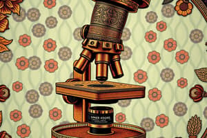
What type of microscope uses multiple lenses?
What type of microscope uses multiple lenses?
The compound light microscope uses multiple lenses to magnify the image of a specimen. It allows you to view small structures, like bacteria, that are invisible to the naked eye.
What is the total magnification of a microscope determined by?
What is the total magnification of a microscope determined by?
Signup and view all the flashcards
What is the ability to distinguish between two closely spaced objects called?
What is the ability to distinguish between two closely spaced objects called?
Signup and view all the flashcards
What microscopy technique uses a dark background to enhance visibility?
What microscopy technique uses a dark background to enhance visibility?
Signup and view all the flashcards
What microscopy technique uses a special condenser and objective lens to enhance contrast?
What microscopy technique uses a special condenser and objective lens to enhance contrast?
Signup and view all the flashcards
What microscopy technique uses fluorescent molecules to make structures appear bright?
What microscopy technique uses fluorescent molecules to make structures appear bright?
Signup and view all the flashcards
Scanning Acoustic Microscopy (SAM)
Scanning Acoustic Microscopy (SAM)
Signup and view all the flashcards
Electron Microscope
Electron Microscope
Signup and view all the flashcards
Transmission Electron Microscope (TEM)
Transmission Electron Microscope (TEM)
Signup and view all the flashcards
Scanning Electron Microscope (SEM)
Scanning Electron Microscope (SEM)
Signup and view all the flashcards
Staining
Staining
Signup and view all the flashcards
Smear
Smear
Signup and view all the flashcards
Simple Stain
Simple Stain
Signup and view all the flashcards
Differential Stains
Differential Stains
Signup and view all the flashcards
Resolving Power (Resolution)
Resolving Power (Resolution)
Signup and view all the flashcards
Darkfield Microscopy
Darkfield Microscopy
Signup and view all the flashcards
Phase-Contrast Microscopy
Phase-Contrast Microscopy
Signup and view all the flashcards
Differential Interference Contrast (DIC) Microscopy
Differential Interference Contrast (DIC) Microscopy
Signup and view all the flashcards
Fluorescence Microscopy
Fluorescence Microscopy
Signup and view all the flashcards
Confocal Microscopy
Confocal Microscopy
Signup and view all the flashcards
Two-Photon Microscopy (TPM)
Two-Photon Microscopy (TPM)
Signup and view all the flashcards
Scanning Acoustic Microscopy
Scanning Acoustic Microscopy
Signup and view all the flashcards
What makes acid-fast bacteria unique?
What makes acid-fast bacteria unique?
Signup and view all the flashcards
What's the purpose of an endospore stain?
What's the purpose of an endospore stain?
Signup and view all the flashcards
How did Ehrlich's observation of acid-fast bacteria influence disinfection?
How did Ehrlich's observation of acid-fast bacteria influence disinfection?
Signup and view all the flashcards
What are the characteristics of Neisseria gonorrhoeae?
What are the characteristics of Neisseria gonorrhoeae?
Signup and view all the flashcards
What are clue cells?
What are clue cells?
Signup and view all the flashcards
Acid-fast staining
Acid-fast staining
Signup and view all the flashcards
Negative staining
Negative staining
Signup and view all the flashcards
Endospore staining
Endospore staining
Signup and view all the flashcards
Flagella staining
Flagella staining
Signup and view all the flashcards
Total magnification
Total magnification
Signup and view all the flashcards
What is a mordant?
What is a mordant?
Signup and view all the flashcards
Decolorizer in Gram staining
Decolorizer in Gram staining
Signup and view all the flashcards
Counterstain
Counterstain
Signup and view all the flashcards
Study Notes
Observing Microorganisms Through a Microscope
- Metric Units: Microorganisms are measured in micrometers (µm) and nanometers (nm).
- Compound Light Microscope: The total magnification is calculated by multiplying the objective lens magnification by the ocular lens magnification. Maximum resolution is 0.2 µm, maximum magnification is 2000x.
- Microscope Types and Uses:
- Brightfield: Used for stained specimens.
- Darkfield: Used to observe small organisms.
- Phase-Contrast: Useful for observing living organisms.
- Differential Interference Contrast (DIC): Produces a three-dimensional image of living cells.
- Fluorescence: Specimens are stained with fluorochromes and illuminated with UV light. This produces bright objects against a dark background, useful for diagnostic procedures.
- Confocal: A specimen is stained with a fluorescent dye and illuminated with short-wavelength light. Produces 2D and 3D images.
- Two-Photon: Live specimen stained with a fluorescent dye and illuminated with long-wavelength light; a computer creates images.
- Scanning Acoustic Microscopy (SAM): Used to study living cells attached to surfaces.
- Electron Microscopy:
- Transmission Electron Microscopy (TEM): Shows thin sections of organisms (10,000-100,000x magnification, 2.5nm resolving power).
- Scanning Electron Microscopy (SEM): Provides three-dimensional views of surfaces (1000–10,000x magnification, 20nm resolving power).
- Scanned-Probe Microscopes: Produce three-dimensional images of surfaces (STM and AFM).
Preparing Specimens
- Preparing Smears: Technique involves fixing the microorganism to the slide so it doesn't move during microscopic examination.
- Staining: Coloring techniques used to make cellular structures more visible.
- Simple Staining: Uses a single basic dye to stain the entire cell.
- Differential Staining: Distinguishes between different types of bacteria. Examples include Gram stain and acid-fast stain. Gram stain differentiates Gram-positive and Gram-negative bacteria. Acid-fast stain is used with Mycobacterium and Nocardia.
- Special Staining: Techniques for specific structures or features. Examples include capsule stain, endospore stain, and flagella stain.
Studying That Suits You
Use AI to generate personalized quizzes and flashcards to suit your learning preferences.
Related Documents
Description
This quiz explores the various types of microscopes used for observing microorganisms and their specific applications. Learn about the principles of magnification, resolution, and the unique features of each microscope type. Test your knowledge on how to measure microorganisms as well.