Podcast
Questions and Answers
During the assessment of a patient with infective endocarditis, which finding would be most indicative of the condition?
During the assessment of a patient with infective endocarditis, which finding would be most indicative of the condition?
- Hypertension and bradycardia
- Peripheral edema and weight gain
- Elevated white blood cell count and decreased heart rate
- A new or changed heart murmur (correct)
A patient is diagnosed with subacute infective endocarditis. Which pre-existing condition is most likely to be present in this patient's history?
A patient is diagnosed with subacute infective endocarditis. Which pre-existing condition is most likely to be present in this patient's history?
- Aortic aneurysm
- Healthy heart valves with rapid onset of symptoms
- History of hypertension
- Preexisting valve disease with a longer clinical course (correct)
A patient with infective endocarditis is receiving IV antibiotic therapy. Which assessment finding requires immediate notification of the provider?
A patient with infective endocarditis is receiving IV antibiotic therapy. Which assessment finding requires immediate notification of the provider?
- Sudden onset of right-sided weakness (correct)
- Slight decrease in appetite
- Low-grade fever that has persisted for 3 days
- Complaints of mild fatigue
When teaching a patient about preventing infective endocarditis, which information should the nurse emphasize?
When teaching a patient about preventing infective endocarditis, which information should the nurse emphasize?
A patient is admitted with possible infective endocarditis. Which laboratory test result is most important in confirming the diagnosis?
A patient is admitted with possible infective endocarditis. Which laboratory test result is most important in confirming the diagnosis?
A patient undergoing treatment for infective endocarditis asks how long the IV antibiotic therapy will last. What is the typical duration the nurse should tell the patient?
A patient undergoing treatment for infective endocarditis asks how long the IV antibiotic therapy will last. What is the typical duration the nurse should tell the patient?
The nurse assesses a client with mitral valve stenosis. Which assessment finding is most closely associated with this condition?
The nurse assesses a client with mitral valve stenosis. Which assessment finding is most closely associated with this condition?
Which pathophysiological change occurs as a result of mitral stenosis that directly impacts pulmonary circulation?
Which pathophysiological change occurs as a result of mitral stenosis that directly impacts pulmonary circulation?
A patient with mitral valve regurgitation is at risk for developing atrial fibrillation. What is the primary reason for this risk?
A patient with mitral valve regurgitation is at risk for developing atrial fibrillation. What is the primary reason for this risk?
Which clinical manifestation is most indicative of acute mitral regurgitation?
Which clinical manifestation is most indicative of acute mitral regurgitation?
A patient with mitral valve prolapse is being discharged. Which lifestyle modification should the nurse include in the teaching plan?
A patient with mitral valve prolapse is being discharged. Which lifestyle modification should the nurse include in the teaching plan?
Which assessment finding in a patient with aortic stenosis indicates a critical reduction in cardiac output?
Which assessment finding in a patient with aortic stenosis indicates a critical reduction in cardiac output?
A patient with aortic regurgitation reports experiencing shortness of breath when lying flat. Which term should the nurse use to document this?
A patient with aortic regurgitation reports experiencing shortness of breath when lying flat. Which term should the nurse use to document this?
A client with symptomatic aortic stenosis is scheduled for valve replacement. Which statement best reflects the rationale for this intervention?
A client with symptomatic aortic stenosis is scheduled for valve replacement. Which statement best reflects the rationale for this intervention?
The nurse clarifies that the focus during the acute phase of rheumatic fever with carditis is on preventing permanent scarring of which structure?
The nurse clarifies that the focus during the acute phase of rheumatic fever with carditis is on preventing permanent scarring of which structure?
The nurse is caring for a patient who is being treated for infective endocarditis. What is a priority nursing intervention for this patient?
The nurse is caring for a patient who is being treated for infective endocarditis. What is a priority nursing intervention for this patient?
A patient is diagnosed with an aortic aneurysm. What is the greatest risk associated with this condition that the nurse should monitor for?
A patient is diagnosed with an aortic aneurysm. What is the greatest risk associated with this condition that the nurse should monitor for?
The nurse is assessing a patient with a known thoracic aortic aneurysm. Which new assessment finding requires immediate action?
The nurse is assessing a patient with a known thoracic aortic aneurysm. Which new assessment finding requires immediate action?
An abdominal aortic aneurysm (AAA) is discovered during a routine physical exam. What action should the nurse avoid?
An abdominal aortic aneurysm (AAA) is discovered during a routine physical exam. What action should the nurse avoid?
A patient post-operative following aortic aneurysm repair, what nursing intervention is critical for assessing the patient's renal status?
A patient post-operative following aortic aneurysm repair, what nursing intervention is critical for assessing the patient's renal status?
Following an endovascular aneurysm repair (EVAR), a patient reports pain in the lower back. What immediate complication should the nurse suspect?
Following an endovascular aneurysm repair (EVAR), a patient reports pain in the lower back. What immediate complication should the nurse suspect?
A patient undergoing aortic aneurysm repair is at risk for developing bowel ischemia. Which assessment finding is indicative of this complication?
A patient undergoing aortic aneurysm repair is at risk for developing bowel ischemia. Which assessment finding is indicative of this complication?
The nurse is providing discharge teaching for a patient who underwent surgical repair of an aortic aneurysm. Which instruction is most important to include?
The nurse is providing discharge teaching for a patient who underwent surgical repair of an aortic aneurysm. Which instruction is most important to include?
A patient is diagnosed with a 'true aneurysm'. What is the defining characteristic of a true aneurysm?
A patient is diagnosed with a 'true aneurysm'. What is the defining characteristic of a true aneurysm?
After open repair of an abdominal aortic aneurysm, a client has absent pedal pulses. Which nursing intervention is most appropriate?
After open repair of an abdominal aortic aneurysm, a client has absent pedal pulses. Which nursing intervention is most appropriate?
Flashcards
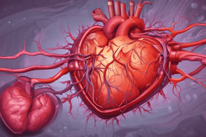
Infective Endocarditis
Infective Endocarditis
Infection of the inner heart layer, including valves; prognosis improved with antibiotics.
Subacute Endocarditis
Subacute Endocarditis
Preexisting valve disease with a longer clinical course.
Acute Endocarditis
Acute Endocarditis
Infective endocarditis affecting healthy valves with a rapidly progressive course.
Infective Endocarditis Symptoms
Infective Endocarditis Symptoms
Signup and view all the flashcards
Valvular Stenosis
Valvular Stenosis
Signup and view all the flashcards
Valvular Regurgitation
Valvular Regurgitation
Signup and view all the flashcards
Mitral Stenosis effects
Mitral Stenosis effects
Signup and view all the flashcards
Main Cause of Mitral Stenosis
Main Cause of Mitral Stenosis
Signup and view all the flashcards
Mitral Stenosis Sounds
Mitral Stenosis Sounds
Signup and view all the flashcards
Mitral Valve Prolapse
Mitral Valve Prolapse
Signup and view all the flashcards
MVP Manifestations
MVP Manifestations
Signup and view all the flashcards
Aortic Valve Stenosis
Aortic Valve Stenosis
Signup and view all the flashcards
Clinical Signs of Aortic Stenosis
Clinical Signs of Aortic Stenosis
Signup and view all the flashcards
Aortic Valve Regurgitation
Aortic Valve Regurgitation
Signup and view all the flashcards
Acute Aortic Regurgitation Signs
Acute Aortic Regurgitation Signs
Signup and view all the flashcards
Chronic Aortic Regurgitation Signs
Chronic Aortic Regurgitation Signs
Signup and view all the flashcards
Meds for Valvular Disease
Meds for Valvular Disease
Signup and view all the flashcards
Valve Repair/Replacement
Valve Repair/Replacement
Signup and view all the flashcards
True Aneurysm
True Aneurysm
Signup and view all the flashcards
False Aneurysm
False Aneurysm
Signup and view all the flashcards
Thoracic Aneurysm Signs
Thoracic Aneurysm Signs
Signup and view all the flashcards
Abdominal Aneurysm Signs
Abdominal Aneurysm Signs
Signup and view all the flashcards
EVAR
EVAR
Signup and view all the flashcards
Ruptured Aneurysm
Ruptured Aneurysm
Signup and view all the flashcards
Postoperative Cardiac Mgmt
Postoperative Cardiac Mgmt
Signup and view all the flashcards
Study Notes
- Nursing 432 Perfusion disorders include:
- Infective Endocarditis
- Cardiac Valvular Disorders
- Cardiomyopathy
- Aortic Aneurysms
Infectious Diseases of the Heart
- Any layer of the heart may be affected by an infectious process
- Diseases are named according to which layer of the heart is affected (epi-, myo-, endo-carditis)
- Diagnosis is based on symptoms and echocardiogram results
- Blood cultures can identify the infectious agent and monitor therapy
- Treatment involves appropriate antimicrobial therapy, requiring the full course
- Patient education is essential for infection prevention and health promotion
Infective Endocarditis (IE)
- IE involves infection of the inner heart layer, including the valves
- Antibiotic therapy has improved the prognosis in the past few years
- IE can be classified as subacute, acute, or by the cause or site of involvement
- Subacute IE involves preexisting valve disease and has a longer clinical course
- Acute IE affects healthy valves and is rapidly progressive
- Organisms causing IE include bacteria such as Strep and Staph, viruses, and fungi
- Key risk factors include age, IV drug use, prosthetic valves and the use of intravascular, cardiac devices, and renal dialysis
Clinical Manifestations of Infective Endocarditis
- Nonspecific signs/symptoms include:
- Low-grade fever (90%)
- Chills
- Weakness
- Malaise
- Fatigue
- Anorexia
- Subacute form signs/symptoms include:
- Arthralgias
- Myalgias
- Back pain
- Abdominal discomfort
- Weight loss
- Headache
- Clubbing of fingers
- Other signs/symptoms include:
- Murmur in most patients
- Heart failure
- Manifestations secondary to embolism include spleen, kidneys, limbs, brain, and lungs
Diagnostic Studies for Infective Endocarditis
- Obtain history
- Laboratory tests:
- Blood cultures (30 min apart; 2 different sites)
- CBC with differential (mild leukocytosis if acute type)
- ESR, C-reactive protein (CRP)
- Echocardiography
- Chest x-ray (to check for cardiomegaly)
- ECG (check 1st or 2nd degree AV block)
- Cardiac catheterization checks valve function, if surgery is needed
- Major diagnostic criteria:
- Requires at least two of the following:
- Two positive blood cultures 12 hours apart
- New or changed heart murmur
- Positive mass or vegetation noted by echocardiogram
- Requires at least two of the following:
Infective Endocarditis: Nursing Implementation
- Administer antibiotic therapy for 4-6 weeks
- Assess home setting
- Monitor lab data, including blood cultures
- Assess IV lines
- Use coping strategies
- Prevent immobility issues: compression stockings, ROM, C&DB q2h
- Patient teaching regarding health promotion includes:
- Disease background
- Avoid people with infections, stress, and fatigue
- Oral hygiene
- Treatment program for IV drug use
- Follow-up care and early treatment of common infections are critical
- Monitor body temperature, notify provider for any recurrent infection symptoms (fever, chills, fatigue)
- Complications include any stroke, pulmonary edema, and heart failure.
Valvular Heart Disease
- The heart has two atrioventricular valves: mitral and tricuspid
- The heart has two semilunar valves: aortic and pulmonic
- Types of valvular heart disease depend on the valve(s) affected
- Types of valvular heart disease depend on the type of functional alteration(s): stenosis or regurgitation
- Study focuses on the valves in the L side of the heart (mitral and aortic)
Valvular Heart Disease - Stenosis
- Stenosis is the constriction or narrowing of valves
- Valve orifice is smaller meaning forward blood flow is impeded
- Pressure differences reflect the degree of stenosis
Valvular Heart Disease - Regurgitation
- Regurgitation is the incompetence/insufficiency of valves
- Incomplete closure of valve leaflets
- Results in backward flow of blood
Mitral Valve Disease - Stenosis
- Adult causes:
- Rheumatic heart disease (scarring of valve leaflets and chordae tendinae, adhesions between commissures of leaflets)
- Decreased blood flow from LA → LV, increased LA pressure and volume, increased pressure in pulmonary BVs
- Risk factor for atrial fibrillation
- Clinical manifestations:
- Exertional dyspnea
- Fatigue, chest pain (perfusion )
- Seizures/stroke (emboli)
- Heart sounds include:
- Loud S₁
- Low-pitched diastolic murmur
- Palpitations
Mitral Valve Disease - Regurgitation
- Damage occurs from MI, IE, mitral valve prolapse, chronic rheumatic heart disease, and ischemic papillary muscle
- Incomplete closure = Backward blood flow
- Acute MR: pulmonary edema
- Chronic MR: L atrial enlargement, ventricular hypertrophy = Decrease in CO
- Acute manifestations: thready peripheral pulses and cool, clammy extremities
- Chronic manifestations: asymptomatic for years until LV failure develops (weakness, fatigue)
Mitral Valve Prolapse
- Mitral valve leaflets prolapse up into left atrium during systole; unknown cause; familial incidence in some
- Usually benign (valve closes effectively); dx with echocardiography; treated with beta blockers or valve surgery if regurgitation
- Clinical manifestations: only 10%
- Systolic murmur (regurgitation)
- Dysrhythmias (palpitations, light-headed, dizzy)
- Infective endocarditis can occur
- Chest pain unresponsive to nitrates
- Teaching: healthy lifestyle (diet, hydration, exercise, avoid stimulants such as caffeine)
Aortic Valve Disease - Stenosis
- Congenital or degenerative
- Obstructs blood flow from LV to aorta, LV hypertrophy and ↑ myocardial O2 consumption, ↓ CO, pulm HTN, and HF
- Clinical manifestations: angina, syncope, DOE, murmur, S4
- Use nitro cautiously (reduces preload and BP, which can worsen chest pain)
Aortic Valve Disease - Regurgitation
- Can be acute or chronic
- Backward blood flow from aorta into LV
- Manifestations: acute AR
- Severe dyspnea
- Chest pain
- Decreased BP, cardiogenic shock; life-threatening emergency
- Manifestations: chronic AR
- Asymptomatic for years
- LV dilation and hypertrophy
- Decreased myocardial contractility
- Pulmonary HTN and R HF
- DOE
- Orthopnea
- PND
- Angina; "water-hammer” pulse if severe, S3 or S4, murmur
Valvular Heart Disease Interprofessional Care
- Diagnosis:
- Patient's history/physical exam
- Chest CT or Xray
- Echocardiogram
- ECG
- Cardiac cath
- Conservative treatment includes:
- Prophylactic antibiotics to prevent recurrent RF, IE
- Depends on valve involved and severity
- Prevent exacerbations of HF, pulmonary edema, thromboembolism, recurrent endocarditis
- Meds to treat, control HF include:
- Vasodilators (e.g., nitrates, ACE inhibitors)
- Positive inotropes (e.g., digoxin)
- Diuretics
- ẞ-Adrenergic blockers
- Antidysrhythmic drugs, including Ca++ channel blockers
Valvular Heart Disease Interprofessional Care - Surgical Therapy
- Valve repair is the surgical procedure of choice
- Lower mortality
- May not restore total valve function
- Includes Commissurotomy (valvulotomy)
- Valvuloplasty: open; minimally invasive
- Annuloplasty
- Valve Replacement can be mechanical or biologic
- Mechanical (artificial) valve
- Last longer; risk of thromboembolism
- Long-term anticoagulation
- Biologic (tissue)
- Bovine, porcine, and human
- No anticoagulation but will need replaced faster
Aortic Aneurysms - Etiology and Risk Factors
- Causes:
- Degenerative
- Congenital
- Mechanical (penetrating or blunt trauma)
- Inflammatory
- Infectious
- Risk Factors:
- Increased age and in men
- HTN and CAD
- Family history
- High cholesterol, ↑ BMI
- Leg PAD, carotid artery disease, previous stroke
- Smoking
- Whites and Native Americans are at ↑ risk than African Americans, Hispanics, Asian
Aortic Aneurysms - Classification
- True aneurysm:
- Wall of artery forms aneurysm
- At least 1 vessel layer is still intact
- Fusiform or saccular
- False aneurysm:
- Also called pseudoaneurysm
- Not an aneurysm
- Disruption of all layers of arterial wall: results in bleeding contained by surrounding
Aortic Aneurysm (AA)- Clinical Manifestations
- Thoracic AAs are:
- Often asymptomatic
- Most common manifestation: deep diffuse chest pain; may extend to interscapular area
- Ascending aorta/aortic arch symptoms include:
- Angina, TIAS
- Coughing and SOB
- Hoarseness +/- dysphagia
- If presses on superior VC: distended neck veins; face and arm edema
- Abdominal AA (AAA)
- Often asymptomatic
- Frequently detected on physical exam or treatment of other problem (i.e., CT scan, abdominal x-ray)
- May mimic pain assoc. with abdominal or back disorders
- May spontaneously embolize Causing “blue toe syndrome”: patchy mottling of feet/toes with palpable pedal pulses
- DON'T PALPATE!
Aortic Aneurysm - Complications
- Rupture serious complication:
- Rupture into retroperitoneal space with bleeding may be tamponaded by surrounding structures, thus preventing exsanguination and death with severe back pain
- May/may not have back/flank ecchymosis (Grey Turner sign)
- Rupture into thoracic or abdominal cavity with massive hemorrhage; most do not survive long enough to get to hospital
Aortic Aneurysm : EVAR: Interprofessional Care
- Endovascular Aneurysm Repair (EVAR) is an alternative to conventional surgery
- Sutureless aortic Dacron graft delivered via sheath and placed into abdominal aorta aneurysm via femoral artery cutdown
- Graft deployed against vessel wall by balloon inflation; anchored to vessel by series of small hooks
- Blood flows through graft; aneurysm shrinks over time since it cannot expand
- Advantages: less risk than surgery, shorter recovery, higher patient satisfaction, less cost
- Disadvantages: endoleak, aneurysm growth or rupture, aortic dissection, bleeding, infection
Aortic Aneurysm : Interprofessional Care
- Open surgical procedure includes:
- If ruptured, emergency surgery with a 90% mortality from AAAS
- Preop: hydration; stabilize electrolytes, coagulation, and hematocrit
- Open aneurysm repair (OAR): incise diseased segment; remove intraluminal thrombus or plaque
- Insert synthetic graft: Dacron or polytetrafluoroethylene (PTFE)
- Suture native aortic wall around graft, which acts as a protective cover.
Nursing Management of AA: Postoperative Care
- Monitor cardiovascular status:
- Continuous ECG monitoring
- Electrolyte monitoring
- ABG monitoring
- O2 administration
- Antidysrhythmic and antihypertensive meds
- Pain control
- Resume cardiac medications
- Monitor infection:
- Antibiotic administration
- Assess body temperature
- Monitor WBC
- Adequate nutrition
- Observe surgical incision
- GI status:
- Record NG tube output
- Assess flatus; bowel sounds; signs of bowel ischemia (ileus, fever, abdominal distention, diarrhea, bloody stools)
- Nurologic
- LOC
- Pupile size and reaction to light
- Facial Symmetry
- Speech
- Movement in extremeties
- Quality of Hand Grasps
- Preripheral Perfusion:
- Pulse Assessment- mark locations with felt tip pen
- Peripheral Perfusion
- Extremity assessment, neurovascular status
- May need doppler to assess
- Renal Perfusion:
- Urinary output
- Fluid intake
- Daily weight
- CVP/PA Pressure
- Blood urea nitrogen/creatinine
Studying That Suits You
Use AI to generate personalized quizzes and flashcards to suit your learning preferences.