Podcast
Questions and Answers
What is the primary role of the neuromuscular junction in the nervous system?
What is the primary role of the neuromuscular junction in the nervous system?
- Connection between central nervous system and muscles (correct)
- Protection of nerve cells
- Integration of autonomic responses
- Transmission of sensory information
Which disorder is primarily associated with a disruption at the neuromuscular junction?
Which disorder is primarily associated with a disruption at the neuromuscular junction?
- Multiple sclerosis
- Amyotrophic lateral sclerosis
- Myasthenia gravis (correct)
- Guillain-Barré syndrome
What characterizes disorders that affect the peripheral nervous system?
What characterizes disorders that affect the peripheral nervous system?
- Nerve pain and reduced motor function (correct)
- Cognitive decline and memory loss
- Enhanced sensory perception
- Increased reflexes and muscle hypertrophy
Which neurotransmitter is primarily involved at the neuromuscular junction?
Which neurotransmitter is primarily involved at the neuromuscular junction?
What symptom might be indicative of a disorder affecting the neuromuscular junction?
What symptom might be indicative of a disorder affecting the neuromuscular junction?
Which of the following correctly describes the implications of disorders affecting the central nervous system?
Which of the following correctly describes the implications of disorders affecting the central nervous system?
What is a common characteristic of disorders related to peripheral nerves?
What is a common characteristic of disorders related to peripheral nerves?
Which option correctly identifies a potential complication of neuromuscular junction disorders?
Which option correctly identifies a potential complication of neuromuscular junction disorders?
How do neuromuscular junction disorders typically affect muscle function?
How do neuromuscular junction disorders typically affect muscle function?
Which statement is true regarding the relationship between peripheral nervous system disorders and motor control?
Which statement is true regarding the relationship between peripheral nervous system disorders and motor control?
Which symptom is most likely associated with disorders of the central nervous system?
Which symptom is most likely associated with disorders of the central nervous system?
What is a potential consequence of disruptions at the neuromuscular junction?
What is a potential consequence of disruptions at the neuromuscular junction?
Which factor could contribute to neuromuscular junction disorders?
Which factor could contribute to neuromuscular junction disorders?
Which type of disorder is most likely to present with sensory symptoms rather than motor symptoms?
Which type of disorder is most likely to present with sensory symptoms rather than motor symptoms?
What could be a characteristic finding in a patient with a disorder of the peripheral nervous system?
What could be a characteristic finding in a patient with a disorder of the peripheral nervous system?
Which of the following symptoms is most commonly associated with disorders of the peripheral nervous system?
Which of the following symptoms is most commonly associated with disorders of the peripheral nervous system?
What is a potential effect of a neuromuscular junction disorder on muscle activity?
What is a potential effect of a neuromuscular junction disorder on muscle activity?
In the context of central nervous system disorders, which of the following might be a common challenge faced by patients?
In the context of central nervous system disorders, which of the following might be a common challenge faced by patients?
Which of these options could be an underlying cause of neuromuscular junction disorders?
Which of these options could be an underlying cause of neuromuscular junction disorders?
Which statement reflects a common misconception about disorders of the central nervous system?
Which statement reflects a common misconception about disorders of the central nervous system?
Flashcards
Neuromuscular Junction Disorders
Neuromuscular Junction Disorders
Conditions affecting the connection between nerves and muscles, impacting muscle function.
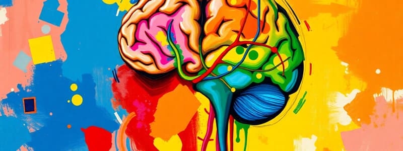
Central Nervous System
Central Nervous System
The brain and spinal cord; the command center of the body.
Peripheral Nervous System
Peripheral Nervous System
Nerves outside of the brain and spinal cord, connecting them to the rest of the body.
Nursing 302
Nursing 302
Signup and view all the flashcards
Disorders
Disorders
Signup and view all the flashcards
Neuromuscular Junction
Neuromuscular Junction
Signup and view all the flashcards
What does the PNS do?
What does the PNS do?
Signup and view all the flashcards
What is the CNS?
What is the CNS?
Signup and view all the flashcards
What is the PNS?
What is the PNS?
Signup and view all the flashcards
What is the neuromuscular junction?
What is the neuromuscular junction?
Signup and view all the flashcards
What are NMJ disorders?
What are NMJ disorders?
Signup and view all the flashcards
What is the focus of Nursing 302?
What is the focus of Nursing 302?
Signup and view all the flashcards
What's the role of the CNS?
What's the role of the CNS?
Signup and view all the flashcards
What happens in NMJ disorders?
What happens in NMJ disorders?
Signup and view all the flashcards
Study Notes
Disorders of the Central and Peripheral and Neuromuscular Junctions
- This is a nursing 302 course topic.
Traumatic Brain Injury (TBI)
- TBI: An alteration in brain function caused by external force (e.g., motor vehicle accidents, falls).
- Primary Injury: Direct impact (focal or diffuse).
- Secondary Injury: Indirect result of the primary injury.
- Most common injury location: Frontal lobe.
- Focal Brain Injury (Observable Injury): Lesion or contusion.
- Open (Penetrating) Trauma: Breaks dura, exposing cranial contents to the environment.
- Closed (Blunt) Trauma: Dura remains intact; brain tissues not exposed.
- Coup Injury: Injury directly below impact point.
- Contrecoup Injury: Opposite side injury from impact.
- Contusions can cause: Epidural, subdural, or intracerebral hematomas.
- Diagnosis: History, GCS score, CT & MRI & PET scan, EEG, ICP.
- Treatment: Goal is to control ICP and manage symptoms; potential surgery.
Hematomas
- Epidural: Bleeding between dura mater and skull (usually due to an artery).
- Typically associated with loss of consciousness on initial injury.
- Medical emergency.
- Symptoms: Worsening headache, vomiting, drowsiness, confusion, seizure, and hemiparesis.
- Diagnosis: CT, MRI.
- Treatment: Monitoring, evaluation, pain management, potential surgery.
- Subdural: Bleeding between dura mater and brain.
- Acts like growing masses; increases ICP, putting pressure on bleeding vessels.
- Symptoms: Headache, drowsiness, restlessness, slowed thought, confusion, loss of consciousness, breathing pattern changes, pupil changes.
- Diagnosis: CT, MRI.
- Treatment: Percutaneous drainage or craniotomy (for chronic bleed).
- Intracerebral: Bleeding within the brain.
- Increases intracranial pressure (ICP) and decreased level of consciousness (LOC).
- Treatment: Decreasing ICP and allowing the hematoma to reabsorb slowly.
Open Brain Injury
- Open Brain Injury: Trauma that penetrates the dura mater.
- Compound Skull Fracture: Opens a path between cranial contents and the environment.
- Basilar Skull Fracture: Fracture to the base of the skull.
- Symptoms: CSF leaking from ear or nose, Battle's sign, periorbital hematoma (racoon eyes).
- Treatment: Bed rest and close observation for meningitis and other complications.
Diffuse Axonal Injury (DAI)
- Widespread brain injury; severity correlates with shearing force to the brainstem.
- Causes: High levels of acceleration/deceleration or rotational forces (e.g., whiplash).
- Results in:
- Axonal damage (shearing, tearing, or stretching of nerve fibers).
- Acute brain swelling.
- Potential long-term neurodegenerative processes.
Concussion (Interchangeable term for TBI)
- Severity is determined by duration of loss of consciousness, GCS, post-traumatic amnesia & brain imaging.
- Goal of treatment: Maintain cerebral perfusion & oxygenation; promote neuroprotection.
- Mild Concussion: No or <30 minutes loss of consciousness; GCS 13-15, confusion (< several minutes), amnesia.
- Moderate Concussion: >30 minutes-6 hours loss of consciousness, GCS 9-12, prolonged confusion &post-traumatic amnesia (>24 hours).
- Severe Concussion: >6 hours loss of consciousness, GCS 3-8, signs of brainstem damage.
Complications of TBI
- Post-concussion syndrome: Weeks to months after, including headache, fatigue, anxiety, irritability, and insomnia.
- Post-traumatic seizures: Can occur within days or up to 5 years after injury.
- Chronic Traumatic Encephalopathy (CTE): Progressive dementing disease that develops with repeated brain injuries (e.g., sports injuries, military-related trauma).
- Symptoms include violent behaviors, loss of control, depression, memory loss, and changes in motor function.
Spinal Cord Injury
- Causes: Motor vehicle accidents, sports activities, falls (>70 years).
- Risk factors: Male, ages 20-39.
- Primary Spinal Cord Injury: Initial mechanical trauma/immediate tissue destruction.
- Secondary Spinal Cord Injury: Pathophysiological cascade of events (edema, ischemia, excitotoxicity, inflammation, cell death) over weeks following injury.
- Life-threatening if swelling occurs in cervical region (C1 to C4).
- Vertebral Injuries: Acceleration/deceleration/deformation forces causing fractures, dislocations & bone fragments that cause compression.
- C1-C4 fracture = aspen collar.
Spinal Cord Injury: Clinical Manifestations
- Spinal Shock: Normal activity of the spinal cord ceases below the injury level; lack of continuous nervous discharges from brain, complete loss of reflex function.
- Neurogenic Shock: Occurs with injuries above T6, caused by absence of sympathetic activity; symptoms include vasodilation, hypotension, bradycardia, & failure of temperature regulation.
- Autonomic Hyperreflexia (Dysreflexia): Massive, uncompensated cardiovascular response to sympathetic nervous system stimulation below the level of injury.
- Usually caused by distended bladder or rectum.
- Diagnosis: Physical exam, imaging studies.
- Treatment: Immobilization to prevent further injury, Surgical fixation/decompression may be necessary, therapeutic hypothermia, nutrition management, lung function, skin integrity, bladder & bowel management, and rehabilitation.
Degenerative Disorders of the Spine
- Low Back Pain: Primary cause of disability worldwide
- Risk factors: Occupations requiring repeated lifting/forward bending, exposure to vibrations, obesity, smoking.
- Degenerative Disc Disease (DDD): Normal aging process, vertical compression of the spine.
- Can lead to disc protrusion, spondylolysis and spinal stenosis.
- Herniated Intervertebral Disc: Displacement of nucleus pulposus or annulus fibrosus.
- Usually caused by trauma, degenerative disc disease, or both.
- Symptoms: Radiculopathy, pain, paresthesia.
Stroke
- Stroke: Interruption in blood flow to brain cells (cerebrovascular accident).
- Third leading cause of death in Canada.
- The sooner circulation returns, the greater the chance of recovery.
- Risk Factors: Poorly controlled hypertension, polycythemia, hyperlipidemia, CHF, PVD, atherosclerosis, arrhythmias (atrials fibrillation), diabetes, lack of exercise, hormonal contraceptive use, and smoking
- Classifications:
- Ischemic (thrombotic or embolic)
- Hemorrhagic
- Transient Ischemic Attacks (TIAs)
Ischemic Stroke
- Pathophysiology: Cerebral infarction—area of brain loses blood due to vascular blockage; prompt infusion of thrombolytics may restore perfusion and prevent necrosis.
- Must be given within 4 hours of onset of symptoms.
- Diagnosis: Cerebral angiography, CT, MRI.
- Treatment: Restoring brain perfusion (tPA), preventing recurrent ischemic events (anticoagulants), physical rehab, and surgery (depending on the extent.)
Hemorrhagic Stroke
- Pathophysiology: Sudden rupture of cerebral artery, leading to diminished blood supply to the affected area.
- Blood accumulates within the brain, causing greater damage, potentially increasing ICP.
- Eventually, reabsorption resolves hemorrhage.
- Diagnosis: Cerebral angiography, CT, MRI.
- Treatment: Stopping or reducing the bleed, controlling blood pressure, and surgery (depending on extent.)
Migraines
- Migraines: Repeated episodic headache lasting 4-72 hours.
- Typically in women 25-55 years old.
- Triggers: altered sleep patterns, overexertion, weather change, stress, hormonal changes, excess sensory stimulation, and chemicals (alcohol or nitrates)
- Phases:
- Premonitory: Tiredness, irritability, inability to concentrate, stiff neck
- Migraine Aura: Visual, sensory, or motor symptoms last up to 1 hour.
- Headache: Throbbing, spreading to the whole head; associated with nausea, vomiting, fatigue, dizziness.
- Recovery: Irritability, fatigue, depression up to days after.
- Diagnosis: History of headaches, medical history (unilateral, throbbing, worsening with movement, moderate or severe).
- Treatment: Avoiding triggers, and pharmacological management.
Headache & Migraines: Cluster Headaches
- Cluster headaches occur in clusters (minutes to hours) followed by a long period of remission.
- Usually in men aged 20-50 years old.
- Symptoms: Trigeminal activation, autonomic dysfunction, unilateral pain, ipsilateral autonomic manifestations (tearing, ptosis, nasal congestion).
- Tension-type headaches are most common. Average onset in the second decade.
- Mild to moderate bilateral pain, tight band sensation. The pain slowly starts.
- Episodes are hours to days in duration.
- At least 15 days per month for 3 months.
Meningitis
- Meningitis: Inflammation of the meninges in brain and spinal cord.
- Blood flow to the brain is reduced, tissues swell causing increased intracranial pressure (ICP).
- Spreads through droplets (bacterial), and quickly due to CSF circulation.
- Causes: Bacteria, viruses, fungi, parasites, or toxins.
- Risk Factors: Malnourishment, immunosuppression, CNS trauma.
- Clinical Manifestations (Children <2 vs. Adults): Fever, anorexia, vomiting, diarrhea, listlessness, and bulging fontanels, cry vs adults (fever, chills, headache, photophobia, neck stiffness, positive Kernig's & Brudzinski sign, tachycardia, LOC deterioration.)
Brain or Spinal Cord Abscess
- Abscess: Localized collection of pus in brain or spinal cord.
- Brain Abscess: Epidural, subdural, or intracerebral.
- Spinal Cord Abscess: Rare; epidural, or intramedullary.
- Risk Factors: Immunosuppressed individuals, IV drug users.
- Pathophysiology: Microorganisms entering the CNS via direct extension along vein walls or via infective emboli.
- Clinical Manifestations: Early (low-grade fever, headache, nausea, vomiting, stiff neck), and then later (confusion, decreased attention span, memory deficits, decreased visual acuity, papilledema, ocular palsy, ataxia, seizures, dementia).
Encephalitis
- Encephalitis: Acute febrile illness usually viral, with nervous system involvement.
- Causes: Mosquito/tick/fly bites, Herpes Simplex type 1, live attenuated vaccines.
- Clinical Manifestations:
- Minor: Malaise, headache, body aches, nausea, and vomiting.
- Major: Fever, delirium, decreased LOC, seizures, paralysis, increased ICP signs.
- Diagnosis: History, CSF examination & culture, WBC count, CT or MRI.
- Treatment: Specific to the type of virus.
Multiple Sclerosis (MS)
- MS: Progressive, inflammatory demyelination of the white matter of the CNS; leading to acquired autoimmune response; loss of myelin disrupts nerve conduction and brain atrophy.
- Characterized by exacerbations and remissions.
- Clinical Manifestations: Vision problems (optic neuritis), paresthesia, emotional lability, dysphagia, muscle weakness/spasticity, hyperreflexia, urinary & bowel problems, gait ataxia, fatigue.
- Diagnosis: History and clinical exam, MRI for lesions, CSF analysis for elevated IgG levels.
- Treatment: Preventing exacerbations, damage, and symptomatic relief (medications like steroids, bed rest, massage, bowel/bladder training, physical therapy).
Myasthenia Gravis
- Myasthenia Gravis: Acquired chronic autoimmune disease producing sporadic/progressive muscular weakness.
- Autoimmune response to acetylcholine receptors.
- Clinical Manifestations: Weakness and fatigue of eye and throat muscles (diplopia, chewing/swallowing problems), drooping jaw, bobbing head.
- May result in myasthenia or cholinergic crisis
- Myasthenia Crisis: Increased symptoms (respiratory distress, tachycardia, decreased gag reflex, visual changes)
- Cholinergic Crisis: Due to anticholinesterase medication toxicity, with symptoms like hypotension, bradycardia, diarrhea, & increased respiratory secretions
- Diagnosis: Tensilon test, Electromyography, Nerve conduction studies.
- Treatment: Anticholinesterase drugs, steroids, and IVIG.
Alterations of Neurological Function in Children
- This is a chapter 17 topic about neurological disorders in children.
Defects of Neural Tube Closure
- Spina Bifida: Vertebrae fail to close; genetic, and folic acid deficiency cause.
- Types: Anencephaly, encephalocele, meningocele, and myelomeningocele.
Encephalopathies
- Encephalopathy: brain tissue abnormality.
- Static encephalopathy: Fixed lesion due to brain injury or disease (not progressing.)
- Causes: Brain malformations, brain injury during gestation/birth, anoxia, trauma, infections, toxins, and metabolic disturbances.
- Cerebral Palsy: Movement and muscle tone disorder occurring due to prenatal or perinatal cerebral hypoxia, hemorrhage, infection or genetic abnormalities, and low birth weight.
- Types: Spasticity, dystonia, ataxia, mixed.
Studying That Suits You
Use AI to generate personalized quizzes and flashcards to suit your learning preferences.


