Podcast
Questions and Answers
Which of the following occupy the intermediate zone of the neural tube?
Which of the following occupy the intermediate zone of the neural tube?
- Ependymal cells
- Neural crest cells
- Primordial white matter tracts
- Primordial neuroblasts (correct)
Which part of the neural tube forms the spinal cord during development?
Which part of the neural tube forms the spinal cord during development?
- Cranial 2/3
- Ventricular zone
- Caudal 1/3 (correct)
- Outer marginal zone
Which part of the developing neural tube gives rise to the white matter tracts?
Which part of the developing neural tube gives rise to the white matter tracts?
- Dorsal gray horns
- Ependymal cells
- Ventricular zone
- Outer marginal zone (correct)
During forebrain development, what do the telencephalic vesicles eventually become?
During forebrain development, what do the telencephalic vesicles eventually become?
What is the function of the notochord in neurulation?
What is the function of the notochord in neurulation?
During neurulation, what structure does the neural tube give rise to?
During neurulation, what structure does the neural tube give rise to?
Where do neural crest cells originate from?
Where do neural crest cells originate from?
What is the future site of the anus according to the provided text?
What is the future site of the anus according to the provided text?
In spinal cord development, what structure forms when the neural folds fuse together?
In spinal cord development, what structure forms when the neural folds fuse together?
Which germ layer is responsible for giving rise to the CNS and PNS?
Which germ layer is responsible for giving rise to the CNS and PNS?
During embryonic folding, what separates the foregut from the stomodeum?
During embryonic folding, what separates the foregut from the stomodeum?
In spinal cord development, what causes lateral folding?
In spinal cord development, what causes lateral folding?
What does the septum transversum develop into during cranial folding?
What does the septum transversum develop into during cranial folding?
At what stage can the neural plate be observed in development?
At what stage can the neural plate be observed in development?
What is the function of the notochord?
What is the function of the notochord?
During neurulation, what structure forms a lumen known as the notochordal canal?
During neurulation, what structure forms a lumen known as the notochordal canal?
What happens after the notochordal process fuses with the endoderm?
What happens after the notochordal process fuses with the endoderm?
Which structure provides a communication channel between the umbilical vesicle and amniotic cavity?
Which structure provides a communication channel between the umbilical vesicle and amniotic cavity?
When does the transformation of the notochordal process into the fully-developed notochord occur?
When does the transformation of the notochordal process into the fully-developed notochord occur?
What is the specific location where mesoderm does not exist between the ectoderm and endoderm of the embryonic disc?
What is the specific location where mesoderm does not exist between the ectoderm and endoderm of the embryonic disc?
Which structure induces the thickening of the embryonic ectoderm to form the neural plate during neurulation?
Which structure induces the thickening of the embryonic ectoderm to form the neural plate during neurulation?
What gives rise to somites by the end of the third week in embryonic development?
What gives rise to somites by the end of the third week in embryonic development?
Which structure initially develops blood vessels in embryonic development, prior to their appearance within the embryo?
Which structure initially develops blood vessels in embryonic development, prior to their appearance within the embryo?
Which structure forms the body wall in the developing embryo?
Which structure forms the body wall in the developing embryo?
What is the main function of the intraembryonic coelom during development?
What is the main function of the intraembryonic coelom during development?
What is the role of somites in embryonic development?
What is the role of somites in embryonic development?
During the 2nd month of development, which body cavity does the intraembryonic coelom develop into?
During the 2nd month of development, which body cavity does the intraembryonic coelom develop into?
What is the significance of the paraxial mesoderm during embryonic development?
What is the significance of the paraxial mesoderm during embryonic development?
Which structure does the tail region project over as the embryo grows?
Which structure does the tail region project over as the embryo grows?
What causes lateral folding during embryonic development?
What causes lateral folding during embryonic development?
What is the role of the oropharyngeal membrane during embryonic development?
What is the role of the oropharyngeal membrane during embryonic development?
Which structure lies ventral to the heart and cranial to the septum transversum due to the head fold?
Which structure lies ventral to the heart and cranial to the septum transversum due to the head fold?
After lateral folding, what is the wide connection between the midgut and umbilical vesicle reduced to?
After lateral folding, what is the wide connection between the midgut and umbilical vesicle reduced to?
During neurulation, what is the function of the neural tube?
During neurulation, what is the function of the neural tube?
What is the primary function of somites during embryonic development?
What is the primary function of somites during embryonic development?
The cloacal membrane and the oropharyngeal membrane are both comprised of two layers with no mesoderm
The cloacal membrane and the oropharyngeal membrane are both comprised of two layers with no mesoderm
The _________ mesoderm eventually gives rise to the the embryonic heart primordia
The _________ mesoderm eventually gives rise to the the embryonic heart primordia
When does the notochordal plate transform to become the notochord?
When does the notochordal plate transform to become the notochord?
What is the significance of the prechordal plate in embryonic development?
What is the significance of the prechordal plate in embryonic development?
Which structure originates from the neural crest cells?
Which structure originates from the neural crest cells?
In embryonic development, which germ layer is responsible for giving rise to the tissues that arise from the neural crest?
In embryonic development, which germ layer is responsible for giving rise to the tissues that arise from the neural crest?
Which structure in the developing spinal cord separates the alar and basal plates?
Which structure in the developing spinal cord separates the alar and basal plates?
During brain development, which primary brain vesicle forms the cerebral hemispheres?
During brain development, which primary brain vesicle forms the cerebral hemispheres?
What gives rise to the sensory epithelia of the eyes, ears, and nose according to germ layer derivatives?
What gives rise to the sensory epithelia of the eyes, ears, and nose according to germ layer derivatives?
During cranial folding, what separates the foregut from the stomodeum?
During cranial folding, what separates the foregut from the stomodeum?
Which structure lies caudal to the heart and develops into the central tendon of the diaphragm?
Which structure lies caudal to the heart and develops into the central tendon of the diaphragm?
Which germ layer gives rise to the epithelial lining of the digestive and respiratory tracts according to germ layer derivatives?
Which germ layer gives rise to the epithelial lining of the digestive and respiratory tracts according to germ layer derivatives?
What is the significance of the neurenteric canal in the development of the notochord?
What is the significance of the neurenteric canal in the development of the notochord?
What is the role of the endoderm in spinal cord development?
What is the role of the endoderm in spinal cord development?
During brain development, what does the telencephalon eventually differentiate into?
During brain development, what does the telencephalon eventually differentiate into?
The primary brain vesicles divide into secondary brain vesicles during week ___
The primary brain vesicles divide into secondary brain vesicles during week ___
Which of the following form the ventral and lateral gray horns?
Which of the following form the ventral and lateral gray horns?
The mesenchyme surrounding the spinal cord forms the __________
The mesenchyme surrounding the spinal cord forms the __________
Which of the following are the primordia of the retinae and the optic nerves?
Which of the following are the primordia of the retinae and the optic nerves?
The___________ divides into the telencephalon and the diencephalon
The___________ divides into the telencephalon and the diencephalon
Which of the following gives rise to the thalami (epithalamus, thalamus, hypothalamus)?
Which of the following gives rise to the thalami (epithalamus, thalamus, hypothalamus)?
Flashcards are hidden until you start studying
Study Notes
Embryonic Development
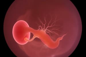
- By the end of the third week, the heart tubes have fused to form a tubular heart that is joined to vessels in the embryo, umbilical vesicle, chorion, and connecting stalk to form a primordial cardiovascular system.
Embryonic Folding
- Embryonic folding is the process by which a relatively "flat" embryonic disk becomes more and more cylindrical in shape.
- Folding occurs in two general planes: • The median plane (cranial-caudal folding): the anterior and posterior ends of the embryo move ventrally. • The horizontal plane (lateral folding): the lateral edges of the embryonic disk move ventrally.
- The edges "roll" ventrally towards the umbilical vesicle.
- Folding begins at the end of the 3rd week and is easy to see in the 4th week.
Cranial Folding
- Part of the endoderm of the umbilical vesicle is incorporated into the embryo as the foregut.
- The foregut lies between the brain and heart.
- Oropharyngeal membrane separates the foregut from the stomodeum (primordium of the mouth).
- Septum transversum lies caudal to the heart, develops into the central tendon of the diaphragm, and separates the abdominal cavity from the thoracic cavity.
- The position of the heart changes due to the head fold: • Heart moves to the ventral surface of the embryo. • Pericardial coelom lies ventral to the heart and cranial to the septum transversum.
Tail Folding
- As the embryo grows, the caudal eminence (tail region) projects over the cloacal membrane (future site of anus).
- Part of the endodermal germ layer is incorporated into the embryo as the hindgut.
- The connecting stalk (primordium of umbilical cord) is now attached to the ventral surface of the embryo, and the allantois is partially incorporated into the embryo.
Lateral Folding
- Lateral folding is caused by the rapidly growing spinal cord and somites.
- As the abdominal walls form, part of the endoderm germ layer is incorporated into the embryo as the midgut.
- Initially, there is a wide connection between the midgut and umbilical vesicle.
- After lateral folding, the connection is reduced to an omphaloenteric duct.
- The region of attachment of the amnion to the ventral surface of the embryo is also reduced to a relatively narrow umbilical region.
Germ Layer Derivatives
- The three germ layers (ectoderm, mesoderm, and endoderm) formed during gastrulation give rise to the primordia of all the tissues and organs.
- Ectoderm: CNS, PNS; sensory epithelia of the eyes, ears, and nose; epidermis and its appendages; mammary glands; subcutaneous glands; enamel of teeth; pituitary gland.
- Mesoderm: connective tissue; cartilage; bone; striated and smooth muscles; heart, blood, and lymphatic vessels; kidneys; ovaries; testes; genital ducts; serous membranes lining the body cavities; spleen; and cortex of suprarenal glands.
- Endoderm: epithelial lining of the digestive and respiratory tracts, parenchyma of the tonsils; thyroid and parathyroid glands; thymus; liver; pancreas; epithelial lining of the urinary bladder and most of the urethra; epithelial lining of the tympanic cavity, tympanic antrum, and eustachian tube.
Development of the Nervous System
- The events of neurulation have already been discussed.
- Neural plate can be seen at day 19.
- Neural plate ! neural groove ! neural tube.
- The neural tube is the primordium of the CNS.
- Neurulation is the process by which the neural tube is formed.
- Neurulation begins with neural plate formation and ends when the tube becomes completely "closed" – no opening at either the caudal or cephalic ends.
- Neurulation is complete at the end of the 4th week.
Neural Crest Cells
- Subset of neuroectodermal cells.
- Originate from the "crest" at the apex of the neural folds.
- Lose affinity to epithelium and neighbouring cells.
- Migrate dorso-laterally on either side of the tube.
- Many migrate widely throughout the mesenchyme.
- Derivatives of the neural crest include: • Ganglia of CN V, VII, IX, X. • Spinal ganglia. • (i.e. dorsal root ganglia).
Brain Development
- Fusion of the neural folds in the cranial region and closure of the rostral (anterior) neuropore form three primary brain vesicles: • Forebrain (prosencephalon). • Midbrain (mesencephalon). • Hindbrain (rhombencephalon).
- During week 5, the prosencephalon partially divides into two secondary brain vesicles: • Telencephalon. • Diencephalon.
- By week 5, the rhombencephalon also partially divides: • Metencephalon. • Myelencephalon.
- Forebrain Development: • Week 5: As closure of the rostral (anterior) neuropore occurs (day 25), two lateral outgrowths-optic vesicles appear one on each side of the forebrain. • Primordia of the retinae and optic nerves. • A second pair of diverticula, the telencephalic vesicles, arise more dorsally and rostrally. • Primordia of the cerebral hemispheres, and their cavities become the lateral ventricles. • Three swellings develop in the lateral walls of the third ventricle, which later become the thalamus, hypothalamus, and the epithalamus.
Notochord
- Roles of the notochord: • Establishes the longitudinal axis of the embryo and gives it some rigidity. • Provides signals for the development of axial MSK structures and the CNS. • Contributes to the intervertebral discs.
- Development of the notochord: • Mesenchymal cells dive into the primitive pit and migrate cephalad. • They form a cord called the notochordal process. • The notochordal process develops a lumen known as the notochordal canal.
- After the notochordal process approaches the prechordal plate, the floor of the process "fuses" with the endoderm.
- The notochordal process is now the notochordal plate.
- The amniotic cavity and the umbilical vesicle can communicate through an opening (the neurenteric canal) – this opening is where the primitive pit opened into the notochordal canal.
- At this point, the notochordal plate cells proliferate and fold inwards, forming the fully-developed notochord.
- No canal is present.
- Notochordal plate ! notochord transition starts cranially and progresses caudally.
- After the notochord is fully-developed, the neurenteric canal is obliterated.### Neurulation
- The neural plate forms on day 19, which later folds into a neural groove and then a neural tube
- The neural tube is the primordium of the Central Nervous System (CNS)
- Neurulation is the process by which the neural tube is formed, which begins with neural plate formation and ends with the closure of the neuropores at day 27
Neural Tube Development
- The cranial 2/3 of the neural tube forms the brain, while the caudal 1/3 forms the spinal cord
- The neural folds fuse at the level of the 5th somite, proceeding cranially and caudally
- The lateral walls of the caudal portion of the neural tube thicken, forming the ventricular zone, intermediate zone, and marginal zone
- The ventricular zone gives rise to all neurons and macroglia, while the intermediate zone becomes populated with primordial neuroblasts
- The marginal zone develops into white matter tracts
Spinal Cord Development
- The alar and basal plates of the developing spinal cord are separated by the sulcus limitans
- Cell bodies in the alar plates form the dorsal gray horns, constituting afferent, or sensory, nuclei
- Cell bodies in the basal plates form the ventral and lateral gray horns, with axons growing out of the spinal cord and forming the ventral roots of the spinal nerves
- Mesenchyme surrounding the spinal cord forms the meninges
Brain Development
- The neural folds in the cranial region fuse and close the rostral (anterior) neuropore, forming three primary brain vesicles: forebrain, midbrain, and hindbrain
- The prosencephalon partially divides into two secondary brain vesicles: telencephalon and diencephalon
- The rhombencephalon also partially divides into metencephalon and myelencephalon
Folding
- Embryonic folding is the process by which the relatively flat embryonic disk becomes more cylindrical in shape
- Folding occurs in two general planes: the median plane (cranial-caudal folding) and the horizontal plane (lateral folding)
- The edges of the embryonic disk "roll" ventrally towards the umbilical vesicle
- Folding begins at the end of the 3rd week and is easily visible in the 4th week
Cranial Folding
- The brain vesicles first begin to appear, and a few somites are formed
- The foregut lies between the brain and heart
- The oropharyngeal membrane separates the foregut from the stomodeum
- The septum transversum lies caudal to the heart, developing into the central tendon of the diaphragm and separating the abdominal cavity from the thoracic cavity
Tail Folding
- The caudal eminence (tail region) projects over the cloacal membrane (future site of anus)
- Part of the endodermal germ layer is incorporated into the embryo as the hindgut
- The connecting stalk (primordium of umbilical cord) is attached to the ventral surface of the embryo, and the allantois is partially incorporated into the embryo
Germ Layer Derivatives
- Ectoderm gives rise to the CNS, PNS, sensory epithelia, epidermis, and its appendages
- Mesoderm gives rise to connective tissue, cartilage, bone, muscles, heart, blood, and lymphatic vessels
- Endoderm gives rise to the epithelial lining of the digestive and respiratory tracts, and other organs such as the liver, pancreas, and thyroid gland### Embryonic Development
- By the end of the third week, the heart tubes have fused to form a tubular heart connected to vessels in the embryo, umbilical vesicle, chorion, and connecting stalk, forming a primordial cardiovascular system.
Embryonic Folding
- Embryonic folding is the process by which the flat embryonic disk becomes cylindrical in shape.
- There are two types of folding:
- Cranial-caudal folding (median plane): the anterior and posterior ends of the embryo move ventrally.
- Lateral folding (horizontal plane): the lateral edges of the embryonic disk move ventrally towards the umbilical vesicle.
Cranial Folding
- The foregut lies between the brain and heart.
- The oropharyngeal membrane separates the foregut from the stomodeum (primordium of the mouth).
- The septum transversum develops into the central tendon of the diaphragm and separates the abdominal cavity from the thoracic cavity.
- The position of the heart changes due to the head fold: the heart moves to the ventral surface of the embryo.
Tail Folding
- The caudal eminence (tail region) projects over the cloacal membrane (future site of anus).
- Part of the endodermal germ layer is incorporated into the embryo as the hindgut.
Lateral Folding
- Lateral folding is caused by the rapidly growing spinal cord and somites.
- As the abdominal walls form, part of the endoderm germ layer is incorporated into the embryo as the midgut.
- The connection between the midgut and umbilical vesicle is reduced to an omphaloenteric duct.
Germ Layer Derivatives
- The three germ layers (ectoderm, mesoderm, and endoderm) formed during gastrulation give rise to the primordia of all tissues and organs.
- Ectoderm derivatives: CNS, PNS, sensory epithelia, epidermis, and its appendages, mammary glands, and more.
- Mesoderm derivatives: connective tissue, cartilage, bone, muscles, heart, blood, and lymphatic vessels, kidneys, ovaries, and more.
- Endoderm derivatives: epithelial lining of digestive and respiratory tracts, thyroid and parathyroid glands, thymus, liver, pancreas, and more.
Development of the Nervous System
- The events of neurulation have already been discussed.
- Neural plate can be seen at day 19.
- The neural plate thickens and a longitudinal neural groove develops, which is flanked by neural folds.
- Fusion of the folds forms the neural tube, the primordium of the CNS.
- Neuroectodermal cells form a neural crest between the surface ectoderm and neural tube.
Week 3 Summary
- At the end of the third week, the embryo is a flat ovoid embryonic disc.
- Mesoderm exists between the ectoderm and endoderm of the disc everywhere except at the oropharyngeal membrane and cloacal membrane.
- The neural plate appears as a thickening of the embryonic ectoderm, induced by the developing notochord.
- The coelom (cavity) within the embryo arises as isolated spaces in the lateral mesoderm and cardiogenic mesoderm.
Studying That Suits You
Use AI to generate personalized quizzes and flashcards to suit your learning preferences.