Podcast
Questions and Answers
What structure separates the frontal lobe from the parietal lobe?
What structure separates the frontal lobe from the parietal lobe?
- Transverse cerebral fissure
- Central sulcus (correct)
- Lateral sulcus
- Parieto-occipital sulcus
Which lobe is located posterior to the parietal lobe?
Which lobe is located posterior to the parietal lobe?
- Frontal lobe
- Temporal lobe
- Insula
- Occipital lobe (correct)
What is the average thickness of the cerebral cortex?
What is the average thickness of the cerebral cortex?
- 10 mm
- 1 mm
- 2-4 mm (correct)
- 5-7 mm
What type of areas in the cerebral cortex are responsible for integrating diverse information?
What type of areas in the cerebral cortex are responsible for integrating diverse information?
Which fissure separates the cerebral hemispheres from the cerebellum?
Which fissure separates the cerebral hemispheres from the cerebellum?
Which type of neurons are primarily found in the cerebral cortex?
Which type of neurons are primarily found in the cerebral cortex?
What is the total number of neurons estimated to be in the human brain?
What is the total number of neurons estimated to be in the human brain?
Which sulcus separates the temporal lobe from the parietal lobe?
Which sulcus separates the temporal lobe from the parietal lobe?
What is one of the primary functions of the cerebellum?
What is one of the primary functions of the cerebellum?
Which structure connects the two hemispheres of the cerebellum?
Which structure connects the two hemispheres of the cerebellum?
Which type of information does the cerebellum use to adjust muscle activity?
Which type of information does the cerebellum use to adjust muscle activity?
What is ataxia, in terms of cerebellar damage?
What is ataxia, in terms of cerebellar damage?
Which of the following roles is NOT associated with the cerebellum?
Which of the following roles is NOT associated with the cerebellum?
What are cerebellar peduncles primarily composed of?
What are cerebellar peduncles primarily composed of?
How does the cerebellum contribute to equilibrium?
How does the cerebellum contribute to equilibrium?
Which part of the brain lies inferior to the occipital lobes?
Which part of the brain lies inferior to the occipital lobes?
What is the primary function of the medulla oblongata?
What is the primary function of the medulla oblongata?
Which structure is involved in reflex actions such as coughing and swallowing?
Which structure is involved in reflex actions such as coughing and swallowing?
What connects the cerebellum to other parts of the central nervous system?
What connects the cerebellum to other parts of the central nervous system?
Which structure is located inferior to the optic chiasma?
Which structure is located inferior to the optic chiasma?
The pons primarily functions in which of the following?
The pons primarily functions in which of the following?
Which part of the brain is the second largest?
Which part of the brain is the second largest?
Which of the following is NOT a function of the medulla oblongata?
Which of the following is NOT a function of the medulla oblongata?
What is the function of the optic nerve?
What is the function of the optic nerve?
What is a primary function of the limbic system?
What is a primary function of the limbic system?
Which area of the brain is mainly involved in maintaining emotional balance?
Which area of the brain is mainly involved in maintaining emotional balance?
What happens to the brain cells as a person ages?
What happens to the brain cells as a person ages?
What is one function of the meninges?
What is one function of the meninges?
The reticular activating system (RAS) is responsible for which of the following?
The reticular activating system (RAS) is responsible for which of the following?
Which protective structure surrounds the central nervous system?
Which protective structure surrounds the central nervous system?
What type of behavior is associated with the functions of the limbic system?
What type of behavior is associated with the functions of the limbic system?
Which statement about the effects of aging on the brain is accurate?
Which statement about the effects of aging on the brain is accurate?
What is the initial condition of a concussion?
What is the initial condition of a concussion?
What happens during memory consolidation?
What happens during memory consolidation?
What occurs in cerebral edema?
What occurs in cerebral edema?
What characterizes Alzheimer’s Disease?
What characterizes Alzheimer’s Disease?
What result follows a cerebrovascular accident (CVA)?
What result follows a cerebrovascular accident (CVA)?
What is the primary function of cerebrospinal fluid?
What is the primary function of cerebrospinal fluid?
Where is cerebrospinal fluid produced?
Where is cerebrospinal fluid produced?
What is true about the blood-brain barrier?
What is true about the blood-brain barrier?
What is a characteristic of slow wave sleep?
What is a characteristic of slow wave sleep?
Which substances can cross the blood-brain barrier?
Which substances can cross the blood-brain barrier?
What is the typical volume of cerebrospinal fluid in the human body?
What is the typical volume of cerebrospinal fluid in the human body?
What role do arachnoid villi play in cerebrospinal fluid management?
What role do arachnoid villi play in cerebrospinal fluid management?
In which type of sleep is the brain most active?
In which type of sleep is the brain most active?
Flashcards
Central sulcus
Central sulcus
Separates the frontal and parietal lobes in the brain.
Lateral sulcus
Lateral sulcus
Separates the temporal lobe from the parietal lobe.
Cerebral cortex
Cerebral cortex
Outer layer of the brain, responsible for higher-level functions like thinking and movement.
Motor areas
Motor areas
Signup and view all the flashcards
Sensory areas
Sensory areas
Signup and view all the flashcards
Association areas
Association areas
Signup and view all the flashcards
Cerebral hemispheres
Cerebral hemispheres
Signup and view all the flashcards
Gray matter
Gray matter
Signup and view all the flashcards
Medulla Oblongata function
Medulla Oblongata function
Signup and view all the flashcards
Medulla Oblongata location
Medulla Oblongata location
Signup and view all the flashcards
Cerebellum size
Cerebellum size
Signup and view all the flashcards
Cerebellum connection
Cerebellum connection
Signup and view all the flashcards
Cerebellum function
Cerebellum function
Signup and view all the flashcards
Brainstem location
Brainstem location
Signup and view all the flashcards
Ascending/Descending Impulses
Ascending/Descending Impulses
Signup and view all the flashcards
Nonvital Reflex Control Centers
Nonvital Reflex Control Centers
Signup and view all the flashcards
Cerebellum Location
Cerebellum Location
Signup and view all the flashcards
Cerebellar Cortex
Cerebellar Cortex
Signup and view all the flashcards
Cerebellar Peduncles
Cerebellar Peduncles
Signup and view all the flashcards
Cerebellar Function - Input
Cerebellar Function - Input
Signup and view all the flashcards
Cerebellar Function - Output
Cerebellar Function - Output
Signup and view all the flashcards
Cerebellar Damage Symptoms
Cerebellar Damage Symptoms
Signup and view all the flashcards
Functional Brain Systems
Functional Brain Systems
Signup and view all the flashcards
Limbic System
Limbic System
Signup and view all the flashcards
Hypothalamus
Hypothalamus
Signup and view all the flashcards
Thalamus
Thalamus
Signup and view all the flashcards
Reticular Activating System (RAS)
Reticular Activating System (RAS)
Signup and view all the flashcards
Meninges
Meninges
Signup and view all the flashcards
Cerebrospinal Fluid (CSF)
Cerebrospinal Fluid (CSF)
Signup and view all the flashcards
Blood-Brain Barrier
Blood-Brain Barrier
Signup and view all the flashcards
Brain Shrinkage
Brain Shrinkage
Signup and view all the flashcards
What are some effects of a concussion?
What are some effects of a concussion?
Signup and view all the flashcards
What is a contusion?
What is a contusion?
Signup and view all the flashcards
What is cerebral edema?
What is cerebral edema?
Signup and view all the flashcards
What is a stroke?
What is a stroke?
Signup and view all the flashcards
What is Alzheimer's disease?
What is Alzheimer's disease?
Signup and view all the flashcards
What produces CSF?
What produces CSF?
Signup and view all the flashcards
CSF: Where does it circulate?
CSF: Where does it circulate?
Signup and view all the flashcards
How is CSF absorbed?
How is CSF absorbed?
Signup and view all the flashcards
CSF: Protective Function
CSF: Protective Function
Signup and view all the flashcards
Blood-Brain Barrier: Permeability
Blood-Brain Barrier: Permeability
Signup and view all the flashcards
Blood-Brain Barrier: Selectivity
Blood-Brain Barrier: Selectivity
Signup and view all the flashcards
Blood-Brain Barrier: What can pass?
Blood-Brain Barrier: What can pass?
Signup and view all the flashcards
REM Sleep: Characteristics
REM Sleep: Characteristics
Signup and view all the flashcards
Study Notes
Objectives
- Outline the development of the Central Nervous System (CNS)
- List the functions of the brain
- Discuss surface anatomy of the brain
- Describe the regions of the Brain
- Describe the protection of the CNS
- Discuss the anatomy and physiology of the Spinal Cord
Introduction
- The central nervous system (CNS) consists of the brain and spinal cord.
- The brainstem connects the brain to the spinal cord.
Parts of the brain...the cerebrum
- Taking up 7/8ths of the brain's weight, the cerebrum governs all sensory and motor activity.
- This includes sensory perception, emotions, consciousness, memory, and voluntary movements.
- It is divided into the left and right hemispheres.
Central Nervous System (CNS)
- CNS develops from the ectoderm surface to produce the embryonic neural tube.
- The neural tube becomes the brain and spinal cord.
- The opening of the neural tube becomes the ventricles.
- Four chambers within the brain are filled with cerebrospinal fluid.
Brain Overview
- Functions of the brain:
- Makes decisions
- Interprets sensations
- Coordinates muscular movements
- Determines perception
- Stores memory
- Reasoning
- Regulates visceral activities
- Determines personality
Surface anatomy
- Gyri (sing gyrus): Elevated ridges, entire surface
- Grooves separate gyri
- Sulci are shallow grooves (sing sulcus)
- Deeper grooves are fissures
Surface anatomy Cont'd
- Longitudinal: separates the cerebral hemispheres
- Transverse: separates cerebrum from cerebellum
- Corpus callosum: connects cerebral hemispheres
Regions of the Brain
- Cerebral hemispheres
- Diencephalon
- Brainstem
- Cerebellum
Lobes of the Cerebrum
- Five (5) lobes bilaterally:
- Frontal lobe
- Parietal lobe
- Temporal lobe
- Occipital lobe
- Insula (buried deep in lateral sulcus)
Cerebral hemispheres
- Divided by longitudinal fissure into right & left sides.
- Central sulcus divides frontal from parietal lobes.
Lateral sulcus
- Separates temporal lobe from parietal lobe.
- Parieto-occipital sulcus divides occipital and parietal lobes.
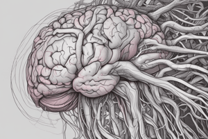
Cerebral cortex
- Executive functioning capability.
- Gray matter: Neuron cell bodies, dendrites, associated glia, blood vessels and short unmyelinated axons.
- 100 billion neurons with an average of 10,000 contacts each.
- No fiber tracts (would be white).
- 2-4 mm thick (about 1/8 inch).
- Brodmann areas (historical: 52 structurally different areas given)
Cerebral cortex
- All the neurons are interneurons.
- By definition confined to the CNS.
- Three kinds of functional areas:
- Motor areas: movement
- Sensory areas: perception
- Association areas: integrate diverse information to enable purposeful action.
Motor Areas (pre-central sulcus)
- Primary motor area
- Frontal lobes
- Control voluntary muscles
- Broca's area
- Anterior to primary motor cortex
- Usually in left hemisphere
- Controls muscles needed for speech
- Frontal eye field
- Above Broca's area
- Controls voluntary movements of eyes & eyelids
Sensory Areas (post-central sulcus)
- Cutaneous sensory area - Parietal lobe: Interprets sensations on skin
- Visual area - Occipital lobe: Interprets vision
- Auditory area - Temporal lobe: Interprets hearing
Association Areas
- Regions that are not primary motor or primary sensory areas.
- Widespread throughout the cerebral cortex.
- Analyze and interpret sensory experiences.
- Provide memory, reasoning, verbalization, judgment, emotions.
- Frontal lobe association areas: concentrating, planning, complex problem-solving
- Parietal lobe association areas: understanding speech, choosing words to express thought
- Temporal lobe association areas: interpret complex sensory experiences, store memories, patterns
- Occipital lobe association areas: analyze and combine visual images and sensory experiences
Functions of the Cerebrum
- Interpreting impulses
- Initiating voluntary movements
- Storing information as memory
- Retrieving stored information
- Reasoning
- Seat of intelligence and personality
The Cerebrum
- The cortex is divided into 4 lobes: frontal lobe specializes in motor activity, personality, and speech; parietal lobe is where language, temperature, pressure, and touch are interpreted; temporal lobe contains centers for hearing, smell, and language input; occipital lobe specializes in vision.
Functions of the Cerebral Lobes
- Frontal lobes: Association areas carry on higher intellectual processes, concentrating, planning, complex problem solving. Motor areas control movements of voluntary skeletal muscles.
- Parietal lobes: Sensory areas provide sensations of temperature, touch, pressure, and pain.
- Temporal lobes: Sensory areas are responsible for hearing, association areas interpret sensory experiences, and remember visual scenes.
- Occipital lobes: Sensory areas are responsible for vision, association areas combine visual images with other sensory experiences.
Homunculus – “little man"
- Body map: human body spatially represented.
- Where on cortex: upside down.
Hemisphere Dominance
- The left hemisphere is dominant in most individuals
- Dominant hemisphere controls:
- Speech
- Writing
- Reading
- Verbal skills
- Analytical skills
- Computational skills
- Nondominant hemisphere controls:
- Nonverbal tasks
- Motor tasks
- Understanding and interpreting musical and visual patterns
- Provides emotional and intuitive thought processes
Diencephalon
- Sits on top of the brain stem and is enclosed by the cerebral hemispheres.
- Made of three parts:
- Thalamus
- Hypothalamus
- Epithalamus
Diencephalon Cont'd
- Thalamus: Gateway for sensory impulses heading to cerebral cortex; sensory relay station; receives all sensory impulses (except smell); channels impulses to appropriate part of cerebral cortex for interpretation.
- Hypothalamus: Maintains homeostasis by regulating visceral activities (like HR, BP, temperature, H₂O & electrolyte balance, hunger, thirst, sleep & wakefulness); links nervous and endocrine systems.
- Epithalamus: Forms the roof of the third ventricle; houses the pineal body (an endocrine gland) that produces melatonin signaling nighttime sleep; includes the choroid plexus which forms cerebrospinal fluid.
Brainstem
- Three parts:
- Midbrain
- Pons
- Medulla Oblongata
Midbrain
- Between diencephalon and pons
- Contains bundles of fibers that join lower parts of brainstem and spinal cord with higher part of brain.
- Cerebral aqueduct: bundles of nerve fibers.
- Corpora quadrigemina: centers for visual and auditory reflexes.
Pons
- Rounded bulge on underside of brainstem
- Between medulla oblongata and midbrain.
- Helps regulate rate and depth of breathing.
- Relays nerve impulses – longitudinally & ventrally.
Medulla Oblongata
- Enlarged continuation of spinal cord
- Conducts ascending and descending impulses between brain and spinal cord.
- Contains cardiac, vasomotor, and respiratory control centers.
- Contains various nonvital reflex control centers like coughing, sneezing, swallowing, and vomiting.
The Cerebellum
- The cerebellum is the second largest part of the brain.
- It contains nerve fibers that connect it to every part of the central nervous system.
- It coordinates voluntary and involuntary patterns of movements.
- It also adjusts muscles to automatically maintain posture
Cerebellum
- Inferior to occipital lobes
- Posterior to pons and medulla oblongata
- Two hemispheres
- Vermis connects hemispheres
- Cerebellar cortex (gray matter)
- Arbor vitae (white matter)
- Cerebellar peduncles (nerve fiber tracts)
- Integrates sensory information concerning position of body parts.
- Coordinates skeletal muscle activity.
Functions of cerebellum
- Smooths, coordinates, and fine-tunes bodily movements.
- Helps maintain body posture.
- Helps maintain equilibrium.
- Gets info from cerebrum re: movements being planned
- Gets info from inner ear re: equilibrium
- Gets info from proprioceptors (sensory receptors) informing where the parts of the body actually are.
- Using feedback, adjustments are made
- Also some role in cognition.
- Damage can cause ataxia, incoordination, wide-based gait, overshooting and proprioception problems.
Major Parts of the Brain
- Detailed breakdown of each part and its functions (Cerebrum, Basal nuclei, Diencephalon, Brainstem, Cerebellum).
Functional Brain Systems
- Networks of distant neurons that function together.
- Limbic system
- Reticular formation
The Limbic System
- Consists of:
- Portions of frontal lobe
- Portions of temporal lobe
- Hypothalamus
- Thalamus
- Basal nuclei (motor control)
- Other deep nuclei
- Functions:
- Controls emotional experiences
- Produces feelings like rage, anger, pleasure
- Survival behaviors
- Interprets sensory impulses associated with smell.
Limbic System Cont'd
- Called the "emotional" brain
- Essential for flexible, stable, and adaptive functioning
- Links different areas so integration can occur
- Processes emotions & allocates attentional resources.
- Necessary for emotional balance and adaptation to environmental demands (including fearful situations).
- Enables meaningful connections with others by interpreting facial expressions and responding appropriately.
Reticular formation
- Runs through central core of medulla, pons, and midbrain
- Reticular activating system (RAS) keeps the cerebral cortex alert and conscious.
- Some motor control.
Lifespan Changes
- Brain cells begin to die before birth.
- Over average lifetime, brain shrinks 10%.
- Most cell death occurs in temporal lobes.
- By age 90, frontal cortex has lost half its neurons.
- Number of dendritic branches decreases.
- Decreased levels of neurotransmitters.
- Fading memory.
- Slowed responses and reflexes.
- Increased risk of falling.
- Changes in sleep patterns that result in fewer sleeping hours.
Protection of the Central Nervous System
- Scalp and skin
- Skull and vertebral column
- Meninges
- Cerebrospinal fluid
- Blood brain barrier
Meninges
- Cover and protect CNS.
- Protect blood vessels.
- Contains CSF.
- Forms partitions in the skull.
Menignes
- Dura mater: 2 layers of fibrous connective tissue (periosteal and meningeal)
- Arachnoid mater: deep to arachnoid is subarachnoid space filled with CSF; Superiorly ,forms arachnoid villi - CSF valves.
- Pia mater is delicate and clings to brain following convolutions
Dura mater - dural partitions
- Falx cerebri (in longitudinal fissure)
- Falx cerebelli (runs vertically along vermis of cerebellum)
- Tentorium cerebelli (sheet in transverse fissure between cerebrum & cerebellum)
Ventricles
- Central cavities expanded, filled with CSF (cerebrospinal fluid).
- Lined by ependymal cells (these cells lining the choroid plexus make the CSF).
- Continuous with each other and central canal of spinal cord.
- Lateral, Third, and Fourth ventricles
Cerebrospinal Fluid
- A colorless fluid produced in the ventricles of the brain.
- Surrounds the brain and spinal cord cushioning them from injury.
- Maintained at a level of around 1/2 - 2/3 cup.
- Secreted by the choroid plexuses of ventricles.
- Filtration of plasma from capillaries through ependymal cells (electrolytes and glucose).
- Circulates in ventricles, central canal, and subarachnoid space.
Lateral ventricles, Third ventricle, Fourth ventricle
- Details of their locations and structures.
Spinal Cord
- Extends downward through vertebral canal.
- Begins at the foramen magnum and terminates at the first and second lumbar vertebrae (L1/L2) interspace.
- Below the interspace is the cauda equina (collection of spinal nerves).
- Enlargements occur in the cervical and lumbar regions.
Spinal Cord Anatomy
- Exterior white matter - conduction tracts
- Internal gray matter - mostly cell bodies
- Dorsal (posterior) horns
- Anterior (ventral) horns
- Central canal filled with cerebrospinal fluid
- Meninges cover the spinal cord
- Nerves leave at the level of each vertebra
- Dorsal root: associated with the dorsal root ganglia - collections of cell bodies outside the central nervous system.
- Ventral root
Functions of Spinal Cord
- Conduit for nerve impulses to and from the brain and brainstem.
- Center for spinal reflexes.
Tracts of the Spinal Cord
- Ascending tracts conduct sensory impulses to the brain while descending tracts conduct motor impulses from the brain to motor neurons reaching muscles and glands
- Detailed list of named tracts
Traumatic Brain Injuries (TBI)
- Concussion: Slight or mild injury, bleeding or tearing of nerve fibers. Recovery likely with some memory loss.
- Contusion: More severe TBI, nervous tissue destruction, may compress and kill brain tissue.
- Cerebral edema: Swelling from the inflammatory response.
Cerebrovascular Accident (CVA)
- Commonly called a stroke
- The result of a ruptured blood vessel supplying a region of the brain.
- Brain tissue supplied with oxygen from that blood source dies.
- Loss of some functions or death may result.
Alzheimer's Disease
- Progressive degenerative brain disease.
- Mostly seen in the elderly, but may begin in middle age.
- Structural changes in the brain include abnormal protein deposits and twisted fibers within neurons.
- Victims experience memory loss, irritability, confusion and ultimately, hallucinations and death.
Types of Sleep
- Slow wave (Non-REM): Person tired, decreasing activity of reticular system, restful, dreamless, reduced blood pressure and respiratory rate, ranges from light to heavy.
- Rapid eye movement (REM): Paradoxical sleep, some areas of brain active, heart and respiratory rates irregular, dreaming occurs.
Memory
- Short-term memory (Working memory): Closed neuronal circuit, circuit is stimulated over & over. When impulse flow ceases, memory does not unless it enters long-term memory via long-term memory consolidation.
- Long-term memory: changes in structure or function of neurons, increases synaptic transmission.
Blood-Brain Barrier
- Tight junctions between endothelial cells of brain capillaries, instead of the usual permeability
- Includes the least permeable capillaries of the body
- Highly selective transport mechanisms excluding many potentially harmful substances, while allowing nutrients, O2, and CO2.
- Not a barrier against uncharged, lipid-soluble molecules; allows alcohol, nicotine, and some drugs including anesthetics.
References
- List of all cited resources.
Studying That Suits You
Use AI to generate personalized quizzes and flashcards to suit your learning preferences.