Podcast
Questions and Answers
Which primary function of the nervous system involves interpreting sensory information?
Which primary function of the nervous system involves interpreting sensory information?
- Motor
- Regulation
- Integration (correct)
- Sensory
The afferent division of the peripheral nervous system is responsible for:
The afferent division of the peripheral nervous system is responsible for:
- Carrying sensory information to the CNS (correct)
- Transmitting motor commands to effectors
- Regulating the activity of smooth muscle
- Controlling glandular secretion
Which of the following is NOT a function of neuroglia?
Which of the following is NOT a function of neuroglia?
- Processing and storage of information (correct)
- Providing physical support to neurons
- Protecting neurons
- Regulating the neuron's environment
Which type of neuroglial cell forms the myelin sheath around axons in the central nervous system?
Which type of neuroglial cell forms the myelin sheath around axons in the central nervous system?
The somatic nervous system primarily innervates:
The somatic nervous system primarily innervates:
Which of the following describes the 'blood-brain barrier'?
Which of the following describes the 'blood-brain barrier'?
Which neuroglia are involved in forming scar tissue in the CNS following injury?
Which neuroglia are involved in forming scar tissue in the CNS following injury?
What is the basic functional unit of the nervous system responsible for processing, transferring, and storing information?
What is the basic functional unit of the nervous system responsible for processing, transferring, and storing information?
Which type of neuron is characterized by having multiple dendrites and a single axon?
Which type of neuron is characterized by having multiple dendrites and a single axon?
Where are true bipolar neurons primarily found?
Where are true bipolar neurons primarily found?
What is the primary function of motor (efferent) neurons?
What is the primary function of motor (efferent) neurons?
What kind of neurons are all interneurons?
What kind of neurons are all interneurons?
Which structural type of neuron lacks clear anatomical distinctions between axons and dendrites, making it difficult to differentiate the processes?
Which structural type of neuron lacks clear anatomical distinctions between axons and dendrites, making it difficult to differentiate the processes?
Where do most sensory neurons have their cell bodies located?
Where do most sensory neurons have their cell bodies located?
What type of synapse occurs between an axon and a dendrite?
What type of synapse occurs between an axon and a dendrite?
What is the purpose of the receptive regions of the neuron called 'dendrites'?
What is the purpose of the receptive regions of the neuron called 'dendrites'?
Which type of cell is responsible for phagocytizing cellular wastes and pathogens in the central nervous system?
Which type of cell is responsible for phagocytizing cellular wastes and pathogens in the central nervous system?
What is the primary function of ependymal cells within the brain and spinal cord?
What is the primary function of ependymal cells within the brain and spinal cord?
In the peripheral nervous system (PNS), what is the primary role of Schwann cells?
In the peripheral nervous system (PNS), what is the primary role of Schwann cells?
What is the significance of the neurilemma formed by Schwann cells?
What is the significance of the neurilemma formed by Schwann cells?
Which type of neuron is responsible for conveying impulses from sensory receptors to the central nervous system (CNS)?
Which type of neuron is responsible for conveying impulses from sensory receptors to the central nervous system (CNS)?
What is the function of motor neurons?
What is the function of motor neurons?
In the context of neuron structure, what are the clusters of cell bodies called in the peripheral nervous system?
In the context of neuron structure, what are the clusters of cell bodies called in the peripheral nervous system?
What is the main functional difference between a dendrite and an axon?
What is the main functional difference between a dendrite and an axon?
What is the primary function of the myelin sheath around an axon?
What is the primary function of the myelin sheath around an axon?
What is the primary difference between white matter and gray matter in the nervous system?
What is the primary difference between white matter and gray matter in the nervous system?
What is a key characteristic of the presynaptic knob?
What is a key characteristic of the presynaptic knob?
Which neuronal circuit is most involved in short-term memory?
Which neuronal circuit is most involved in short-term memory?
What is the function of the synaptic cleft?
What is the function of the synaptic cleft?
What best describes a diverging neuronal circuit?
What best describes a diverging neuronal circuit?
Which of the following structures is NOT a component of a typical synapse?
Which of the following structures is NOT a component of a typical synapse?
In a neuronal circuit, what does the term 'converging' refer to?
In a neuronal circuit, what does the term 'converging' refer to?
What is the function of receptors on the postsynaptic membrane?
What is the function of receptors on the postsynaptic membrane?
What is the primary function of a parallel-after-discharge neuronal circuit?
What is the primary function of a parallel-after-discharge neuronal circuit?
What is the primary function of the perineurium in a peripheral nerve?
What is the primary function of the perineurium in a peripheral nerve?
Which of the following structures is characteristic of 'white matter'?
Which of the following structures is characteristic of 'white matter'?
Where are the cell bodies of post-synaptic neurons found in the autonomic nervous system?
Where are the cell bodies of post-synaptic neurons found in the autonomic nervous system?
What is the role of the neurilemma or sheath of Schwann?
What is the role of the neurilemma or sheath of Schwann?
Where do synapses between pre-synaptic and post-synaptic neurons occur in the autonomic nervous system?
Where do synapses between pre-synaptic and post-synaptic neurons occur in the autonomic nervous system?
In the context of neural tissue organization, what is the function of an endoneurium?
In the context of neural tissue organization, what is the function of an endoneurium?
What is the main difference between ganglia and nuclei/centers in the nervous system?
What is the main difference between ganglia and nuclei/centers in the nervous system?
What is the structural characteristic of the Nodes of Ranvier?
What is the structural characteristic of the Nodes of Ranvier?
Which of the following best describes the function of the LMN system?
Which of the following best describes the function of the LMN system?
The autonomic nervous system (ANS) is primarily responsible for regulating which of the following activities?
The autonomic nervous system (ANS) is primarily responsible for regulating which of the following activities?
What is a key difference in the neural pathways between the somatic and autonomic nervous systems?
What is a key difference in the neural pathways between the somatic and autonomic nervous systems?
Which type of nerve fiber is characteristic of a preganglionic neuron in the autonomic nervous system?
Which type of nerve fiber is characteristic of a preganglionic neuron in the autonomic nervous system?
What is the concept of 'dual innervation' within the autonomic nervous system?
What is the concept of 'dual innervation' within the autonomic nervous system?
Where do the preganglionic cell bodies for the sympathetic nervous system primarily originate?
Where do the preganglionic cell bodies for the sympathetic nervous system primarily originate?
In contrast to the sympathetic nervous system, where do the preganglionic cell bodies for the parasympathetic nervous system originate?
In contrast to the sympathetic nervous system, where do the preganglionic cell bodies for the parasympathetic nervous system originate?
Sympathetic ganglia are found in which of the following locations?
Sympathetic ganglia are found in which of the following locations?
What is the function of white rami in the sympathetic nervous system?
What is the function of white rami in the sympathetic nervous system?
Which of the following is an example of an effector that is controlled by postganglionic fibers of the sympathetic nervous system?
Which of the following is an example of an effector that is controlled by postganglionic fibers of the sympathetic nervous system?
Flashcards
What are the three main functions of the nervous system?
What are the three main functions of the nervous system?
The nervous system has three main roles: receiving sensory information, processing it, and sending out motor responses.
What are the two main anatomical divisions of the nervous system?
What are the two main anatomical divisions of the nervous system?
The central nervous system (CNS) consists of the brain and spinal cord, which act as the control center of the body. The peripheral nervous system (PNS) includes all the nerves that extend outside the CNS.
What is the afferent division of the PNS responsible for?
What is the afferent division of the PNS responsible for?
The afferent division of the PNS carries sensory information from the body to the CNS.
What is the efferent division of the PNS responsible for?
What is the efferent division of the PNS responsible for?
Signup and view all the flashcards
What does the somatic nervous system control and what kind of information does it carry?
What does the somatic nervous system control and what kind of information does it carry?
Signup and view all the flashcards
What does the autonomic nervous system control and what are its branches?
What does the autonomic nervous system control and what are its branches?
Signup and view all the flashcards
What are astrocytes and what are their main functions?
What are astrocytes and what are their main functions?
Signup and view all the flashcards
What are oligodendrocytes and what is their function?
What are oligodendrocytes and what is their function?
Signup and view all the flashcards
Synapses
Synapses
Signup and view all the flashcards
Dendrites - Receptive Regions
Dendrites - Receptive Regions
Signup and view all the flashcards
Tracts in White Matter
Tracts in White Matter
Signup and view all the flashcards
Peripheral Nerves in the PNS
Peripheral Nerves in the PNS
Signup and view all the flashcards
Multipolar Neuron
Multipolar Neuron
Signup and view all the flashcards
Bipolar Neuron
Bipolar Neuron
Signup and view all the flashcards
Unipolar (Pseudounipolar) Neuron
Unipolar (Pseudounipolar) Neuron
Signup and view all the flashcards
Sensory (Afferent) Neurons
Sensory (Afferent) Neurons
Signup and view all the flashcards
Schwann cells
Schwann cells
Signup and view all the flashcards
Satellite cells
Satellite cells
Signup and view all the flashcards
Myelin sheath
Myelin sheath
Signup and view all the flashcards
Nodes of Ranvier
Nodes of Ranvier
Signup and view all the flashcards
Neuron
Neuron
Signup and view all the flashcards
Axon regeneration
Axon regeneration
Signup and view all the flashcards
Microglia
Microglia
Signup and view all the flashcards
Ependymal cells
Ependymal cells
Signup and view all the flashcards
Sensory neuron
Sensory neuron
Signup and view all the flashcards
Motor neuron
Motor neuron
Signup and view all the flashcards
Presynaptic knob
Presynaptic knob
Signup and view all the flashcards
Synaptic cleft
Synaptic cleft
Signup and view all the flashcards
Postsynaptic membrane
Postsynaptic membrane
Signup and view all the flashcards
Diverging circuit
Diverging circuit
Signup and view all the flashcards
Converging circuit
Converging circuit
Signup and view all the flashcards
Reverberating circuit
Reverberating circuit
Signup and view all the flashcards
Parallel-after-discharge circuit
Parallel-after-discharge circuit
Signup and view all the flashcards
NSF (N-ethylmaleimide sensitive factor)
NSF (N-ethylmaleimide sensitive factor)
Signup and view all the flashcards
Parallel-after-discharge
Parallel-after-discharge
Signup and view all the flashcards
Nerves
Nerves
Signup and view all the flashcards
Tracts/Pathways
Tracts/Pathways
Signup and view all the flashcards
Ganglia
Ganglia
Signup and view all the flashcards
Nuclei/Centers
Nuclei/Centers
Signup and view all the flashcards
Epineurium
Epineurium
Signup and view all the flashcards
Perineurium
Perineurium
Signup and view all the flashcards
Endoneurium
Endoneurium
Signup and view all the flashcards
Rubrospinal, Vestibulospinal, Reticulospinal, and Tectospinal Tracts
Rubrospinal, Vestibulospinal, Reticulospinal, and Tectospinal Tracts
Signup and view all the flashcards
Autonomic Nervous System
Autonomic Nervous System
Signup and view all the flashcards
Somatic Nervous System
Somatic Nervous System
Signup and view all the flashcards
Autonomic Nervous System
Autonomic Nervous System
Signup and view all the flashcards
Preganglionic vs. Postganglionic Neurons
Preganglionic vs. Postganglionic Neurons
Signup and view all the flashcards
Sympathetic vs. Parasympathetic Nervous System
Sympathetic vs. Parasympathetic Nervous System
Signup and view all the flashcards
Sympathetic (Thoracolumbar) vs Parasympathetic (Craniosacral) Divisions
Sympathetic (Thoracolumbar) vs Parasympathetic (Craniosacral) Divisions
Signup and view all the flashcards
Locations of Autonomic Ganglia
Locations of Autonomic Ganglia
Signup and view all the flashcards
Structures of the Sympathetic Nervous System
Structures of the Sympathetic Nervous System
Signup and view all the flashcards
Two Neuron Pathway in the ANS
Two Neuron Pathway in the ANS
Signup and view all the flashcards
Study Notes
Fundamentals of the Nervous System
- The nervous system has three major functions: sensory, integration, and motor.
- Sensory monitors internal and external environments via receptors.
- Integration interprets sensory information and processes complex functions.
- Motor responds to processed information to stimulate effectors (muscle contraction/glandular secretion).
General Organization of the Nervous System
- Two anatomical divisions: central nervous system (CNS) and peripheral nervous system (PNS).
- CNS includes the brain in the cranial cavity and spinal cord in the spinal canal.
- PNS is all neural tissue outside the CNS.
- Two functional divisions: somatic and autonomic nervous systems.
- Somatic: provides sensory and motor innervation to all body parts except viscera, smooth muscle, and glands.
- Autonomic: sympathetic, parasympathetic, and enteric—provides involuntary motor innervation and sensory innervation to viscera (pain and autonomic reflexes).
Somatic vs. Visceral Pain
- Somatic pain involves nociceptors in skin, muscle, tendons, and bones. Often well localized and described as throbbing or aching.
- Visceral pain involves hollow organ and smooth muscle nociceptors. It is associated with poorly localized, vague, and diffuse pain, often referred to different locations. Associated with autonomic symptoms.
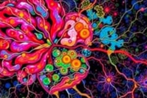
Histology of Neural Tissue
- Two types of neural cells: neurons and neuroglia.
- Neurons process, transfer, and store information. They consist of a cell body and processes of varying length.
- Neuroglia support, regulate, and protect neurons. They are non-conducting.
Neuroglia (Glial Cells)
- CNS neuroglia: astrocytes, oligodendrocytes, microglia, and ependymal cells.
- PNS neuroglia: Schwann cells (neurolemmocytes) and satellite cells in ganglia.
Astrocytes
- Supportive role, regulate extracellular pH and K+ concentration, induce blood-brain barrier formation.
- Involved in structural support, repair, and regulating the environment around neurons.
- Two types: fibrous astrocytes (prominent in white matter) and protoplasmic astrocytes (prominent in grey matter).
- Special types include Bergmann glial cells in cerebellum and Muller cells in retina.
Oligodendrocytes
- Create myelin sheaths around axons in CNS.
- Myelinated axons transmit impulses faster than unmyelinated axons.
Microglia
- Brain macrophages that phagocytize cellular wastes and pathogens.
Ependymal Cells
- Line ventricles of brain and central canal of spinal cord.
- Produce, monitor, and help circulate cerebrospinal fluid (CSF).
Schwann Cells
- Surround all axons in the PNS, creating a neurilemma crucial for damaged axon regeneration.
- Create myelin sheaths around most PNS axons.
- Satellite cells support groups of neuronal cell bodies within ganglia of PNS.
Myelin in the Peripheral and Central Nervous Systems
- Myelin in both PNS and CNS is formed by different glial cells. PNS uses Schwann cells whereas CNS uses oligodendrocytes to create myelin sheaths around axons.
- Myelin speeds up the transmission of action potentials, allowing impulses to travel faster down axons.
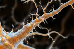
Neuron Structure
- Neurons: structural and functional unit of nervous system
- Sensory neurons convey impulses to CNS. Includes somatic afferent for surface pain, temperature, touch, and proprioception, and visceral afferent from mucosae, glands, blood vessels.
- Motor neurons convey impulses from CNS or ganglia to effector cells (somatic efferent : voluntary impulses to skeletal muscle, visceral efferent : involuntary impulses to cardiac muscle, smooth muscle, glands).
- Interneurons communicate and integrate sensory and motor neurons within CNS.
Neuron Classification
- Structural classification: anaxonic, bipolar, pseudounipolar, and multipolar neurons, based on the number of processes emanating from the neuron cell body.
- Functional classification: sensory (afferent), motor (efferent), and interneurons, based on information type and transmission direction.
Synapses
- Specialized junctions between neurons or between axons and target cells
- Most synapses in the nervous system are chemical synapses, involving neurotransmitter release.
- The presynaptic knob contains synaptic vesicles filled with neurotransmitters.
- The synaptic cleft is the space between the presynaptic and postsynaptic neurons.
- The postsynaptic membrane contains receptors for the neurotransmitter.
Neuronal Circuits
- Diverging: one neuron stimulates many other neurons. This is prevalent in motor pathways.
- Converging: many neurons stimulate a single neuron. This is seen in sensory and integrative pathways
- Reverberating: impulses from later cells repeatedly stimulate early cells, involved in short-term memory.
- Parallel-after-discharge: a single cell stimulates a group of cells that all stimulate a common postsynaptic cell (problem-solving, complex processing).
Anatomical Organization of Neurons
- Neurons group together into organized bundles, forming nerves (PNS) and tracts/pathways (CNS).
- Cell bodies are clustered into ganglia (PNS) or nuclei/centers (CNS); these are unmyelinated and part of "gray matter"
- Axons are bundled together in myelinated structures ("white matter").
Peripheral Nerves
- Bundles of nerve fibers held together by connective tissue (epineurium, perineurium, and endoneurium).
- Individual neurons are surrounded by the neurilemma (or sheath of Schwann).
The Autonomic Nervous System
- Regulates activity of smooth muscle, cardiac muscle, and certain glands.
- Two neuron system: preganglionic neurons (in CNS) and postganglionic neurons (outside CNS).
- Two major divisions: sympathetic and parasympathetic. Sympathetic = fight-or-flight; Parasympathetic = rest-and-digest.
Sympathetic and Dorsal Root Ganglia
- Clusters of neuronal cell bodies outside the CNS. Sensory ganglia carry impulses to CNS; autonomic ganglia contain cell bodies of post-synaptic neurons.
Spinal Cord
- The spinal cord runs through the vertebral column, from the foramen magnum to the second lumbar vertebra.
- It has regions divided into cervical, thoracic, lumbar, and sacral.
- The spinal cord has an outer white matter and an inner gray matter.
- The gray matter is H-shaped, containing cell bodies of neurons and nerve fibers.
- The white matter surrounds the gray matter, containing myelinated axons organized into columns to carry information up and down the column.
- The dorsal roots carry sensory neurons, and the ventral roots carry motor neurons.
Meninges
- Connective tissue membranes covering the spinal cord: dura mater, arachnoid mater, and pia mater.
- Spaces include subdural (serous fluid) and subarachnoid (CSF).
- The meninges provide cushioning, protection and anchor the spinal cord.
Control of Autonomic Nervous System
- The hypothalamus plays a key role in regulating autonomic functions.
- The hypothalamus integrates sensory input, including emotions, and visceral information from the body, triggering outputs to nuclei in the brainstem and spinal cord.
- Posterior and lateral portions primarily control sympathetic activity, while anterior and medial regions primarily control parasympathetic activity.
ANS Neurotransmitters
- Cholinergic neurons release acetylcholine (ACh).
- Adrenergic neurons release norepinephrine (NE).
Parasympathetic Cranial Nerves
- Oculomotor, Facial, Glossopharyngeal, and Vagus nerves convey parasympathetic signals.
Parasympathetic Sacral Nerve Fibers
- Form pelvic splanchnic nerves that innervate smooth muscles and glands in the distal digestive system, urinary, and reproductive organs.
Studying That Suits You
Use AI to generate personalized quizzes and flashcards to suit your learning preferences.