Podcast
Questions and Answers
How do astrocytes contribute to neuronal function within the brain?
How do astrocytes contribute to neuronal function within the brain?
- By forming the myelin sheath around axons in the central nervous system.
- By forming the myelin sheath around axons in the peripheral nervous system.
- By regulating the chemical environment around neurons and providing metabolic support. (correct)
- By removing cellular debris and pathogens through phagocytosis.
What is the primary function of the myelin sheath that surrounds certain neuronal axons?
What is the primary function of the myelin sheath that surrounds certain neuronal axons?
- To decrease the speed of action potential transmission.
- To increase the energy consumption of the neuron.
- To insulate the axon and increase the speed of signal transmission. (correct)
- To facilitate direct ionic exchange between the intracellular and extracellular environment.
How does the sodium-potassium pump (Na+/K+ ATPase) contribute to maintaining the resting membrane potential of a neuron?
How does the sodium-potassium pump (Na+/K+ ATPase) contribute to maintaining the resting membrane potential of a neuron?
- By actively transporting Na+ out of the cell and K+ into the cell, maintaining concentration gradients. (correct)
- By preventing the movement of any ions across the cell membrane, ensuring a stable charge.
- By passively allowing sodium ions to flow into the cell and potassium ions to flow out.
- By equally distributing sodium and potassium ions across the cell membrane.
During the generation of an action potential, what event is primarily responsible for the depolarization phase?
During the generation of an action potential, what event is primarily responsible for the depolarization phase?
What is the role of voltage-gated calcium channels in neurotransmitter release at chemical synapses?
What is the role of voltage-gated calcium channels in neurotransmitter release at chemical synapses?
Which of the following best describes the function of interneurons?
Which of the following best describes the function of interneurons?
How do metabotropic receptors differ from ionotropic receptors in synaptic transmission?
How do metabotropic receptors differ from ionotropic receptors in synaptic transmission?
What is the absolute refractory period, and what causes it?
What is the absolute refractory period, and what causes it?
How does the Nernst equation contribute to understanding the resting membrane potential?
How does the Nernst equation contribute to understanding the resting membrane potential?
What is the functional consequence of spatial summation in neurons?
What is the functional consequence of spatial summation in neurons?
Flashcards
Neurophysiology
Neurophysiology
The study of the function of the nervous system, incorporating biophysics, molecular biology, and anatomy to study electrical and chemical processes.
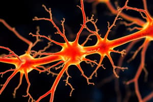
Neurons
Neurons
Fundamental structural and functional units of the nervous system, specialized for electrical and chemical signaling.
Glial cells
Glial cells
Cells that support neuronal function by providing structural and metabolic support.
Neuron Doctrine
Neuron Doctrine
Signup and view all the flashcards
Sensory Neurons
Sensory Neurons
Signup and view all the flashcards
Motor Neurons
Motor Neurons
Signup and view all the flashcards
Interneurons
Interneurons
Signup and view all the flashcards
Resting Membrane Potential
Resting Membrane Potential
Signup and view all the flashcards
Action Potentials
Action Potentials
Signup and view all the flashcards
Neurotransmitter Binding
Neurotransmitter Binding
Signup and view all the flashcards
Study Notes
- Neurophysiology involves studying the function of the nervous system
- It uses biophysics, molecular biology, and anatomy techniques
- To understand the electrical and chemical processes of neuronal signaling and behavior
Cells of the Nervous System
- Neurons are primary units for signaling
- Glial cells support the function of neurons
Neurons
- Neurons specialize in electrical and chemical signals
- Neurons utilize unique structures to transmit information rapidly and precisely
Neuron Doctrine
- The neuron is the fundamental unit of the nervous system (structural and functional)
- Neurons are discrete, not continuous cells
- Neurons include the cell body (soma), dendrites, and axon
- Synapses transmit signals via chemical messengers
Neuron Structure
- Soma contains the nucleus and organelles
- Dendrites are branching extensions that receive signals
- Axon is a single extension that transmits signals
- The axon hillock is where signals are generated
- Myelin sheath insulates the axon, increasing signal speed
- Nodes of Ranvier are gaps in the myelin sheath exposing the axon
- Axon terminals are specialized endings that release neurotransmitters
Types of Neurons
- Sensory neurons transmit information from receptors to the central nervous system (afferent)
- Motor neurons transmit information from the central nervous system to muscles/glands (efferent)
- Interneurons connect sensory and motor neurons in the central nervous system, mediating reflexes and higher-order processing
Glial Cells
- Glial cells support neurons structurally and metabolically
- Types of glial cells: astrocytes, oligodendrocytes, microglia, and Schwann cells
Astrocytes
- Most abundant glial cells in the brain
- Regulate the chemical environment by removing excess neurotransmitters and ions
- Provide metabolic support by supplying nutrients
- Contribute to the blood-brain barrier
Oligodendrocytes
- Form the myelin sheath around CNS axons
- One oligodendrocyte myelinates multiple axons
Microglia
- Immune cells of the central nervous system
- Remove debris and pathogens through phagocytosis
- Release cytokines and other signaling molecules for inflammation and immune responses
Schwann Cells
- Form the myelin sheath around PNS axons
- One Schwann cell myelinates a single axon
Electrical Signals in Neurons
- Neurons use electrical signals for transmitting information
- These signals result from ion flow changes across the neuron's cell membrane
Membrane Potential
- The membrane potential is the electrical charge difference inside versus outside the cell
- It is typically negative at rest
Resting Membrane Potential
- The resting membrane potential is the stable potential of a non-signaling neuron
- It is typically around -70 mV
- Maintained by unequal ion distribution (Na+, K+, Cl-, A-)
- The sodium-potassium pump (Na+/K+ ATPase) maintains concentration gradients by transporting Na+ out and K+ into the cell
- Ion channels allow selective ion movement, contributing to the membrane potential
Ion Channels
- Ion channels are transmembrane proteins that allow specific ions to flow
- Types of ion channels: voltage-gated, ligand-gated, or mechanically gated
Voltage-Gated Channels
- Voltage-gated channels open/close in response to membrane potential changes
Ligand-Gated Channels
- Ligand-gated channels open/close in response to neurotransmitter or signaling molecule binding
Mechanically-Gated Channels
- Mechanically-gated channels open/close in response to cell membrane deformation
Nernst Equation
- The Nernst equation calculates the equilibrium potential for an ion
- Based on its concentration gradient, determining each ion's contribution to resting membrane potential
Goldman-Hodgkin-Katz (GHK) Equation
- The GHK equation calculates membrane potential using permeability and concentration gradients of multiple ions
- It is a more comprehensive model than the Nernst equation
Action Potentials
- Action potentials are rapid, transient changes in membrane potential
- They travel along the axon
- They are the primary means of long-distance communication in the nervous system
Action Potential Generation
- When the membrane potential reaches a threshold (around -55 mV), voltage-gated Na+ channels open
- This causes a rapid Na+ influx, depolarizing the membrane
- Voltage-gated K+ channels then open, allowing K+ to flow out
- K+ efflux repolarizes the membrane
- Voltage-gated Na+ channels inactivate shortly after opening
- The membrane potential may briefly hyperpolarize due to continued K+ efflux
- The Na+/K+ ATPase restores ion gradients
Action Potential Propagation
- Action potentials propagate along the axon without decrement
- The influx of Na+ depolarizes adjacent regions, triggering more voltage-gated Na+ channels to open
- This continues down the axon
Refractory Period
- After an action potential, there is a brief refractory period
- The absolute refractory period: no stimulus can generate another action potential due to inactivated Na+ channels
- The relative refractory period: a stronger stimulus is required due to persistent K+ efflux and inactivated Na+ channels
Myelination and Saltatory Conduction
- In myelinated axons, action potentials "jump" from one node of Ranvier to the next
- This process is called saltatory conduction
- Myelination increases speed by reducing capacitance and increasing membrane resistance
Synaptic Transmission
- Synaptic transmission is the process by which neurons communicate at synapses
- Synapses are specialized junctions
- There are two main types: chemical and electrical
Chemical Synapses
- Chemical synapses use neurotransmitters for signal transmission
- The presynaptic neuron releases neurotransmitters into the synaptic cleft
- These neurotransmitters bind to receptors on the postsynaptic neuron
Neurotransmitter Release
- When an action potential reaches the axon terminal, voltage-gated Ca2+ channels open
- Ca2+ influx triggers neurotransmitter-containing vesicles to fuse with the presynaptic membrane
- Neurotransmitters are released into the synaptic cleft by exocytosis
Neurotransmitter Binding
- Neurotransmitters diffuse across the synaptic cleft and bind to postsynaptic receptors
- Receptors can be ionotropic (ligand-gated ion channels) or metabotropic (G protein-coupled receptors)
Ionotropic Receptors
- Ionotropic receptors are ligand-gated ion channels
- These receptors open/close upon neurotransmitter binding
- They mediate fast synaptic transmission
Metabotropic Receptors
- Metabotropic receptors are G protein-coupled receptors
- They activate intracellular signaling pathways upon neurotransmitter binding
- They mediate slower, longer-lasting synaptic transmission
Postsynaptic Potentials
- Neurotransmitter binding causes changes in the postsynaptic membrane potential
- Excitatory postsynaptic potentials (EPSPs) depolarize the membrane, increasing the likelihood of an action potential
- Inhibitory postsynaptic potentials (IPSPs) hyperpolarize the membrane, decreasing the likelihood of an action potential
Neurotransmitter Removal
- Neurotransmitters are removed from the synaptic cleft by:
- Reuptake into the presynaptic neuron or glial cells
- Enzymatic degradation
- Diffusion away from the synapse
Electrical Synapses
- Electrical synapses use gap junctions to connect cytoplasm between two neurons directly
- Ions and small molecules flow directly from one neuron to another
- Electrical synapses mediate very fast transmission
Synaptic Integration
- Synaptic integration is the summation of synaptic inputs to determine action potential firing
- Temporal summation occurs when multiple EPSPs/IPSPs occur rapidly at the same synapse
- Spatial summation occurs when EPSPs/IPSPs occur simultaneously at different synapses
Neurotransmitters
- Neurotransmitters are chemical messengers for transmitting signals from one neuron to another
- Examples: acetylcholine, glutamate, GABA, dopamine, norepinephrine, and serotonin
Acetylcholine
- Acetylcholine is a neurotransmitter involved in muscle contraction, learning, and memory
Glutamate
- Glutamate is the primary excitatory neurotransmitter in the brain
GABA
- GABA is the primary inhibitory neurotransmitter in the brain
Dopamine
- Dopamine is a neurotransmitter involved in reward, motivation, and motor control
Norepinephrine
- Norepinephrine is a neurotransmitter involved in arousal, attention, and stress responses
Serotonin
- Serotonin is a neurotransmitter involved in mood, sleep, and appetite
Studying That Suits You
Use AI to generate personalized quizzes and flashcards to suit your learning preferences.