Podcast
Questions and Answers
What is the primary function of dendrites in a neuron?
What is the primary function of dendrites in a neuron?
- Collect information from other neurons (correct)
- Conduct electrical signals to the soma
- Synthesize macromolecules
- Transmit information to other neurons
Which part of a neuron is responsible for integrating electrical signals?
Which part of a neuron is responsible for integrating electrical signals?
- Dendrites
- Soma (cell body) (correct)
- Axon terminals
- Myelin sheath
What role does the myelin sheath play in neuronal function?
What role does the myelin sheath play in neuronal function?
- Serves as a channel for neurotransmitter release
- Provides structural support to the soma
- Acts as a barrier preventing signal transmission
- Increases the speed of signal transmission along the axon (correct)
Which of the following best describes the axon of a neuron?
Which of the following best describes the axon of a neuron?
What is a characteristic feature of axon terminals?
What is a characteristic feature of axon terminals?
What is the role of the cytoskeleton in neurons?
What is the role of the cytoskeleton in neurons?
Which neuronal structure is primarily involved in the synthesis of macromolecules?
Which neuronal structure is primarily involved in the synthesis of macromolecules?
The node of Ranvier is essential for which aspect of neuronal function?
The node of Ranvier is essential for which aspect of neuronal function?
Which ventricle is located above the cerebral aqueduct?
Which ventricle is located above the cerebral aqueduct?
What structure separates the inner meningeal layer from the outer periosteal layer of the dura mater?
What structure separates the inner meningeal layer from the outer periosteal layer of the dura mater?
What is the role of the anterior communicating artery?
What is the role of the anterior communicating artery?
Which space is normally filled with cerebrospinal fluid (CSF)?
Which space is normally filled with cerebrospinal fluid (CSF)?
Which artery primarily supplies the medial aspect of the brain?
Which artery primarily supplies the medial aspect of the brain?
What structure follows the dura mater and is separated from it by the subarachnoid space?
What structure follows the dura mater and is separated from it by the subarachnoid space?
Which of the following does not constitute a component of the brain's vascular supply?
Which of the following does not constitute a component of the brain's vascular supply?
Where is the superior sagittal sinus located?
Where is the superior sagittal sinus located?
Which artery does not arise from the internal carotid artery?
Which artery does not arise from the internal carotid artery?
The pia mater is best described as:
The pia mater is best described as:
What part of the lateral ventricle is described as resembling butterfly wings from a posterior view?
What part of the lateral ventricle is described as resembling butterfly wings from a posterior view?
What are the arachnoid trabeculae primarily responsible for?
What are the arachnoid trabeculae primarily responsible for?
Which space is considered a potential space between the dura and calvaria?
Which space is considered a potential space between the dura and calvaria?
What primarily differentiates the distribution of gray and white matter between the brain and spinal cord?
What primarily differentiates the distribution of gray and white matter between the brain and spinal cord?
Which anatomical structure helps facilitate communication between the two cerebral hemispheres?
Which anatomical structure helps facilitate communication between the two cerebral hemispheres?
What is the primary function of the choroid plexus found within the ventricular system?
What is the primary function of the choroid plexus found within the ventricular system?
What separates the frontal lobe from the parietal lobe?
What separates the frontal lobe from the parietal lobe?
Which of the following best describes the anatomical location of the insula?
Which of the following best describes the anatomical location of the insula?
What structure divides the anterior limb of the internal capsule from the genu?
What structure divides the anterior limb of the internal capsule from the genu?
Which of the following lobes is located posteriorly in the cerebral hemispheres?
Which of the following lobes is located posteriorly in the cerebral hemispheres?
What anatomical feature is primarily responsible for increasing the surface area of the brain?
What anatomical feature is primarily responsible for increasing the surface area of the brain?
What connects the lateral ventricles to the third ventricle?
What connects the lateral ventricles to the third ventricle?
What does the term 'operculum' refer to in the context of the cerebral hemispheres?
What does the term 'operculum' refer to in the context of the cerebral hemispheres?
Which gray matter structure is not directly part of the cerebral cortex?
Which gray matter structure is not directly part of the cerebral cortex?
In which region of the brain are the anterior and inferior horns of the lateral ventricles located?
In which region of the brain are the anterior and inferior horns of the lateral ventricles located?
What is the primary anatomical orientation of the posterior limb of the internal capsule?
What is the primary anatomical orientation of the posterior limb of the internal capsule?
What divides the parietal and occipital lobes laterally?
What divides the parietal and occipital lobes laterally?
Which structure is located at the bottom of the retracted portion of the insular cortex?
Which structure is located at the bottom of the retracted portion of the insular cortex?
What is the function of the foramen of Monro?
What is the function of the foramen of Monro?
Which meningeal layer is also described as tough and fibrous?
Which meningeal layer is also described as tough and fibrous?
Where does the fourth ventricle send cerebrospinal fluid (CSF)?
Where does the fourth ventricle send cerebrospinal fluid (CSF)?
What structure separates the cerebral hemispheres within the cranial cavity?
What structure separates the cerebral hemispheres within the cranial cavity?
Which of the following describes the path CSF takes from the third to fourth ventricle?
Which of the following describes the path CSF takes from the third to fourth ventricle?
What is one of the functions of the meninges?
What is one of the functions of the meninges?
What component surrounds the third ventricle?
What component surrounds the third ventricle?
Which potential space is located superficial to the periosteal layer of the dura mater?
Which potential space is located superficial to the periosteal layer of the dura mater?
What structure lies between the cerebellum and the pons?
What structure lies between the cerebellum and the pons?
What does the term 'septum' refer to in the context of cerebral meninges?
What does the term 'septum' refer to in the context of cerebral meninges?
Where are the dural venous sinuses primarily formed?
Where are the dural venous sinuses primarily formed?
Which structure is part of the internal capsule located in the brain?
Which structure is part of the internal capsule located in the brain?
Which artery does NOT arise from the vertebrobasilar system?
Which artery does NOT arise from the vertebrobasilar system?
What is the primary function of the posterior communicating artery?
What is the primary function of the posterior communicating artery?
Where does the inferior sagittal sinus join to form the straight sinus?
Where does the inferior sagittal sinus join to form the straight sinus?
How are the cerebral hemispheres drained before reaching the internal jugular vein?
How are the cerebral hemispheres drained before reaching the internal jugular vein?
Which sinus drains into the transverse sinus?
Which sinus drains into the transverse sinus?
What structure acts as a drainage point for both the superior petrosal and inferior petrosal sinuses?
What structure acts as a drainage point for both the superior petrosal and inferior petrosal sinuses?
Which artery does NOT contribute to the vascular supply of the inferior and medial temporal and occipital lobes?
Which artery does NOT contribute to the vascular supply of the inferior and medial temporal and occipital lobes?
Which of the following statements about the sinuses is incorrect?
Which of the following statements about the sinuses is incorrect?
Which of the following veins primarily drains into the cavernous sinus from the temporal lobe?
Which of the following veins primarily drains into the cavernous sinus from the temporal lobe?
What is the confluence of the sinuses?
What is the confluence of the sinuses?
What role does the basilar artery play in the vascular system?
What role does the basilar artery play in the vascular system?
What role does the thalamus play in information processing within the brain?
What role does the thalamus play in information processing within the brain?
Which arteries arise from the internal carotid artery?
Which arteries arise from the internal carotid artery?
Which structure connects the two thalamic masses across the midline of the third ventricle?
Which structure connects the two thalamic masses across the midline of the third ventricle?
What defines the inferior margin of the falx cerebri?
What defines the inferior margin of the falx cerebri?
Which of the following is NOT a part of the major functional classes of thalamic nuclei?
Which of the following is NOT a part of the major functional classes of thalamic nuclei?
What is a primary function of the thalamus in relation to sensory information?
What is a primary function of the thalamus in relation to sensory information?
Which artery does NOT branch off from the basilar artery?
Which artery does NOT branch off from the basilar artery?
What type of veins do superficial and deep veins connect to before reaching the internal jugular vein?
What type of veins do superficial and deep veins connect to before reaching the internal jugular vein?
Which vein connects perpendicularly to the superficial middle cerebral vein and drains into the superior sagittal sinus?
Which vein connects perpendicularly to the superficial middle cerebral vein and drains into the superior sagittal sinus?
Which part of the brain does the pericallosal artery supply?
Which part of the brain does the pericallosal artery supply?
Which area of the brain's venous drainage is primarily associated with the internal cerebral veins?
Which area of the brain's venous drainage is primarily associated with the internal cerebral veins?
What is primarily received by the superior sagittal sinus in the context of venous drainage?
What is primarily received by the superior sagittal sinus in the context of venous drainage?
How are thalamic nuclei categorized based on their structural relationship to the internal medullary lamina?
How are thalamic nuclei categorized based on their structural relationship to the internal medullary lamina?
Which nerve is NOT present within the cavernous sinus?
Which nerve is NOT present within the cavernous sinus?
What is the primary role of deep veins in comparison to superficial veins?
What is the primary role of deep veins in comparison to superficial veins?
Which of these structures is located immediately lateral to the thalamus?
Which of these structures is located immediately lateral to the thalamus?
What primarily limits the variability of the superficial veins in the brain?
What primarily limits the variability of the superficial veins in the brain?
Which anatomical feature is located just above the sphenoidal air sinus?
Which anatomical feature is located just above the sphenoidal air sinus?
Which vein serves as a connection for venous drainage from the temporal lobe to the transverse sinus?
Which vein serves as a connection for venous drainage from the temporal lobe to the transverse sinus?
Flashcards are hidden until you start studying
Study Notes
Neuron Structure
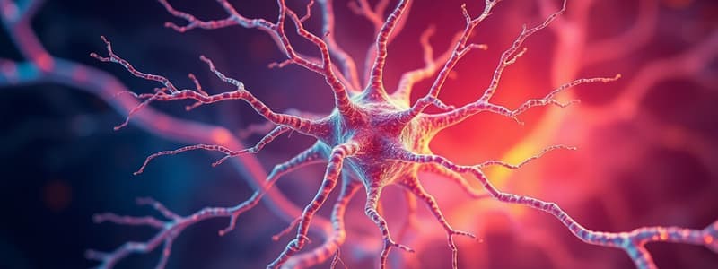
- Dendrites are tapered extensions of the cell body that collect information from other neurons. They contain cytoskeleton, mitochondria, and other important organelles.
- Soma (cell body) is the main part of the neuron, with a nucleus, Golgi apparatus, Nissl substance, cytoskeleton, and mitochondria. It synthesizes macromolecules and integrates electrical signals.
- Axon is a single, cylindrical extension that can be many centimeters long. It conducts information to other neurons and may be myelinated or unmyelinated. It contains cytoskeleton, mitochondria, and transport vesicles.
- Axon terminals (synaptic endings) are vesicle-filled structures that transmit information to other neurons. They typically connect to dendrites or the soma of other neurons, but other configurations exist.
Nervous System Organization
- The nervous system is divided into the somatic nervous system and the visceral nervous system.
- Somatic nervous system controls voluntary movements and conscious sensations.
- Visceral nervous system regulates autonomic functions (e.g., heart rate, breathing, digestion).
Brain Structure
- The cerebral hemispheres are the largest part of the brain and are covered by the cerebral cortex, which consists of six layers of gray matter.
- Gray matter is composed of cell bodies, while white matter contains myelinated axons.
- The cerebral cortex is divided into four lobes: frontal, parietal, occipital, and temporal.
- Frontal lobe is responsible for planning, movement, and complex thought.
- Parietal lobe processes sensory information, including touch, temperature, and pain.
- Occipital lobe is responsible for vision.
- Temporal lobe processes auditory information and memory.
- Internal capsule is a V-shaped structure consisting of axons connecting the cerebral cortex to other brain regions.
- Corpus callosum is a thick band of white matter connecting the two hemispheres and enabling communication between them.
Ventricular System
- The ventricular system is a network of cavities within the brain filled with cerebrospinal fluid (CSF).
- Lateral ventricles are the largest cavities and are located within the cerebral hemispheres.
- Third ventricle is located in the diencephalon and connects to the lateral ventricles through the interventricular foramina (of Monro).
- Fourth ventricle is located below the cerebellum and connects to the third ventricle through the cerebral aqueduct (of Sylvius).
- Choroid plexus is a network of modified ependymal cells that produce CSF.
Meninges
- Meninges are three layers of connective tissue that surround the brain and spinal cord.
- Dura mater, the outermost layer, is tough and fibrous and has two layers: periosteal layer and meningeal layer.
- Arachnoid mater, the middle layer, has a web-like structure and is separated from the dura mater by the subdural space.
- Pia mater, the innermost layer, is a delicate membrane that closely adheres to the brain and spinal cord.
- The subarachnoid space is located between the arachnoid mater and the pia mater and contains CSF.
Cerebral Vasculature
-
Blood supply to the brain comes from the internal carotid arteries and vertebral arteries.
-
Internal carotid arteries provide blood to the anterior circulation and branch into the anterior cerebral arteries (ACAs) and middle cerebral arteries (MCAs).
-
Anterior communicating artery connects the two ACAs.
-
Vertebral arteries provide blood to the posterior circulation.
-
Posterior communicating arteries connect the internal carotid arteries to the posterior cerebral arteries (PCAs), which are branches of the vertebral arteries.
-
The circle of Willis is a ring-shaped network of arteries that supplies blood to the brain and provides an alternative route for blood flow if one artery is blocked.### Arterial Supply to the Brain
-
The posterior cerebral cortex receives blood supply from the vertebrobasilar system, which starts with the vertebral arteries branching off from the subclavian arteries.
-
The vertebral arteries ascend through the foramen transversarium of the cervical vertebrae before entering the foramen magnum and joining to form the basilar artery.
-
The basilar artery runs along the ventral brainstem and gives rise to the posterior cerebral artery (PCA) at the midbrain level.
-
The PCA perfuses the inferior and medial temporal and occipital lobes and gives rise to the posterior communicating artery, which connects it to the internal carotid artery.
Venous Drainage of the Brain
- Superficial and deep veins of the cerebral hemispheres drain into the dural venous sinuses before reaching the internal jugular vein.
- The superior sagittal sinus runs along the superior edge of the falx cerebri, continuing posteriorly to drain into the transverse sinuses bilaterally.
- Each transverse sinus becomes the sigmoid sinus, which exits the jugular foramen to become the internal jugular vein.
- The inferior sagittal sinus, located along the inferior margin of the falx cerebri, joins the great vein of Galen to form the straight sinus.
- The confluence of the sinuses, where the straight sinus, superior sagittal sinus, and occipital sinus meet, drains into the transverse sinuses.
- The cavernous sinus, located on either side of the hypophysial fossa, receives drainage from other sinuses and the ophthalmic veins.
- The cavernous sinus drains via the superior petrosal sinus to the transverse sinus and the inferior petrosal sinuses to the internal jugular vein.
Superficial Veins
- Three main superficial veins drain the cerebral cortex:
- Superficial middle cerebral vein: runs parallel to the lateral fissure and drains the temporal lobe into the cavernous sinus.
- Superior Anastomotic Vein (of Trolard): travels perpendicularly to the superficial middle cerebral vein across the parietal lobe and drains into the superior sagittal sinus.
- Inferior Anastomotic Vein (of Labbé): travels perpendicularly to the superficial middle cerebral vein and drains the temporal lobe into the transverse sinus.
Deep Veins
- Deep veins drain into the great cerebral vein (of Galen) before reaching dural venous sinuses.
- The anterior cerebral vein and deep middle cerebral vein travel adjacent to the ACA and MCA.
- These veins join to form the basal vein (of Rosenthal), which encircles the lateral aspect of the midbrain.
- The internal cerebral veins, formed at the interventricular foramen by the septal and thalamostriate veins, join the basal veins to form the great cerebral vein (of Galen).
Thalamus
- The thalamus is a large, egg-shaped mass of gray matter derived from the diencephalon.
- It primarily serves as a relay center for pathways projecting to the cerebral cortex and also acts as a gatekeeper, regulating information transfer.
- Sensory, motor, limbic, and modulatory signals synapse within thalamic nuclei.
- The thalamus is adjacent to the interventricular foramen, hypothalamus, posterior limb of the internal capsule, body of the lateral ventricle, and the midbrain.
- The two thalamic masses are connected by the interthalamic adhesion.
Thalamic Nuclei
- Thalamic nuclei are classified into four groups based on their location relative to the internal medullary lamina: anterior, medial, lateral, and intralaminar.
- Thalamic nuclei are also divided into three functional classes: relay, intralaminar, and reticular.
- Relay nuclei, which have reciprocal excitatory connections with the cortex, comprise most of the thalamus.
- Relay nuclei are further classified as specific or nonspecific based on their projections to specific areas of the primary sensory and motor cortex or more diffuse cortical projections.
- The specific relay nuclei, primarily located in the lateral thalamus, are involved in sensory modalities (except olfaction) before reaching the primary cortical targets.
- Intralaminar nuclei are associated with arousal and attention.
- The reticular nucleus, located at the periphery of the thalamus, acts as an inhibitory gatekeeper.
- The thalamus receives vascular supply from penetrating branches of the ACA, anterior choroidal artery, lenticulostriate arteries of the MCA, and thalamoperforator arteries of the PCAs.
Studying That Suits You
Use AI to generate personalized quizzes and flashcards to suit your learning preferences.


