Podcast
Questions and Answers
Which of the following is the primary function of dendrites in a neuron?
Which of the following is the primary function of dendrites in a neuron?
- To insulate the axon and speed up signal transmission
- To conduct electrical impulses away from the cell body
- To release neurotransmitters into the synaptic cleft
- To receive messages from other neurons and transmit them to the cell body (correct)
What initiates an action potential in a neuron?
What initiates an action potential in a neuron?
- A strong enough signal that causes a change in the neuron's membrane potential (correct)
- The release of neurotransmitters from the axon terminals
- The opening of voltage-gated potassium channels
- The binding of neurotransmitters to receptors on the axon
Where are neurotransmitters released during synaptic transmission?
Where are neurotransmitters released during synaptic transmission?
- Into the synaptic cleft, the gap between neurons (correct)
- Into the myelin sheath surrounding the axon
- Into the cell body of the neuron
- Directly onto the dendrites of the next neuron
What is the primary role of the sodium-potassium pump in neuron function?
What is the primary role of the sodium-potassium pump in neuron function?
Which type of neuron is responsible for carrying impulses from the body's receptors to the brain or spinal cord?
Which type of neuron is responsible for carrying impulses from the body's receptors to the brain or spinal cord?
During which phase of the action potential do voltage-gated potassium channels open, allowing potassium ions to flow out of the cell?
During which phase of the action potential do voltage-gated potassium channels open, allowing potassium ions to flow out of the cell?
What is the absolute refractory period?
What is the absolute refractory period?
Which of the following describes the location of the spinal cord in relation to its vertebral protection?
Which of the following describes the location of the spinal cord in relation to its vertebral protection?
What is the cauda equina?
What is the cauda equina?
Which of the following describes the arrangement of meninges around the spinal cord, from superficial to deep?
Which of the following describes the arrangement of meninges around the spinal cord, from superficial to deep?
What is the primary function of the ascending tracts in the spinal cord?
What is the primary function of the ascending tracts in the spinal cord?
Which of the following best describes mixed nerves?
Which of the following best describes mixed nerves?
What is the primary function of the stretch (myotatic) reflex?
What is the primary function of the stretch (myotatic) reflex?
Which of the following is a function of the cervical plexus?
Which of the following is a function of the cervical plexus?
What is the significance of the corpus callosum in the brain?
What is the significance of the corpus callosum in the brain?
Which of the following is the function of the cerebellum?
Which of the following is the function of the cerebellum?
The forebrain divides into which two structures?
The forebrain divides into which two structures?
Which of the following structures contains the choroid plexus?
Which of the following structures contains the choroid plexus?
What is the primary function of the blood-brain barrier?
What is the primary function of the blood-brain barrier?
What is the general function of the medulla oblongata?
What is the general function of the medulla oblongata?
Lesions in the cerebellum are most likely to result in difficulties with which of the following?
Lesions in the cerebellum are most likely to result in difficulties with which of the following?
Which of the following is primarily regulated by the hypothalamus?
Which of the following is primarily regulated by the hypothalamus?
What is the function of the hippocampus?
What is the function of the hippocampus?
During which stage of sleep do sleep spindles occur?
During which stage of sleep do sleep spindles occur?
What is the state of the body during REM sleep?
What is the state of the body during REM sleep?
Which area of the brain is responsible for formulating phrases according to learned rules of grammar when planning to speak?
Which area of the brain is responsible for formulating phrases according to learned rules of grammar when planning to speak?
What type of aphasia is characterized by slow speech and difficulty in choosing words?
What type of aphasia is characterized by slow speech and difficulty in choosing words?
Damage to which cranial nerve would most likely result in impaired sense of smell?
Damage to which cranial nerve would most likely result in impaired sense of smell?
Which cranial nerve is responsible for controlling muscles that turn the eyeball up, down, and medially, as well as controlling the iris, lens, and upper eyelid?
Which cranial nerve is responsible for controlling muscles that turn the eyeball up, down, and medially, as well as controlling the iris, lens, and upper eyelid?
Flashcards
Cell Body (Soma)
Cell Body (Soma)
The metabolic center and contains the nucleus of the neuron.
Dendrites
Dendrites
Branch-like structures receiving messages from other neurons, transmitting them to the cell body.
Axon
Axon
A long, slender projection that conducts electrical impulses away from the cell body.
Action potentials
Action potentials
Signup and view all the flashcards
Synapse
Synapse
Signup and view all the flashcards
Neurotransmitters
Neurotransmitters
Signup and view all the flashcards
Sensory Neurons
Sensory Neurons
Signup and view all the flashcards
Motor Neurons
Motor Neurons
Signup and view all the flashcards
Interneurons
Interneurons
Signup and view all the flashcards
Resting Membrane Potential
Resting Membrane Potential
Signup and view all the flashcards
Stimulus
Stimulus
Signup and view all the flashcards
Sodium Influx
Sodium Influx
Signup and view all the flashcards
Action Potential Peak
Action Potential Peak
Signup and view all the flashcards
Potassium Efflux
Potassium Efflux
Signup and view all the flashcards
Overshoot
Overshoot
Signup and view all the flashcards
Absolute Refractory Period
Absolute Refractory Period
Signup and view all the flashcards
Relative Refractory Period
Relative Refractory Period
Signup and view all the flashcards
Ion Redistribution
Ion Redistribution
Signup and view all the flashcards
Conduction
Conduction
Signup and view all the flashcards
Neural Integration
Neural Integration
Signup and view all the flashcards
Locomotion
Locomotion
Signup and view all the flashcards
Reflexes
Reflexes
Signup and view all the flashcards
Medullary cone
Medullary cone
Signup and view all the flashcards
Cauda equina
Cauda equina
Signup and view all the flashcards
Meninges
Meninges
Signup and view all the flashcards
Gray Matter
Gray Matter
Signup and view all the flashcards
White Matter
White Matter
Signup and view all the flashcards
Ascending Tracts
Ascending Tracts
Signup and view all the flashcards
Descending Tracts
Descending Tracts
Signup and view all the flashcards
Ganglion
Ganglion
Signup and view all the flashcards
Study Notes
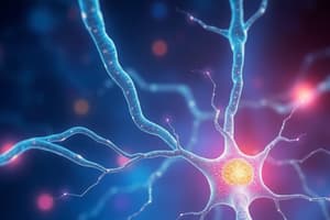
Neuron Structure
- Neurons have dendrites, which are branch-like structures that receive messages from other neurons and transmit them to the cell body.
- The cell body (soma) contains the nucleus and is the metabolic center of the neuron.
- The axon is a long, slender projection that conducts electrical impulses away from the cell body to other neurons, muscles, or glands.
Action Potential
- Neurons use electrical impulses called action potentials to communicate.
- When a neuron receives a strong enough signal, it triggers an action potential that travels down the axon to the axon terminals.
Synapse
- When the action potential reaches the axon terminals, neurotransmitters are released into the synaptic cleft (the gap between neurons).
- These neurotransmitters bind to receptors on the dendrites of the next neuron, continuing the signal transmission.
- Different neurotransmitters have varying effects, such as exciting or inhibiting the next neuron, coordinating various functions.
Types of neurons
- Sensory neurons carry impulses from the body's receptors to the brain or spinal cord.
- Motor neurons carry impulses from the brain or spinal cord to the body's muscles, organs, and glands.
- Interneurons connect sensory neurons to motor neurons and are the most common type of neuron.
Resting Membrane Potential
- When a neuron is at rest, it has a stable negative charge inside the cell (around -70 mV) due to the distribution of ions (mainly sodium (Na+) and potassium (K+)) across the membrane.
Depolarization
- A stimulus causes the neuron to reach its threshold potential (around -55 mV), triggering the opening of voltage-gated sodium channels.
- Sodium ions rush into the cell, causing the inside to become more positive
Action Potential Peak
- Membrane potential reaches a peak (around +30 mV), and the sodium channels begin to close.
Repolarization
- Voltage-gated potassium channels open, allowing potassium ions to flow out of the cell, restoring the negative charge inside.
Hyperpolarization
- Sometimes the membrane potential becomes more negative than the resting potential before stabilizing.
Refractory Period
- During the absolute refractory period, the neuron cannot fire another action potential.
- During the relative refractory period, the neuron can fire again but needs a stronger stimulus.
- The sodium-potassium pump helps restore the original ion distribution, returning the neuron to its resting state.
Spinal Cord Functions
- Conduction: Nerve fibers conduct information up and down the cord, connecting different levels of the trunk to each other and the brain.
- Neural integration: Input from multiple sources is integrated, and output is executed.
- Locomotion: Repetitive, coordinated contractions of muscle groups occur in the limbs.
- Reflexes: Involuntary, stereotyped responses to stimuli.
Spinal Cord
- The spinal cord ends at L1.
- There are 31 pairs of spinal nerves.
- Longitudinal grooves are on anterior and posterior sides.
- There are anterior median fissure and posterior median sulcus.
- Medullary cone (conus medullaris) tapers the cord to a point inferior to lumbar enlargement.
- Cauda equina is a bundle of nerve roots that occupy the vertebral canal from L2 to S5.
- Meninges are three fibrous connective tissue membranes that enclose the brain and spinal cord.
- From superficial to deep: dura mater, arachnoid mater, and pia mater.
- Cerebrospinal fluid fills the space between the arachnoid and pia mater.
Spinal Cord Defects and Anatomy
- Spina bifida: Congenital defect where one or more vertebrae fail to form a complete vertebral arch.
- Folic acid (a B vitamin) as part of a healthy diet for all women of childbearing age reduces risk.
- Gray matter consists of neuron cell bodies with little myelin and is butterfly-shaped.
- It is the site of information processing and synaptic integration.
- White matter is made of abundantly myelinated axons.
- It carries signals from one part of the CNS to another.
Horns of the Spinal cord
- Posterior (dorsal) root of the spinal nerve carries only sensory fibers.
- Anterior (ventral) root of the spinal nerve carries only motor fibers.
- The gray commissure connects right and left sides.
- A central canal lined with ependymal cells with CSF is punctured.
- The lateral horn is visible from T2 through L1.
- Contains neurons of the sympathetic nervous system.
Tracts of the Spinal Cord
- Ascending tracts carry sensory information up the spinal cord.
- Descending tracts carry motor information down the spinal cord.
- All nerve fibers in a given tract have a similar origin, destination, and function.
- Upper motor neurons originate in the cerebral cortex or brainstem and terminate on a lower motor neuron.
- Lower motor neurons are located in the brainstem or spinal cord.
Nerves of the Spinal cord
- Mixed nerves contain both afferent (sensory) and efferent (motor) fibers.
- Nerves of the peripheral nervous system are ensheathed in Schwann cells.
- Myelin sheath surrounds the axon.
- Sensory (afferent) nerves carry signals from sensory receptors to the CNS.
- Motor (efferent) nerves carry signals from the CNS to muscles and glands.
- Mixed nerves consist of both afferent and efferent fibers and conduct signals in two directions.
- Ganglion is a cluster of neurosomas outside the CNS.
- There are 31 pairs of spinal nerves (mixed nerves).
Plexuses of the Spinal Cord
- Cervical plexus is in the neck, C1 to C5.
- Supplies neck and phrenic nerve to the diaphragm.
- Lumbar plexus is in the lower back, L1 to L4.
- Supplies abdominal wall, anterior thigh, and genitalia.
- Sciatica is sharp pain that travels from the gluteal region along the posterior side of the thigh and leg to ankle.
- Reflexes are quick, involuntary, stereotyped reactions of glands or muscle to stimulation.
- Stretch (myotatic) reflex occurs when a muscle is stretched, and it "fights back" and contracts, maintaining increased tonus and making it stiffer than unstretched muscle.
- It helps maintain equilibrium and posture.
- Head starts to tip forward as you fall asleep.
- Conduction involves nerve fibers that conduct information up and down the cord, connecting different levels of the trunk.
- Neural integration is the input from multiple sources, integrated, and executed output.
- Locomotion refers to repetitive, coordinated contractions of several muscle groups in the limbs.
- Reflexes are involuntary stereotyped responses to stimuli.
Brain Anatomy
- Rostral: Toward the forehead.
- Caudal: Toward the spinal cord.
- Cerebrum is 83% of brain volume, consists of cerebral hemispheres, gyri and sulci, longitudinal fissure, and corpus callosum.
- Cerebellum contains 50% of the neurons and is the second largest brain region in the posterior cranial fossa.
- Brainstem is the portion of the brain remaining if the cerebrum and cerebellum are removed; includes the diencephalon, midbrain, pons, and medulla oblongata.
- Longitudinal fissure separates cerebral hemispheres.
- Gyri are thick folds.
- Sulci are shallow grooves.
- Corpus callosum is a thick nerve bundle at the bottom of the longitudinal fissure that connects hemispheres.
- The nervous system develops from the ectoderm (outermost tissue layer of the embryo).
- Gray matter is the seat of neuron cell bodies, dendrites, and synapses.
- White matter is bundles of axons.
Brain Development
- Forebrain: (prosencephalon)
- Midbrain: (mesencephalon)
- Hindbrain: (rhombencephalon)
- Forebrain divides into two
- Telencephalon: becomes cerebral hemispheres
- Diencephalon: has optic vesicles that become the retina of the eye
- Midbrain remains undivided: Mesencephalon
- Hindbrain divides into two vesicles
- Metencephalon
- Myelencephalon
Protection of the Brain
- Meninges are three connective tissue membranes that envelop the brain.
- Arachnoid mater is a transparent membrane over the brain surface.
- The subarachnoid space separates it from the pia mater below.
- The subdural space separates it from the dura mater above in some places.
- Pia mater is a very thin membrane that follows the contours of the brain, even dipping into sulci; not usually visible without a microscope.
- Meningitis is the inflammation of the meninges and is a serious disease of infancy and childhood.
- Ventricles are four internal chambers within the brain.
- Choroid plexus is a spongy mass of blood capillaries on the floor of each ventricle.
- CSF is reabsorbed by arachnoid villi, which are cauliflower-shaped extensions of the arachnoid meninx.
- These protrude through the dura mater into the superior sagittal sinus.
- CSF penetrates the walls of the villi and mixes with the blood in the sinus.
Importance of Oxygen and Glucose in the Brain
- Neurons have a high demand for ATP and thus, oxygen and glucose. Therefore, a constant supply of blood is critical to the nervous system.
- The blood-brain barrier protects blood capillaries throughout brain tissue.
- It consists of tight junctions between endothelial cells that form the capillary walls.
- The blood barrier system is highly permeable to water, glucose, and lipid-soluble substances
- Oxygen, carbon dioxide, alcohol, caffeine, nicotine, and anesthetics.
- The embryonic myelencephalon becomes the medulla oblongata.
- The metencephalon develops into the pons and cerebellum.
- The pons is an anterior bulge in the brainstem, rostral to the medulla.
- Cerebral peduncles connect the cerebellum to the pons and midbrain.
- The mesencephalon becomes one mature brain structure, the midbrain.
Components of the Brain
- The largest part of the hindbrain and the second largest part of the brain, as a whole, is the Cerebellum
- Consists of right and left cerebellar hemispheres connected by the vermis
- The forebrain consists of two parts: the Diencephalon and the Telencephalon
- Diencephalon encloses the third ventricle and is the most rostral part of the brainstem and has three major embryonic derivatives
- Thalamus
- Hypothalamus
- Epithalamus
- Telencephalon develops chiefly into the cerebrum
- The thalamus is an ovoid mass on each side of the brain perched at the superior end of the brainstem beneath the cerebral hemispheres
Hypothalamus and Lobes of the Brain
- The hypothalamus forms part of the walls and floor of the third ventricle.
- It plays an essential role in the homeostatic regulation of all body systems.
- It plays a role in food and water intake, and emotional behavior.
- The frontal lobe is for voluntary motor functions.
- The parietal lobe receives and integrates general sensory information, taste, and some visual processing.
- The occipital lobe is the primary visual center of the brain.
- The temporal lobe is areas for hearing, smell, learning, memory, and some aspects of vision and emotion.
Limbic System and Sleep
- The limbic system is an important center of emotion and learning.
- Hippocampus is in the medial temporal lobe and is important for memory.
- The Amygdala is immediately rostral to the hippocampus and is important for emotion
- Sleep occurs in cycles called circadian rhythms, and is a temporary state of unconsciousness from which one can awaken when stimulated.
- Stage 1: Feel drowsy/close eyes/begin to relax, feel drifting sensation/easily awakened sleep, alpha waves dominate
- Stage 2 transition into light sleep, and EEG declines in frequency, sleep spindles occur
- Stage 3 is moderate to deep sleep, 20 minutes after initial stage. theta and delta waves appear, muscles relax and vitals fall
- Stage 4 Called slow-wave sleep dominated by low-frequency/high-amplitude delta waves, muscles completely relaxed, its more difficult to awaken person
REM Sleep and Amygdala
- During rapid eye movement (REM) sleep, the eyes oscillate back and forth.
- During REM sleep, muscles are paralyzed, and body temperature, blood pressure, and heart rate and respiratory rate increase.
- The suprachiasmatic nucleus (SCN) is an important control center for sleep.
- Amygdala receives input from sensory systems and plays a role in food intake, sexual behavior, and attention to novel stimuli.
- Primary sensory cortex is where sensory input is first received and consciousness is reached of a stimulus
- Primary visual cortex is bordered by visual association area
Vision and Hearing
- Visual primary cortex is located in the far posterior region of the occipital lobe.
- A significant portion of the inferior temporal lobe deals with facial recognition and other familiar objects.
- The primary auditory cortex is located in the superior region of the temporal lobe and insula and processes spoken words.
- Signals for balance and sense of motion project mainly to the cerebellum and several brainstem nuclei concerned with head.
Taste and Smell
- Gustatory (taste) signals are received by the primary gustatory cortex at the inferior end of the postcentral gyrus of the parietal lobe and the anterior region of the insula.
- Olfactory (smell) signals are received by the primary olfactory cortex in the medial surface of the temporal lobe and the inferior surface of the frontal lobe.
- Sensory homunculus diagram of the primary somesthetic cortex. It resembles an upside-down sensory map of the contralateral side of the body.
Wernicke and Broca areas
- Motor homunculus has a distorted look because the amount of cortex devoted to a given body region is proportional to the number of muscles and motor units
- Wernicke area permits the recognition of spoken and written language and creates a plan of speech
- When intent is to speak, Wernicke area formulates phrases according to learned rules of grammar
- Broca area generates the motor program for the muscles of the larynx, tongue, cheeks, and lips and transmits program to primary motor cortex for commands to the lower motor neurons
- Aphasia any language deficit from lesions in the same hemisphere (usually left) containing the Wernicke and Broca Area Nonfluent (Broca) aphasia
- Lesion in Broca area
- slow speech, difficulty in choosing words
Aphasia and Cranial Nerves
- Fluent (Wernicke) aphasia
- Lesion in Wernicke’s area
- speech normal but senseless jargon filled
- Cannot comprehend written and spoken words
- Olfactory Nerve (I) governs the sense of smell and damage to which causes impaired sense of smell.
- Optic Nerve (II) provides vision and damage to it causes blindness in part or all of visual field.
- Oculomotor Nerve (III) controls muscles that turn the eyeball up, down, and medially, as well as controlling the iris, lens, and upper eyelid. Damage to which causes drooping eyelid, dilated pupil, double vision, difficulty focusing, and inability to move eye in certain directions.
- Trochlear Nerve (IV) governs eye movement (superior oblique muscle) and damage of eye Inferolaterally causes double vision and inability to rotate eye.
- Trigeminal Nerve (V) is the largest of the cranial nerves and the most important sensory nerve of the face.
- Abducens Nerve (VI) provides eye movement (lateral rectus m.).
More Cranial Nerves
- Facial Nerve (VII) is the major motor nerve of facial muscles and sensory nerve for taste on the anterior two-thirds of tongue. Damage causes the sagging of facial muscles and disturbed sense of taste (no sweet and salty).
- Vestibulocochlear Nerve (VIII) governs hearing and equilibrium and damage to it produces deafness, dizziness, nausea, loss of balance. Glossopharyngeal Nerve (IX) is involved in swallowing, salivation, gagging, control of BP and respiration, and sensation of posterior one-third of the tongue. Damage for its impairment results in the loss of bitter and sour taste and impaired swallowing.
- Vagus Nerve (X) has the most extensive distribution of any cranial nerve and plays a major role in the control of cardiac, pulmonary, digestive, and urinary function. Also includes: swallowing, speech, and regulation of viscera.
- Accessory Nerve (XI) controls swallowing/head, neck, and shoulder movement and damage causes impaired head, neck, shoulder movement; head turns toward injured side.
- Hypoglossal Nerve (XII) controls tongue movements for speech, food manipulation, and swallowing.
Studying That Suits You
Use AI to generate personalized quizzes and flashcards to suit your learning preferences.